. 2016 Dec 22:6:39668.
doi: 10.1038/srep39668.

The differentiated airway epithelium infected by influenza viruses maintains the barrier function despite a dramatic loss of ciliated cells

Affiliations

The differentiated airway epithelium infected by influenza viruses maintains the barrier function despite a dramatic loss of ciliated cells

Nai-Huei Wu et al. Sci Rep. .

Abstract

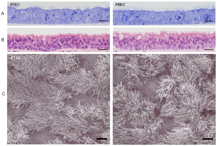
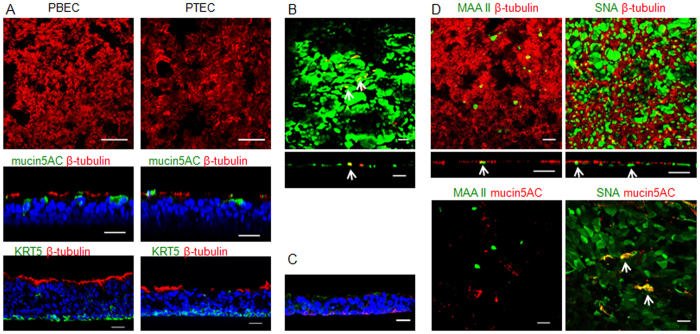
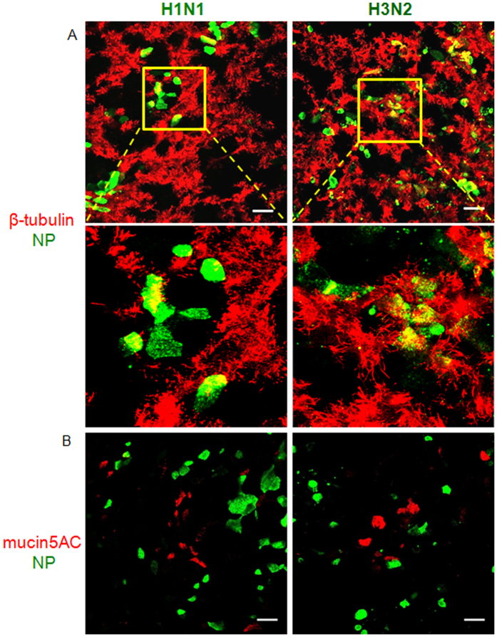
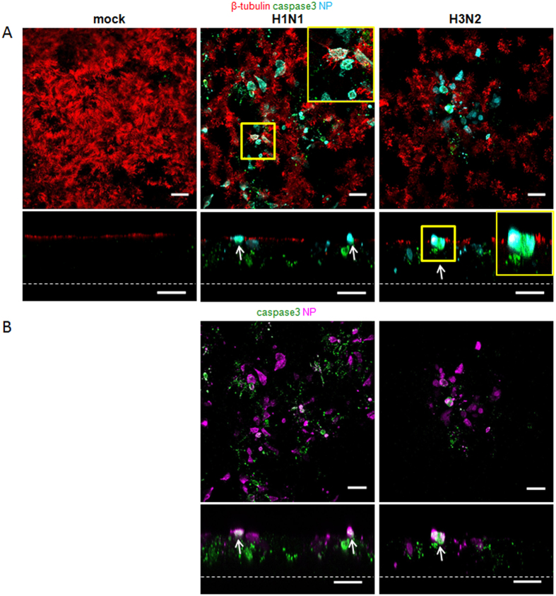
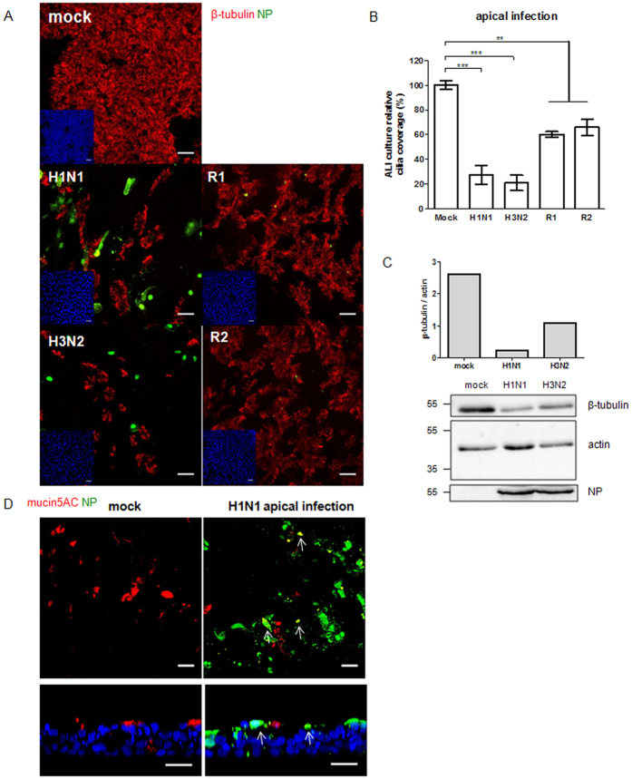
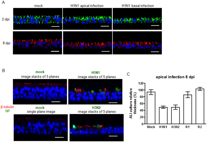
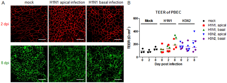
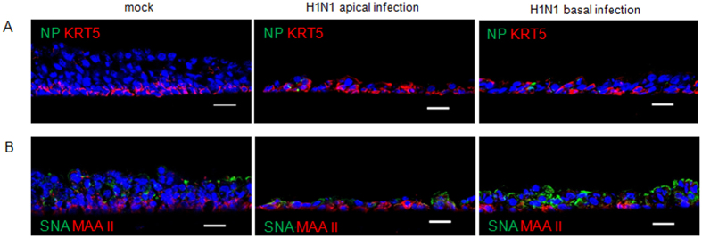
Virus-host interactions in the respiratory epithelium during long term influenza virus infection are not well characterized. Therefore, we developed an air-liquid interface culture system for differentiated porcine respiratory epithelial cells to study the effect of virus-induced cellular damage. In our well-differentiated cells, α2,6-linked sialic acid is predominantly expressed on the apical surface and the basal cells mainly express α2,3-linked sialic acid. During the whole infection period, release of infectious virus was maintained at a high titre for more than seven days. The infected epithelial cells were subject to apoptosis resulting in the loss of ciliated cells together with a thinner thickness. Nevertheless, the airway epithelium maintained trans-epithelial electrical resistance and retained its barrier function. The loss of ciliated cells was compensated by the cells which contained the KRT5 basal cell marker but were not yet differentiated into ciliated cells. These specialized cells showed an increase of α2,3-linked sialic acid on the apical surface. In sum, our results help to explain the localized infection of the airway epithelium by influenza viruses. The impairment of mucociliary clearance in the epithelial cells provides an explanation why prior viral infection renders the host more susceptible to secondary co-infection by another pathogen.


Figures

Figure 1
Figure 1. Morphological examination of porcine well-differentiated airway epithelial cell cultures.
(A) PTEC and PBEC cultures were grown under ALI conditions for more than 4 weeks. The semi-thin sections followed by toluidine blue staining were performed. (B) Epithelia from porcine trachea and primary bronchus were collected, followed by histological sectioning and H&E staining for the morphological comparison. The histological examination was evaluated by light microscopy and the representative histological sections (40x magnification) are shown. (C) The micrograph of the scanning electron microscopy illustrates the apical surface of PTEC and PBEC. The ciliated epithelial cells are the predominant cell type. Scale bars, 20 μm (A,B), 5 μm (C).
Figure 2
Figure 2. Characterization of porcine well-differentiated airway epithelial cell cultures.
PTEC and PBEC were cultured under ALI conditions for at least 4 weeks and analyzed by immunofluorescence. (A) Immunofluorescent staining of whole-filter cultures (top and middle panels) or cryosections (lower panels) of PTEC and PBEC. The cilia are stained in red by using anti-β-tubulin antibody (top panels in horizontal sections and middle panels in vertical sections). More than half of the PTEC and PBEC surface was covered by cilia. The positive staining of mucus (green, mucin 5AC monoclonal antibody, middle panels in vertical sections) indicated the presence of mucus-producing cells. The basal cells were stained by antibody against cytokeratin 5 (KRT 5, green, lower panels) and were located above the filter support. (B) Detection of sialic acid on the apical surface of wdPBEC. Antibodies against SNA (green) and MAA II (red) were used to recognize α2,6- and α2,3-linked sialic acids, respectively. The images are shown in horizontal (top) or vertical (lower) sections. (C) Detection of sialic acids on basal cells in wdPBEC. Cryosections of PBEC cultures were stained by SNA (green) and MAA II (red). (D) The distribution of sialic acids in wdPBEC. PBEC stained for SNA or MAA II (green) were co-stained for the presence of cilia or mucus (red). The images are shown in vertical (middle panels) or horizontal (others) sections. The pseudo-colour was applied in red (mucin 5AC) and green (MAA II) by using LAS AF Lite software for image comparison (lower left panel). The arrows show co-localization. Scale bars, 50 μm (A, top), 25 μm (others).
Figure 3
Figure 3. Replication of influenza A viruses in porcine well-differentiated airway epithelial cell cultures.
(A) Replication kinetics of R1 and R2 viruses in wdPBEC. (B) Replication kinetics of swIAV in wdPBEC. WdPBEC were inoculated with IAV from apical (left panels) or basolateral (right panels) sides at an MOI of 0.25. Viruses released from the apical side were harvested at different time points and titrated by focus-forming assay in MDCK cells. The results were shown as means ± SEM of nine PBECs from three independent donors (swIAV) or six PBECs from two donors (R1 and R2). Each sample was processed with two technical replicates. It should be noted that some error bars are too small to be printed. Statistical analysis was performed with two-tailed unpaired Student’s t-test (***P < 0.001, **P < 0.01, *P < 0.05).
Figure 4
Figure 4. Differences in tropism of swIAV to distinct types of differentiated airway cells.
WdPBECs were infected with swIAV H1N1 or H3N2 from the apical surface at an MOI of 0.25 and fixed at 1 dpi, followed by immunofluorescent staining to detect viral nucleoprotein (green), cilia (A, red) and mucus (B, red). Magnifications of squared areas are presented in the lower panels of A. Scale bars, 25 μm.
Figure 5
Figure 5. Immunofluorescent staining of apoptotic cells in PBEC at 2 days post infection.
WdPBECs were infected by swIAV from the apical surface at an MOI of 0.25 and fixed at 2 dpi. The cilia (red) and the viral nucleoprotein (A, cyan or B, magenta) were stained. The apoptotic cells were detected by visualizing cleaved caspase-3 (green). Confocal images are shown in horizontal (top panels) or vertical (lower panels) sections. Magnifications of squared areas are presented on the top-right corner (A). The pseudo-colour was applied in magenta (viral nucleoprotein) by using LAS AF Lite software for image comparison (B). The arrows show co-localization, and the dashed lines indicate the location of the supporting membrane. Scale bars: 25 μm.
Figure 6
Figure 6. Immunofluorescent staining of porcine well-differentiated airway epithelial cells at 8 days post infection.
WdPBECs were inoculated with IAV from the apical side at an MOI of 0.25 and fixed at 8 dpi. (A) PBEC cultures were stained for viral nucleoprotein (green) and cilia (red). (B) Quantification of the ciliated area at 8 dpi. Results are shown as percentages (means ± SEM) compared to mock-infected cultures. For each infection, six PBECs from three independent donors were measured, and three fields per culture were evaluated as technical replicates. (C) Western blot analysis of β-tubulin expression level in PBECs after swIAV infection. The relative expression level of β-tubulin was normalized to actin expression. The viral NP could be detected in the infected culture. (D) Immunofluorescent staining for viral nucleoprotein (green) and mucin (red). The nuclei were stained by DAPI (blue) (A and D). The arrows show co-localization. Scale bars, 25 μm.
Figure 7
Figure 7. Decreased thickness of porcine well-differentiated airway epithelial cell cultures after IAV infection.
WdPBECs were inoculated by IAV from the apical (middle panels of A; B and C) or basolateral side (right panels of A) at an MOI of 0.25. (A) wdPBEC were inoculated with swIAV H1N1 and fixed at 2 or 8 dpi, followed by immunofluorescent staining with antibody against the adherens junction protein β-catenin (green at 2 dpi; red at 8 dpi). Confocal images are shown in vertical sections. (B) WdPBECs were fixed at 8 dpi and stained for viral nucleoprotein (green) and cilia (red) (vertical sections). To measure the thickness accurately, the vertical image stacks of 5 planes (distance of 1.0 μm per plane) were merged. It should be noted that the epithelium forms a pseudostratified layer in the single plane image. The nuclei were stained by DAPI (blue) (A&B). (C) Quantification of wdPBEC thickness at 8 dpi. Results are shown as percentages (means ± SEM) compared to mock-infected ALI cultures. For each infection, numbers of six PBECs from three independent donors were measured. Additionally, three fields per culture were evaluated as technical replicates. Scale bars, 25 μm.
Figure 8
Figure 8. Porcine well-differentiated airway epithelial cell cultures preserve tight junctions after swIAV infection.
(A) WdPBECs were inoculated with swIAV H1N1 from the apical (middle panels) or basolateral (right panels) side at an MOI of 0.25. ALI cultures were fixed at 2 dpi (top panels) and 8 dpi (lower panels), followed by staining with anti-ZO-1 antibody (red in top panels; green in lower panels) to detect tight junction. Scale bars, 25 μm. (B) WdPBECs were inoculated by swIAV from the apical or basolateral side. The trans-epithelial electrical resistance (TEER) values of mock-infected and swIAV-infected PBEC were determined at the indicated time points. The results are shown as three PBECs from three independent donors. Each sample was performed with 3 technical replicates.
Figure 9
Figure 9. Sialic acid expression on PBEC after swIAV infection.
WdPBECs were inoculated by swIAV H1N1 from the apical (middle panels) or basolateral (right panels) side at an MOI of 0.25. Cryosections were prepared at 8 dpi. (A) Immunofluorescent staining for KRT 5 (red, basal cells) and viral nucleoprotein (green). (B) Immunofluorescent staining to detect α2,3- and α2,6-linked sialic acid using MAA II and SNA lectins, respectively. The nuclei were stained by DAPI (blue) (A&B). Scale bars: 25 μm.

Similar articles

Cited by

References

    1. Tugizov S. M., Herrera R. & Palefsky J. M. Epstein-Barr virus transcytosis through polarized oral epithelial cells. J Virol 87, 8179–8194, doi: 10.1128/jvi.00443-13 (2013). - DOI - PMC - PubMed
    1. Ludlow M., Allen I. & Schneider-Schaulies J. Systemic spread of measles virus: overcoming the epithelial and endothelial barriers. Thrombosis and haemostasis 102, 1050–1056, doi: 10.1160/th09-03-0202 (2009). - DOI - PubMed
    1. Lemon K. et al.. Early target cells of measles virus after aerosol infection of non-human primates. PLoS Pathog 7, e1001263, doi: 10.1371/journal.ppat.1001263 (2011). - DOI - PMC - PubMed
    1. Munkholm M. & Mortensen J. Mucociliary clearance: pathophysiological aspects. Clinical physiology and functional imaging 34, 171–177, doi: 10.1111/cpf.12085 (2014). - DOI - PubMed
    1. Ganesan S., Comstock A. T. & Sajjan U. S. Barrier function of airway tract epithelium. Tissue barriers 1, e24997, doi: 10.4161/tisb.24997 (2013). - DOI - PMC - PubMed

Publication types

MeSH terms