

Bmi1-positive cells in the lingual epithelium could serve as cancer stem cells in tongue cancer
- PMID: 28004815
- PMCID: PMC5177893
- DOI: 10.1038/srep39386
Bmi1-positive cells in the lingual epithelium could serve as cancer stem cells in tongue cancer
Abstract
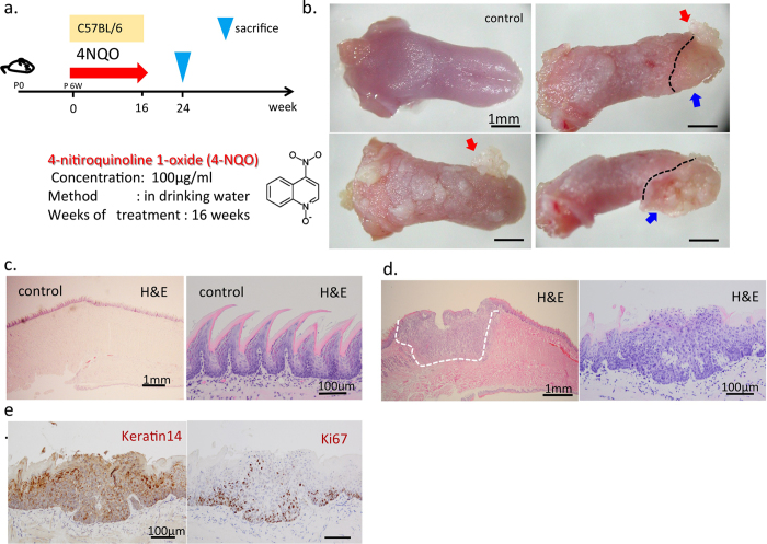
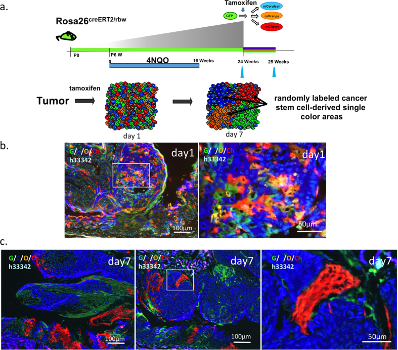
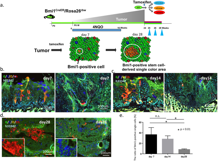
We recently reported that the polycomb complex protein Bmi1 is a marker for lingual epithelial stem cells (LESCs), which are involved in the long-term maintenance of lingual epithelial tissue in the physiological state. However, the precise role of LESCs in generating tongue tumors and Bmi1-positive cell lineage dynamics in tongue cancers are unclear. Here, using a mouse model of chemically (4-nitroquinoline-1-oxide: 4-NQO) induced tongue cancer and the multicolor lineage tracing method, we found that each unit of the tumor was generated by a single cell and that the assembly of such cells formed a polyclonal tumor. Although many Bmi1-positive cells within the tongue cancer specimens failed to proliferate, some proliferated continuously and supplied tumor cells to the surrounding area. This process eventually led to the formation of areas derived from single cells after 1-3 months, as determined using the multicolor lineage tracing method, indicating that such cells could serve as cancer stem cells. These results indicate that LESCs could serve as the origin for tongue cancer and that cancer stem cells are present in tongue tumors.
Figures
Similar articles
-
Intestinal cancer stem cells marked by Bmi1 or Lgr5 expression contribute to tumor propagation via clonal expansion.Sci Rep. 2017 Feb 8;7:41838. doi: 10.1038/srep41838. Sci Rep. 2017. PMID: 28176811 Free PMC article.
-
Establishment of a novel lingual organoid culture system: generation of organoids having mature keratinized epithelium from adult epithelial stem cells.Sci Rep. 2013 Nov 15;3:3224. doi: 10.1038/srep03224. Sci Rep. 2013. PMID: 24232854 Free PMC article.
-
Identification of stem cells that maintain and regenerate lingual keratinized epithelial cells.Nat Cell Biol. 2013 May;15(5):511-8. doi: 10.1038/ncb2719. Epub 2013 Apr 7. Nat Cell Biol. 2013. PMID: 23563490
-
Lingual Epithelial Stem Cells and Organoid Culture of Them.Int J Mol Sci. 2016 Jan 28;17(2):168. doi: 10.3390/ijms17020168. Int J Mol Sci. 2016. PMID: 26828484 Free PMC article. Review.
-
Role of BMI1, a stem cell factor, in cancer recurrence and chemoresistance: preclinical and clinical evidences.Stem Cells. 2012 Mar;30(3):372-8. doi: 10.1002/stem.1035. Stem Cells. 2012. PMID: 22252887 Review.
Cited by
-
Mechanisms of Cisplatin Resistance in HPV Negative Head and Neck Squamous Cell Carcinomas.Cells. 2022 Feb 5;11(3):561. doi: 10.3390/cells11030561. Cells. 2022. PMID: 35159370 Free PMC article. Review.
-
HPV+ve/-ve oral-tongue cancer stem cells: A potential target for relapse-free therapy.Transl Oncol. 2021 Jan;14(1):100919. doi: 10.1016/j.tranon.2020.100919. Epub 2020 Oct 24. Transl Oncol. 2021. PMID: 33129107 Free PMC article. Review.
-
Emerging Role of MicroRNA-200 Family in Dentistry.Noncoding RNA. 2021 Jun 11;7(2):35. doi: 10.3390/ncrna7020035. Noncoding RNA. 2021. PMID: 34208375 Free PMC article. Review.
-
Spatial transcriptomics reveals molecular cues underlying the site specificity of the adult mouse oral mucosa and its stem cell niches.Stem Cell Reports. 2024 Dec 10;19(12):1706-1719. doi: 10.1016/j.stemcr.2024.10.007. Epub 2024 Nov 27. Stem Cell Reports. 2024. PMID: 39547226 Free PMC article.
-
Polycomb Repressive Complex 1 Controls Maintenance of Fungiform Papillae by Repressing Sonic Hedgehog Expression.Cell Rep. 2019 Jul 2;28(1):257-266.e5. doi: 10.1016/j.celrep.2019.06.011. Cell Rep. 2019. PMID: 31269445 Free PMC article.
References
-
- Kamangar F., Dores G. M. & Anderson W. F. Patterns of cancer incidence, mortality, and prevalence across five continents: defining priorities to reduce cancer disparities in different geographic regions of the world. J Clin Oncol 24, 2137–2150 (2006). - PubMed
-
- Leemans C. R., Braakhuis B. J. & Brakenhoff R. H. The molecular biology of head and neck cancer. Nat Rev Cancer 11, 9–22 (2011). - PubMed
-
- Silva P. et al.. Clinical and biological factors affecting response to radiotherapy in patients with head and neck cancer: a review. Clin Otolaryngol 32, 337–345 (2007). - PubMed
-
- Petersen P. E. Oral cancer prevention and control–the approach of the World Health Organization. Oral Oncol 45, 454–460 (2009). - PubMed
-
- Ganly I., Patel S. & Shah J. Early stage squamous cell cancer of the oral tongue--clinicopathologic features affecting outcome. Cancer 118, 101–111 (2012). - PubMed
Publication types
MeSH terms
Substances
LinkOut - more resources
Full Text Sources
Other Literature Sources
Molecular Biology Databases
