. 2016 Dec 22:6:39735.
doi: 10.1038/srep39735.

The citrus flavonoid naringenin confers protection in a murine endotoxaemia model through AMPK-ATF3-dependent negative regulation of the TLR4 signalling pathway

Affiliations

The citrus flavonoid naringenin confers protection in a murine endotoxaemia model through AMPK-ATF3-dependent negative regulation of the TLR4 signalling pathway

Xin Liu et al. Sci Rep. .

Abstract

Excessive activation of the TLR4 signalling pathway is critical for inflammation-associated disorders, while negative regulators play key roles in restraining TLR4 from over-activation. Naringenin is a citrus flavonoid with remarkable anti-inflammatory activity, but the mechanisms underlying its inhibition of LPS/TLR4 signalling are less clear. This study investigated the molecular targets and therapeutic effects of naringenin in vitro and in vivo. In LPS-stimulated murine macrophages, naringenin suppressed the expression of TNF-α, IL-6, TLR4, inducible NO synthase (iNOS), cyclo-oxygenase-2 (COX2) and NADPH oxidase-2 (NOX2). Naringenin also inhibited NF-κB and mitogen-activated protein kinase (MAPK) activation. However, it did not affect the IRF3 signalling pathway or interferon production, which upregulate activating transcription factor 3 (ATF3), an inducible negative regulator of TLR4 signalling. Naringenin was demonstrated to directly increase ATF3 expression. Inhibition of AMPK and its upstream calcium-dependent signalling reduced ATF3 expression and dampened the anti-inflammatory activity of naringenin. In murine endotoxaemia models, naringenin ameliorated pro-inflammatory reactions and improved survival. Furthermore, it induced AMPK activation in lung tissues, which was required for ATF3 upregulation and the enhanced anti-inflammatory activity. Overall, this study reveals a novel mechanism of naringenin through AMPK-ATF3-dependent negative regulation of the LPS/TLR4 signalling pathway, which thereby confers protection against murine endotoxaemia.


Figures

Figure 1
Figure 1. Chemical structure of naringenin.
Figure 2
Figure 2. Naringenin inhibits the upregulation of proinflammatory mediators induced by LPS in RAW 264.7 cells.
(A) TNF-α and IL-6 production affected by naringenin in series concentrations. Cells were treated with 80 μM NG, LPS or LPS with NG (2.5, 5, 10, 20, 40 and 80 μM) for 12 h. Supernatant TNF-α and IL-6 levels were detected by ELISA (n = 3). *P < 0.05, **P < 0.01 vs LPS (TNF-α), #P < 0.05; ##P < 0.01 vs LPS (IL-6). (B) Time-dependent analysis of TNF-α and IL-6 production upon naringenin treatment. Cells were treated for 12 h with LPS or with NG, which was added 2 and 1 h before LPS or 0, 1 or 2 h post-LPS stimulation. Supernatant TNF-α and IL-6 levels were detected by ELISA (n = 3). *P < 0.05, **P < 0.01 vs LPS. (C) TNF-α and IL-6 mRNA expression affected by series concentrations of naringenin. Cells were treated with LPS or with NG (10–80 μM) for 4 h. TNF-α and IL-6 mRNA was detected by real-time PCR (n = 3). **P < 0.01 vs LPS (TNF-α), ##P < 0.01 vs LPS (IL-6). (D) Inhibition of mRNA expression of TLR4, iNOS, COX-2 and NOX-2 by naringenin. Cells were treated with LPS alone or with NG for 4 h. mRNA expression of TLR4, iNOS, COX-2 and NOX2 was detected by real-time PCR (n = 3). *P < 0.05 vs LPS. Naringenin is abbreviated as NG. The concentrations of LPS and NG were 100 ng/ml and 80 μM unless indicated.
Figure 3
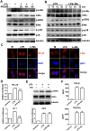
Figure 3. Naringenin inhibits MAPK and NF-κB pathways in LPS-treated RAW 264.7 cells.
(A,B) Time- and dose- dependent inhibition on MAPK and NF-κB activation by naringenin. Cells were treated with LPS alone or with 20, 40 and 80 μM NG for 30 min (A). Cells were treated with LPS alone or with NG for 0, 5, 15 and 30 min (B). Protein levels of pIκBα, IκBα, pERK, ERK, p-p38, p38 and tubulin were detected by western blot. (C) Inhibition of nuclear translocation of NF-Kb p65 and c-fos by naringenin. Cells were treated with LPS alone or with NG for 2 h. Nuclear translocation of NF-κB p65 and c-fos were detected by immunofluorescence. (D) Inhibition of reporter activity of NF-kB and AP-1 by naringenin. Cells transfected with NF-κB and AP-1 reporter plasmids and treated with LPS alone or together with NG for 6 h. Relative reporter activity was detected by the luciferase assay (n = 3). **P < 0.01 vs LPS. (E) Activity of IRF3 detection. Cells were treated with LPS alone or together with NG; IRF3 phosphorylation was detected by western blot (upper), and the relative reporter activity of IRF3 was detected by a luciferase assay (lower). (F) Production of supernatant IFN-β and RANTES levels. Cells were treated as in (E) for 12 h and supernatant IFN-β and RANTES levels were detected by ELISA (n = 3). Naringenin is abbreviated as NG. The concentrations of LPS and NG were 100 ng/ml and 80 μM unless indicated.
Figure 4
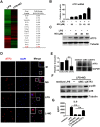
Figure 4. Naringenin upregulates ATF3 expression in RAW 264.7 cells, which is required for its anti-inflammatory action.
(A) ATF3 expression as demonstrated in the transcriptome assay. Cells were treated with LPS or LPS plus NG for 4 h. The gene expression ratio of LPS + NG/LPS was analysed by a transcriptome assay. (B) Naringenin induces ATF3 mRNA expression. Cells were treated with NG, LPS and LPS plus NG (20, 40 and 80 μM) for 4 h. ATF3 mRNA was detected by real-time PCR (n = 3). (C) Naringenin induces ATF3 protein expression. Cells were treated with LPS alone or with NG (20, 40 and 80 μM) for 4 h. The protein level of ATF3 was detected by western blot. (D) Naringenin enhances cellular staining of ATF3. Cells were treated with NG, LPS or LPS plus NG for 4 h. Intracellular ATF3 was detected by immunofluorescence. Uncropped images are presented in Supplementary Figure S8A. (E) ATF3 knockdown by siRNA. Cells were transfected with control siRNA (siNC) or ATF3 siRNA (siATF3) for 24 h. Then, ATF3 mRNA was detected by PCR (n = 3, **P < 0.01). The protein level of ATF3 was detected by western blot. (F,G) ATF3 knockdown dampens the anti-inflammatory activity of naringenin. Wild-type cells and siNC- or siATF3-pretreated cells were treated with LPS alone or LPS plus NG. Plasma protein was collected 30 min after LPS treatment, and p-p38, p38 and tubulin were detected by western blot (F). Uncropped images are presented in Supplementary Figure S8A. Supernatants were collected 12 h after LPS treatment, and the IL-6 level was detected by ELISA (G). n = 3, **P < 0.01. Naringenin is abbreviated as NG. The concentrations of LPS and NG were 100 ng/ml and 80 μM unless indicated.
Figure 5
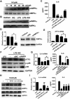
Figure 5. Naringenin activates AMPKα to mediate ATF3 upregulation in RAW 264.7 cells.
(A) Dose-dependent induction of AMPK phosphorylation by naringenin. Cells were treated with NG (0, 10, 20, 40 and 80 μM) or 1 mM AICAR for 1 h. The phosphorylation of AMPKα was detected by western blot. (B) Naringenin enhances AMPK phosphorylation with LPS. Cells were treated with NG, LPS or LPS plus NG for 1 h. AMPKα phosphorylation was detected by western blot. (C) Modulation of AMPK affects IL-6 suppression by naringenin. Cells were treated with NG alone or in combination with 1 mM AICAR or 2 μM compound C before stimulation with LPS. Supernatant IL-6 was detected by ELISA (n = 3). **P < 0.01 vs LPS; #P < 0.05, ##P < 0.01 vs LPS + NG (D) AMPK knockdown by siRNA. Cells were transfected with control siRNA (siNC) or AMPKα siRNA (siAMPKα) for 24 h. Then, AMPKα mRNA was detected by PCR (n = 3, *P < 0.05). The protein level of ATF3 was detected by western blot. Uncropped images are presented in Supplementary Figure S8B. (EG) ATF3 expression and anti-inflammatory activity affected by AMPKα siRNA. Wild-type cells and siNC- or siAMPKα-transfected cells were treated with LPS alone or LPS plus NG. The mRNA expression of ATF3 was detected by RT-PCR (E) (n = 3, *P < 0.05). Then, ATF3, p-p38, p38 and tubulin were detected by western blot (F). The mRNA and protein levels of IL-6 were detected by RT-PCR and ELISA (n = 3, *P < 0.05, **P < 0.01). (H–I) Effects of calcium and CaMKKβ inhibition on cytokine production and ATF3/AMPKα activation. Cells were pretreated with 5 mM EGTA, 10 μM LiCl3 (Li3+) or 1 μM STO-609 before treating with NG alone or in combination with LPS. mRNA expression of IL-6 was detected by RT-PCR. The supernatant level of IL-6 was detected by ELISA n=3, **P < 0.01 vs LPS, ##P < 0.01 vs LPS + NG. ATF3, p-AMPK, AMPK and GAPDH were detected by western blot. Uncropped images are presented in Supplementary Figure S8B. Naringenin is abbreviated as NG. The concentrations of LPS and NG were 100 ng/ml and 80 μM unless indicated.
Figure 6
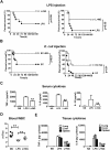
Figure 6. Naringenin improves survival and ameliorates systemic and tissue inflammatory reactions in endotoxaemia mice.
(A) Survival analysis of the LPS-injected mice. Mice were intraperitoneally injected with 10 mg/kg (left) or 20 (right) mg/kg LPS (■) or in combination with NG (L + NG, □). Survival was observed for 7 days (n = 10). (B) Survival analysis in the bacteria injection model. Mice were intraperitoneally injected with 1.0 × 1010 CFU/kg (left) or 2.0 × 1010CFU/kg (right) heat-killed E. coli (■) or in combination with NG (L + NG, □). Survival was observed for 7 days (n = 16). (CE) Detection of cytokines and cell counts in blood or tissue samples. Mice were injected with normal saline (NS), LPS or LPS with NG (L + NG). Samples from blood, lung, liver and spleen tissues were obtained 12 h after injection. Serum levels of TNF-α, IL-6 and IL-1β were detected by ELISA (C). Blood WBCs were detected using haematological analysers (D). **P < 0.01 vs LPS. The levels of TNF-α and IL-6 per gram protein (E) were detected in homogenates of the lung, liver and spleen (n = 3). **, ## and §§represent P < 0.01 vs LPS for lung, liver and spleen, respectively. Naringenin is abbreviated as NG. The doses of LPS and NG were both 10 mg/kg unless indicated.
Figure 7
Figure 7. Naringenin upregulates ATF3 expression in lung tissues of LPS-challenged mice, which is AMPK dependent and required for limiting proinflammatory reactions.
(A,B) Detection of cytokines and cell counts in BALF and Western blot assays for signalling molecules in murine lung tissues. Mice were injected with NS, LPS or LPS with NG. BALF or lung tissues were collected 12 h after LPS injection. TNF-α, IL-6 and IL-10 levels and WBC counts (n = 3) in the BALF were measured (A). Protein levels of p-IκBα, IκBα, ERK, pERK, p-p38, p38 and tubulin were detected by western blot (B). (CE) Detection of histological changes, ATF3 expression and AMPK activation in murine lung tissues. Mice were injected with NS, NG, LPS or LPS with NG, and lung tissues were collected 12 h after injection. Histopathological changes (C) were observed, and the protein levels of ATF3 (D) or pAMPKα and AMPKα (E) were detected by western blot. Uncropped images are presented in Supplementary Figure S8C,D. (F,G) Effects of the co-injection of AMPK modulators on the levels of ATF3 and proinflammatory cytokines. Mice were injected with NS, NG, LPS, LPS plus NG, LPS plus NG and compound C (1 mg/kg) or LPS plus naringenin and AICAR (100 mg/kg). Blood, lung tissues and BALF were collected 12 h after injection. ATF3 expression in the lung tissues were detected by western blot (F). TNF-α and IL-6 levels in the serum or in the BALF (G) were detected by ELISA (n = 3). *P < 0.05, **P < 0.01 vs LPS; #P < 0.05; ##P < 0.01 vs LPS+NG. Naringenin is abbreviated as NG. Broncho-alveolar lavage fluid is abbreviated as BALF. The doses of LPS and NG were both 10 mg/kg unless indicated.
Figure 8
Figure 8. A schematic diagram describing the anti-inflammatory action of naringenin through AMPK-dependent upregulation of ATF3.

Similar articles

Cited by

References

    1. Molteni M., Gemma S. & Rossetti C. The Role of Toll-Like Receptor 4 in Infectious and Noninfectious Inflammation. Mediators Inflamm 2016, 6978936 (2016). - PMC - PubMed
    1. Akira S. & Takeda K. Toll-like receptor signalling. Nat Rev Immunol 4, 499–511 (2004). - PubMed
    1. Broz P. & Monack D. M. Newly described pattern recognition receptors team up against intracellular pathogens. Nat Rev Immunol 13, 551–565 (2013). - PubMed
    1. Mills K. H. TLR-dependent T cell activation in autoimmunity. Nat Rev Immunol 11, 807–822 (2011). - PubMed
    1. Gay N. J., Symmons M. F., Gangloff M. & Bryant C. E. Assembly and localization of Toll-like receptor signalling complexes. Nat Rev Immunol 14, 546–558 (2014). - PubMed

Publication types

MeSH terms