. 2017 Jan;12(1):195-207.
doi: 10.1038/nprot.2016.170. Epub 2016 Dec 22.

Generation of nephron progenitor cells and kidney organoids from human pluripotent stem cells

Affiliations

Generation of nephron progenitor cells and kidney organoids from human pluripotent stem cells

Ryuji Morizane et al. Nat Protoc. 2017 Jan.

Abstract

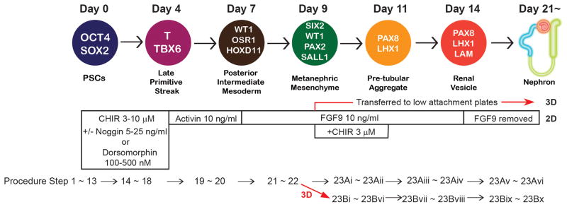
A variety of protocols have been developed that demonstrate the capability to differentiate human pluripotent stem cells (hPSCs) into kidney structures. Our goal was to develop a high-efficiency protocol to generate nephron progenitor cells (NPCs) and kidney organoids to facilitate applications for tissue engineering, disease modeling and chemical screening. Here, we describe a detailed protocol resulting in high-efficiency production (80-90%) of NPCs from hPSCs within 9 d of differentiation. Kidney organoids were generated from NPCs within 12 d with high reproducibility using 96-well plates suitable for chemical screening. The protocol requires skills for culturing hPSCs and careful attention to morphological changes indicative of differentiation. This kidney organoid system provides a platform for studies of human kidney development, modeling of kidney diseases, nephrotoxicity and kidney regeneration. The system provides a model for in vitro study of kidney intracellular and intercompartmental interactions using differentiated human cells in an appropriate nephron and stromal context.


Conflict of interest statement

J.V.B. is a coinventor on KIM-1 patents, which have been licensed by Partners Healthcare to several companies. He has received royalty income from Partners Healthcare. J.V.B. or his family has received income for consulting from companies interested in biomarkers: Sekisui, Millennium, Johnson & Johnson, and Novartis.

Figures

Figure 1
Figure 1. The differentiation protocols into kidney organoids from hPSCs
The diagram shows markers for each step of differentiation in a sequential pattern identifying days of differentiation. OCT4: POU class 5 homeobox 1. SOX2: SRY-box 2. T: brachyury. WT1: Wilms tumor 1. OSR1: odd-skipped related transcription factor 1. HOXD11: homeobox D11. SIX2: SIX2 homeobox 2. PAX2: paired box 2. SALL1: spalt like transcription factor 1. PAX8: paired box 8. LHX1: LIM homeobox 1. LAM: laminin. The concentration of each growth factor and small molecule necessary for each stage of differentiation is shown as well as corresponding procedural step numbers. This figure is modified from the one published previously .
Figure 2
Figure 2. Morphological changes of hPSCs at each step of differentiation
Representative bright field imaging at each step of differentiation. Day 0, undifferentiated hPSCs (H9) when differentiation is initiated. Day 4, late primitive streak stage. Day 9, nephron progenitor stage. Day 14, renal vesicle stage. Day 21, nephron stage. The optimal morphology of cells to proceed to activin A treatment on day 4 is the visual presence of loosely dense clusters. Representative bright field imaging of “too loose” or “too dense” clusters on day 4 is also shown. Scale bar: 100 μm. The scale bar is representative of all panels.
Figure 3
Figure 3. Immunostaining for NPCs and nephrons
(a) Immunocytochemistry for SIX2 at day 9 of differentiation revealing NPCs. Scale bar: 50 μm. (b) Immunocytochemistry to identify nephron segments in 2D culture on day 21 of the differentiation. Scale bar: 50 μm. (c) Immunohistochemistry to identify nephron segments in 3D culture with frozen sections on day 21 of differentiation. Scale bar: 50 μm. (d) Whole mount staining for nephrons in 3D culture on day 28 (left: high magnification, scale bar: 50 μm) and 21 (right: low magnification, scale bar: 100 μm). The inset on the right shows DAPI (4′,6-diamidino-2-phenylindole) staining. PODXL: podocalyxin (a podocyte marker). LTL: lotus tetragonolobus lectin (a proximal tubule marker). CDH1: cadherin1 (also known as E-cadherin) (a loop of Henle and distal tubule marker). (e) Bright field imaging of an organoid in 3D culture on day 21. Arrows indicate a glomerular structure. Scale bar: 100 μm. (c) and the left panel of (d) were originally published in ref .
Figure 4
Figure 4. Nephrotoxicity assay
Immunohistochemical staining for CDH1 (cadherin1), KIM1 (kidney injury molecule1), and LTL (lotus tetragonolobus lectin) in kidney organoids after 24 hours treatment with cisplatin 5 μM. LTL+ tubules expressed KIM1 after the treatment, which is a marker for proximal tubular injury. Kidney organoids generated in 3D culture were treated with cisplatin 5 μM for 24 hours from day 23 to 24 of the differentiation. Organoids were fixed and analyzed on day 24. Scale bars: 50 μm. The scale bars are representative of the corresponding right panels.

Similar articles

Cited by

References

    1. Takahashi K, et al. Induction of pluripotent stem cells from adult human fibroblasts by defined factors. Cell. 2007;131:861–872. doi: 10.1016/j.cell.2007.11.019. - DOI - PubMed
    1. Takahashi K, Yamanaka S. Induction of pluripotent stem cells from mouse embryonic and adult fibroblast cultures by defined factors. Cell. 2006;126:663–676. doi: 10.1016/j.cell.2006.07.024. - DOI - PubMed
    1. Takasato M, et al. Directing human embryonic stem cell differentiation towards a renal lineage generates a self-organizing kidney. Nature cell biology. 2014;16:118–126. doi: 10.1038/ncb2894. - DOI - PubMed
    1. Lam AQ, et al. Rapid and efficient differentiation of human pluripotent stem cells into intermediate mesoderm that forms tubules expressing kidney proximal tubular markers. Journal of the American Society of Nephrology: JASN. 2014;25:1211–1225. doi: 10.1681/ASN.2013080831. - DOI - PMC - PubMed
    1. Taguchi A, et al. Redefining the in vivo origin of metanephric nephron progenitors enables generation of complex kidney structures from pluripotent stem cells. Cell Stem Cell. 2014;14:53–67. doi: 10.1016/j.stem.2013.11.010. - DOI - PubMed

MeSH terms