. 2017 Jun;12(2):314-326.
doi: 10.1007/s11481-016-9719-0. Epub 2016 Dec 22.

HIV-1 Glycoprotein 120 Enhancement of N-Methyl-D-Aspartate NMDA Receptor-Mediated Excitatory Postsynaptic Currents: Implications for HIV-1-Associated Neural Injury

Affiliations

HIV-1 Glycoprotein 120 Enhancement of N-Methyl-D-Aspartate NMDA Receptor-Mediated Excitatory Postsynaptic Currents: Implications for HIV-1-Associated Neural Injury

Yan Zhou et al. J Neuroimmune Pharmacol. 2017 Jun.

Abstract

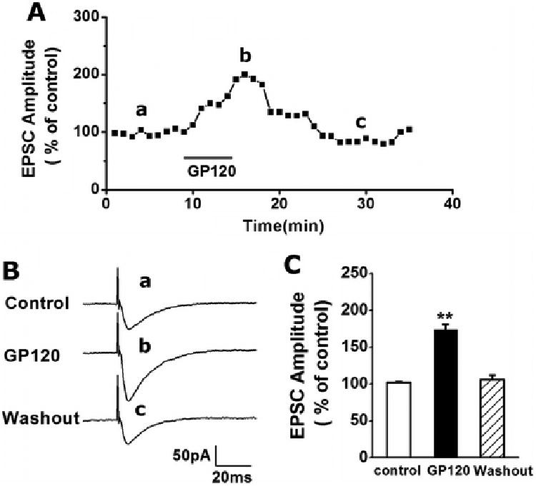
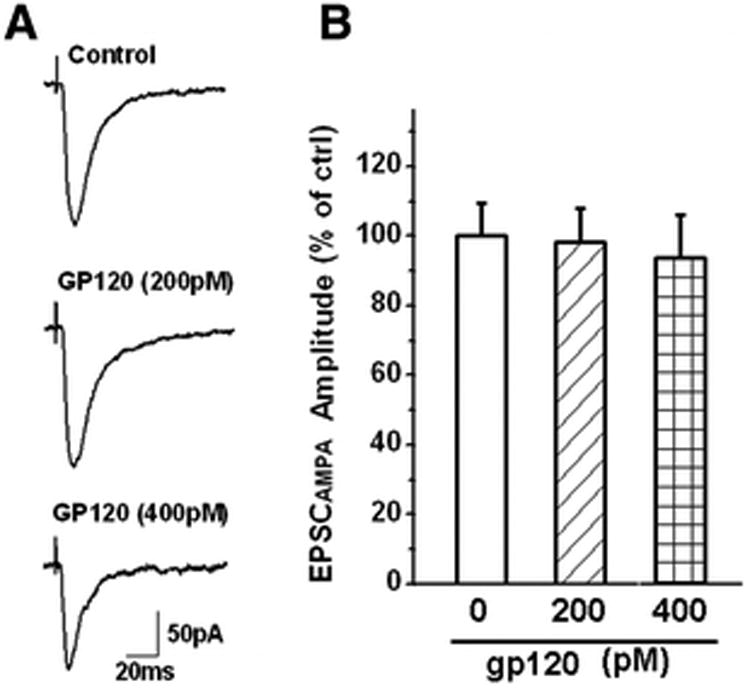
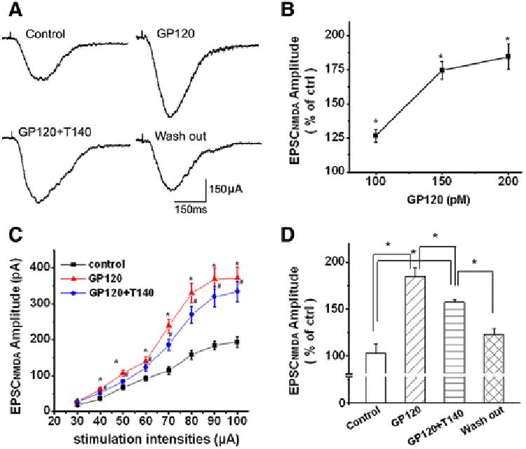
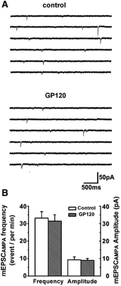
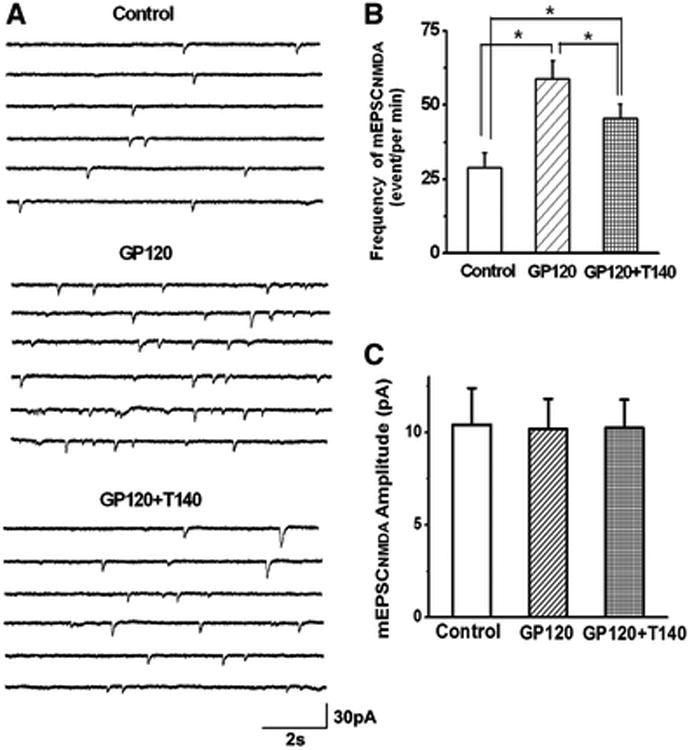
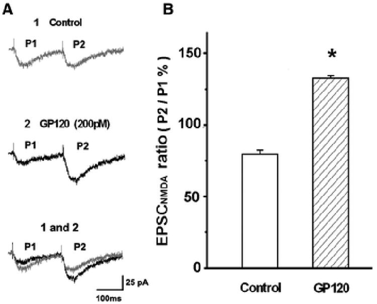
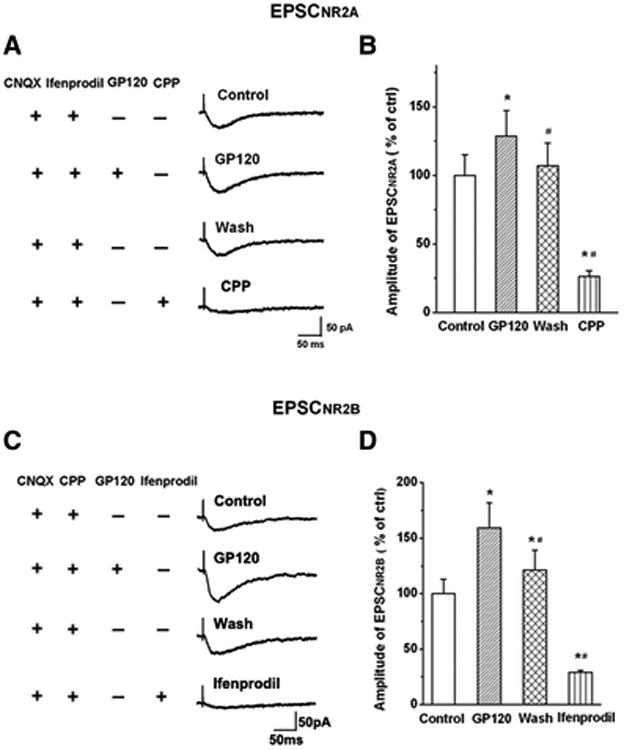
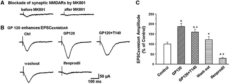
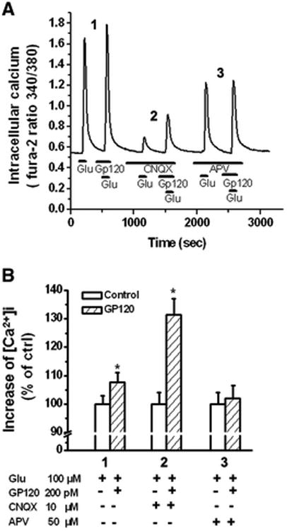
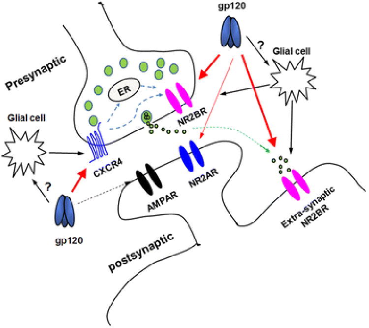
It is widely accepted that human immunodeficiency virus type 1 (HIV-1) envelope glycoprotein 120 (gp120) plays an important role in HIV-1-induced neural injury and pathogenesis of HIV-1-associated dementia (HAND). Multiple pathways have been proposed for gp120-induced neurotoxicity, amongst is the activation of N-Methyl-D-Aspartate receptors (NMDARs). It has been shown that gp120 causes neuronal injury or death and gp120 transgenic mice exhibit neurological similarity to that of HAND, all of which can be blocked or attenuated by NMDAR antagonists. Several lines of evidence indicate the subtype and location of activated NMDARs are key determinants of the nature of NMDAR physiology. To examine the subtype and the location of NMDARs affected by gp120, we studied gp120 on subtype NMDAR-mediated EPSCs in the CA1 region of rat hippocampal slices through "blind" whole-cell patch recordings. Our results showed bath application of gp120 increased both NR2A- and NR2B-mediated EPSCs possibly via a presynaptic mechanism, with much stronger effect on NR2B-mediated EPSCs. In contrast, gp120 failed on enhancing AMPA receptor-mediated EPSCs. Ca2+ imaging studies revealed that gp120 potentiated glutamate-induced increase of intracellular Ca2+ concentration in rat hippocampal neuronal cultures which were blocked by a NMDAR antagonist, but not by an AMPA receptor antagonist, indicating gp120 induces Ca2+ influx through NMDARs. Further investigations demonstrated that gp120 increased the EPSCs mediated by extrasynaptic NR2BRs. Taken together, these results demonstrate that gp120 interacts with both NR2A and NR2B subtypes of NMDARs with a predominant action on the extrasynaptic NR2B, implicating a role NR2B may play in HIV-1-associated neuropathology.

Keywords: CA1; Glutamate receptors; HIV; Neurodegeneration; Synaptic transmission.


Figures

Figure 1
Figure 1. Gp120 enhancement of EPSCs in the CA1 region of rat hippocampal slices
A. A representative time course illustrating bath application of gp120 (200pM), indicated by a horizontal bar, increased the EPSC amplitudes recorded in a CA1 neuron. The graph plots normalized amplitudes of the EPSCs evoked in response to constant current stimulation of Schaffer-collateral fibers (120μA, 20μs, 0.05Hz). Each point in this graph is an average of 3 consecutive EPSCs. B. Representative individual EPSCs taken from different time points as indicated by letters a, b and c. C. Summary data (n=8) showing average EPSC peak amplitudes before (control), during (gp120) and after (washout) bath application of gp120. Note gp120 significantly increased amplitude of EPSCs. ** p < 0.001 vs. Ctrl.
Figure 2
Figure 2. GP120 has no significant effect on EPSCAMPAR
EPSCAMPAR was recorded in the presence of a specific NMDA receptor antagonist APV (50μM) in the perfusate. The recorded EPSCAMPAR can be blocked by addition of a specific AMPAR antagonist CNQX (10 μM) to the bath. A. Representative EPSCAMPAR current traces recorded from a CA1 pyramidal neuron in a rat hippocampal slice showing the amplitudes of EPSCAMPAR were not altered by bath application of gp120 at a concentration of 200pM (n=9), or even at 400pM (n=5). Each trace shown was an average of consecutive 10 evoked EPSCAMPAR. B. A summary bar graph illustrating average EPSCAMPAR amplitudes measured before (Ctrl) and during bath perfusion of gp120 (gp120). Note that gp120 failed to alter the EPSCAMPAR amplitudes at either 200pM (n=9) or 400pM (n=5).
Figure 3
Figure 3. Gp120 enhancement of EPSCNMDAR via CXCR4
A. Exemplary whole-cell current traces illustrating gp120 (200pM) increase of EPSCNMDAR recorded from a CA1 neuron in a rat hippocampal slice before (Ctrl), during bath perfusion of gp120 (gp120), gp120+T140 (50nM) (gp120+T140), or after washout of gp120+T140 (washout). Note that bath application of gp120 produced an increase of EPSCNMDAR (upper right), addition of T140 to the bath partially inhibited gp120-induced increase of EPSCNMDAR (lower left) and the EPSCNMDAR returned to the control level after washout of gp120 +T140 (lower right). B. A dose-responsive curve showing that gp120 increased EPSCNMDAR in a dose-dependent manner. C. The input/output curve shows that gp120 significantly increased the amplitude of EPSCNMDAR evoked by an electric stimulation on Schaffer collateral fibers with different intensities (30μA∼100μA) and that the gp120-induced increase of the EPSCNMDAR amplitudes were partially inhibited by T140 (n=9, * p<0.05 vs ctrl, #p<0.05 vs gp120. D. Bar graph shows the average of normalized EPSCNMDAR amplitudes measured before (Ctrl), during bath application of gp120 (gp120), during bath application of gp20+T140 (GP120+T140), or after washout of gp120+T140 (washout). Values are the means ± SEM, n=9, *p<0.05 as determined by one-way ANOVA.
Figure 4
Figure 4. Gp120 failure on alteration of spontaneous mini EPSCAMPAR (mEPSCAMPAR)
A. Representative traces of mEPSCAMPAR recorded from a CA1 neuron in a rat hippocampal slice in the presence of NMDA receptor antagonist AP-V (50μM) in the perfusate. Addition of gp120 to the perfusate failed to alter mEPSCAMPAR. B. Bar graph showing average event frequency and amplitude of mEPSCAMPAR were not significantly changed after addition og gp120 (200pM) to the perfusate (n=9, p> 0.05 vs control.
Figure 5
Figure 5. Gp120 increase of frequency, but not amplitude, of mEPSCNMDAR
A. Representative traces of mEPSCNMDAR were recorded before (Ctrl), during bath perfusion of 200pM (gp120), and during bath perfusion of gp120 and T140 (gp120+T140). Data showed that the frequency of mEPSCNMDAR was increased by bath application of gp120, which was partially blocked by addition of T140 to the bath. B-C. Bar graph illustrates that gp120 increased the mean frequency of mEPSCNMDAR, but not the mean amplitude, suggesting a presynaptic site for gp120 action. The gp120-associated increase of mean frequency was attenuated by T140, a specific CXCR4 antagonist, indicating gp120 may interacts with chemokine receptor CXCR4. Graphed values are the means ± SEM, n=9, *p<0.05 as determined by one-way ANOVA test.
Figure 6
Figure 6. gp120 increased paired-pulse facilitation (PPF) of EPSCNMDAR
Paired-pulse facilitation of EPSCNMDAR was generated by paired-pulse stimulations with inter-stimulus pulse interval of150ms. The paired-pulse ratio was calculated by dividing the first EPSCNMDAR peak amplitude from the second EPSCNMDAR peak amplitude. A. Representative PPF traces of EPSCNMDAR were recorded from a CA1 neuron in a rat hippocampal slice in the presence of glycine (1μM) and AMPA receptor antagonist CNQX (10μM) before (Ctrl) and during bath perfusion of gp120 (gp120). B. Summarized bar graph illustrating averaged paired-pulse ratio of EPSCNMDAR were significantly elevated by bath application of gp120 (200pM). n=8 * p<0.05 vs Ctrl
Figure 7
Figure 7. Gp120 enhancement of EPSCNR2AR and EPSCNR2BR
A. EPSCNR2AR was isolated by addition of AMPAR antagonist CNQX (10μM) and NR2BR antagonist ifenprodil (10μM) to the perfusate. Exemplary EPSCNR2AR traces were recorded in a CA1 neuronal cell before (Ctrl), during (gp120), after (washout) bath perfusion of gp120, and subsequent pharmacological confirmation of EPSCNR2AR by a specific NR2AR blocker R-CPP (CPP). Note that bath perfusion of gp120 increased EPSCNR2A and the EPSCNR2A returned to control level after washout of gp120. Addition of R-CPP almost completely blocked EPSCNR2A, demonstrating the pharmacologically isolated EPSCs were EPSCNR2AR. B. EPSCNR2BR was isolated with the addition of AMPAR antagonist CNQX (10μM) and NR2AR antagonist R-CPP (1μM) to the perfusate. Exemplary EPSCNR2BR traces were recorded from another CA1 neuronal cell before (Ctrl), during (gp120), after (washout) bath perfusion of gp120, and pharmacological confirmation of EPSCNR2BR by a specific NR2BR blocker ifenprodil (Ifenprodil). B and D are summary bar graphs showing average amplitudes (% of control) of EPSCnr2ar (B) and EPSCNR2BR (D) recorded under various experimental conditions as indicated, respectively. Note that gp120 enhanced both EPSCNR2AR (128.61±18.64% of control, n=11, M±SD) and EPSCNR2BR (159.25±22.77% of control, n=8, M±SD) with a significant stronger effect (t(17)=0.0046; p<0.01) on enhancing EPSCNR2BR and EPSCNR2AR. * p<0.05 vs control; # p<0.05 vs gp120 group.
Figure 8
Figure 8. gp120 interacts with extrasynaptic NR2BRs (exNR2BRs)
A. Blockade of synaptic EPSCNMDAR by MK801 (70μM, an open NMDAR channel blocker)combined with a 10min low frequency (0.05Hz) stimulation of Schaffer-collateral fiber protocol. B. Exemplary EPSCNMDAR traces recorded in different experimental conditions after synaptic NMDARs were blocked by MK801 as shown in A. Note addition of gp120 enhanced EPSCNMDAR which was most likely mediated via an interaction with extrasynaptic NMDARs. Since NMDARs in the hippocampus are composed mainly of NR2ARs and NR2BRs, the enhancement of EPSCNMDAR by gp120 under the blockade of synaptic NMDARs (predominantly NR2ARSs) suggests that gp120 may act most on exNR2BRs. This suggestion was supported by the experimental results that addition of ifenprodil, a specific NR2BR antagonist, blocked EPSCNMDAR, demonstrating gp120 enhancement of EPSCexNR2BR. The gp120-mediated enhancement of EPSCexNR2BR was attenuated by T140, indicating gp120 interacts with CXCR4. C. Average EPSCexNR2BR recorded under different experimental conditions as indicated in panel B. Note a significant enhancement of EPSCexNR2BR by gp120 and its attenuation by a CXCR4 receptor blocker T140. These results illustrate that gp120 acts on exNR2BRs. n=8, *p<0.05 vs control, #p<0.05 vs gp120.
Figure 9
Figure 9
gp120 potentiation of glutamate-induced increase of [Ca2+]I via NMDARs. A. An example of Ca2+ imaging study showing gp120 potentiation of glutamate (Glu)-induced Ca2+ influx via NMDARs in a cultured rat hippocampal neuron. Note that gp120 produced an increase of [Ca2+ ]I in the presence of an AMPAR antagonist CNQX (10μM). The gp120-associated potentiation disappeared when NMDARs were blocked by addition of a specific NMDAR antagonist APV, demonstrating that gp120 interacts with NMDARs. B. Bar graph shows average [Ca2+ ]I in different experimental conditions and illustrates gp120 potentiation of Glu-induced increase of [Ca2+ ]I via NMDARs. Experiments were done in triplicates. n=12, * p < 0.05 vs control.
Figure 10
Figure 10
A schematic diagram illustrating the sites of gp120 action in the hippocampal slices. As shown in big red arrows, gp120 acts on presynaptic CXCR4 and on pre- and post-synaptic NR2BRs. It is also possible that gp120 acts on glial cells, resulting in glial cell release of neurotoxic molecules including, but not limited to, cytokines, chemokines, amino acids, etc, which in turn act on CXCR4 and NMDARs (black arrows). Gp120-induced enhancement of EPSCNMDAR can be blocked by a CXCR4 blocker T140, suggesting the existence a functional coupling between CXCR4 and NMDARs as proposed by Anna Pittaluga and her colleagues (Di Prisco et al., 2016).

Similar articles

Cited by

References

    1. Acquas E, Bachis A, Nosheny RL, Cernak I, Mocchetti I. Human immunodeficiency virus type 1 protein gp120 causes neuronal cell death in the rat brain by activating caspases. Neurotox Res. 2004;5:605–615. - PubMed
    1. Alfahad TB, Nath A. Update on HIV-associated neurocognitive disorders. Curr Neurol Neurosci Rep. 2013;13:387. - PMC - PubMed
    1. Anderson ER, Gendelman HE, Xiong H. Memantine protects hippocampal neuronal function in murine human immunodeficiency virus type 1 encephalitis. J Neurosci. 2004;24:7194–7198. - PMC - PubMed
    1. Antinori A, et al. Updated research nosology for HIV-associated neurocognitive disorders. Neurology. 2007;69:1789–1799. - PMC - PubMed
    1. Banerjee A, Larsen RS, Philpot BD, Paulsen O. Roles of Presynaptic NMDA Receptors in Neurotransmission and Plasticity. Trends Neurosci. 2016;39:26–39. - PMC - PubMed

Publication types

MeSH terms

Substances

LinkOut - more resources