. 2017 Apr;56(4):465-476.
doi: 10.1165/rcmb.2016-0104OC.

Tensin 1 Is Essential for Myofibroblast Differentiation and Extracellular Matrix Formation

Affiliations

Tensin 1 Is Essential for Myofibroblast Differentiation and Extracellular Matrix Formation

Ksenija Bernau et al. Am J Respir Cell Mol Biol. 2017 Apr.

Abstract

Myofibroblasts, the primary effector cells that mediate matrix remodeling during pulmonary fibrosis, rapidly assemble an extracellular fibronectin matrix. Tensin (TNS) 1 is a key component of specialized cellular adhesions (fibrillar adhesions) that bind to extracellular fibronectin fibrils. We hypothesized that TNS1 may play a role in modulating myofibroblast-mediated matrix formation. We found that TNS1 expression is increased in fibroblastic foci from lungs with idiopathic pulmonary fibrosis. Transforming growth factor (TGF)-β profoundly up-regulates TNS1 expression with kinetics that parallel the expression of the myofibroblast marker, smooth muscle α-actin. TGF-β-induced TNS1 expression is dependent on signaling through the TGF-β receptor 1 and is Rho coiled-coiled kinase/actin/megakaryoblastic leukemia-1/serum response factor dependent. Small interfering RNA-mediated knockdown of TNS1 disrupted TGF-β-induced myofibroblast differentiation, without affecting TGF-β/Smad signaling. In contrast, loss of TNS1 resulted in disruption of focal adhesion kinase phosphorylation, focal adhesion formation, and actin stress fiber development. Finally, TNS1 was essential for the formation of fibrillar adhesions and the assembly of nascent fibronectin and collagen matrix in myofibroblasts. In summary, our data show that TNS1 is a novel megakaryoblastic leukemia-1-dependent gene that is induced during pulmonary fibrosis. TNS1 plays an essential role in TGF-β-induced myofibroblast differentiation and myofibroblast-mediated formation of extracellular fibronectin and collagen matrix. Targeted disruption of TNS1 and associated signaling may provide an avenue to inhibit tissue fibrosis.

Keywords: extracellular matrix; fibrillar adhesion; megakaryoblastic leukemia-1; myofibroblast; tensin 1.


Figures

Figure 1.
Figure 1.
Tensin (TNS) 1 is up-regulated in myofibroblasts and in idiopathic pulmonary fibrosis (IPF) lung. (A) Western blot of human lung fibroblast (HLF) lysates after 1 ng/ml transforming growth factor (TGF)-β stimulation at indicated time points against TNS1 and smooth muscle α-actin (ACTA2). (B) TNS1 mRNA expression in normal HLFs after 6- or 24-hour treatment with 1 ng/ml TGF-β and compared with time 0 controls (unstimulated). Mixed-effect ANOVA (*P < 0.05) was used for statistical analysis. (C) Immunohistochemical staining of normal and IPF lung samples against TNS1. Scale bars, 100 μm. (D) Total TNS1 mRNA in normal and IPF lung homogenates. Student’s t test (*P < 0.05) was used for statistical analysis. (E and F) Total TNS1 mRNA (E) or protein (F) in lysates from normal and IPF-derived lung fibroblasts. Student’s t test (*P < 0.05) was used for statistical analyses. Data are presented as means (±SD). Ctrl, control.
Figure 2.
Figure 2.
TGF-β–induced TNS1 expression requires signaling through TGF-β receptor I but not Smads. (A) Western blot of HLF lysates after stimulation with TGF-β (1 ng/ml) at indicated time points against phosphorylated (p) Smad2, pSmad3, and phosphorylated extracellular signal-regulated kinases 1 and 2 (pErk1/2). (B) HLFs were treated with 1 ng/ml TGF-β for 30 minutes in the presence of 10 μM SB531542 or vehicle control, followed by Western blotting for the indicated (phospho) proteins. (C and D) HLFs were treated with 1 ng/ml TGF-β for 24 hours in the presence of 10 μM SB531542 or vehicle control, followed by Western blotting for TNS1 (C) and subsequent densitometry (D). (E and F) HLFs were transfected with Smad2 or Smad3 small interfering (si)RNA or scrambled control followed by treatment with 1 ng/ml TGF-β for 24 hours and Western (E) blotting and densitometry (F) for the indicated proteins. Mixed-effect ANOVA (*P < 0.05; NS, not significant) was used for all statistical analyses. Data are presented as means (± SD). scram., scrambled.
Figure 3.
Figure 3.
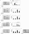
TGF-β–induced TNS1 expression is mediated by Rho coiled-coiled kinase (ROCK)/actin/megakaryoblastic leukemia (MKL)-1. (A and B) HLFs were treated with 1 ng/ml TGF-β for 72 hours in the presence of the ROCK inhibitor Y27632 (10 μM), the actin filament disruptor latrunculin B (500 nM), the formin inhibitor 1-(3-bromophenyl)-5-(2-furanylmethylene)dihydro-2-thioxo-4,6(1H,5H)-pyrimidinedione (SMIFH2) (20 μM), or vehicle control, followed by Western blotting for TNS1 and ACTA2 (A) and densitometry of the indicated bands and compared with time 0 controls (unstimulated) (B). (CF) HLFs were transiently transfected with MKL1 siRNA or scrambled control before a 14-hour treatment with either the MKL1 actin disruptor cytochalasin D (2 μM [C and D]), or the actin filament stabilizer jasplakinolide (300 nM [E and F]), followed by Western blotting and densitometry. (G and H) Western blot analysis and densitometry of TNS1 and MKL1 expression in HLF lysates after transient transfection with siRNA against MKL1 or scrambled control, and subsequent treatment with TGF-β (1 ng/ml) for 24 hours. (I and J) HLFs were treated with 1 ng/ml TGF-β for 24 hours in the presence of the MKL1 inhibitor N-[2-[4(4-chlorophenyl)amino]-1-methyl-2-oxoethoxy]-3,5-bis(trifluoromethyl)-benzamide (CCG-1423) (10 μM) or vehicle control, followed by Western blotting and densitometry of TNS1. (K) HLFs were treated with 0.5 U/μl human α-thrombin for 24 hours in the presence of the MKL1 inhibitor CCG-1423 (10 μM) or vehicle control, followed by Western blotting against TNS1 and ACTA2. Mixed-effect ANOVA (* and #P < 0.05) was used for all statistical analyses. * and # symbols designate significant difference between the indicated conditions. Data are presented as means (±SD). TUBA, α-tubulin.
Figure 4.
Figure 4.
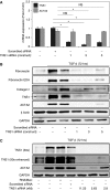
TNS1 is essential for myofibroblast differentiation. (A) Total TNS1 and ACTA2 mRNA expression in HLFs upon TNS1 knockdown using three different siRNA constructs (or treatment with scrambled control). Mixed-effect ANOVA (*P < 0.05; NS, not significant) was used for statistical analysis. (B) HLFs were transfected with the indicated TNS1 siRNAs or scrambled control, followed by treatment with 1 ng/ml TGF-β for 72 hours and Western blotting for the indicated proteins. Arrow indicates the expected TNS1 band. (C) Dose–response of TGF-β–induced (1 ng/ml for 72 h) ACTA2 expression to varying concentrations of TNS1 siRNA. The top TNS1 blot was exposed for a typical length of time (30 s) without saturation of the bands. The lower TNS1 blot was overexposed with increased contrast to visualize low-level TNS1 expression under low concentrations of TNS1 siRNA. Data are presented as means (±SD). EDA, extra domain A.
Figure 5.
Figure 5.
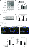
TNS1 is dispensable for TGF-β signaling but required for focal adhesion–dependent signals. (A and B) HLFs were transfected with TNS1 siRNA or scrambled control followed by treatment with 1 ng/ml TGF-β for 30 minutes and Western blotting and densitometry for the indicated (phospho) proteins. Mixed-effect ANOVA (*P < 0.05; NS, not significant) was used for statistical analysis. (C and D) HLFs were transfected with TNS1 siRNA or scrambled control, followed by treatment with 1 ng/ml TGF-β for 24 hours, Western blotting, and densitometry for the phosphorylated tyrosine 397 residue of focal adhesion kinase (FAK). Mixed-effect ANOVA (* and #P < 0.05) was used for statistical analysis. (E) Merged immunocytochemistry images of HLFs stained against phalloidin (green) and vinculin (red) upon TNS1 knockdown using TNS1 siRNA (or treatment with scrambled control) and treatment with 1 ng/ml TGF-β for 24 hours. Scale bar, 50 μm. Insets show digital magnifications of vinculin staining for each condition. Scale bar, 50 μM. (F) Quantitation of number and length of focal (vinculin-containing) adhesions in TGF-β–induced (1 ng/ml for 24 h) myofibroblasts under siRNA-mediated TNS1 knockdown or scrambled control. Student’s t test (*P < 0.05) was used for statistical analysis. * and # symbols designate significant difference between the indicated conditions. Data are presented as means (±SD). DAPI, 4′,6-diamidino-2-phenylindole.
Figure 6.
Figure 6.
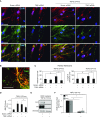
TNS1 is required for fibrillar adhesion formation and extracellular matrix (ECM) deposition by myofibroblasts. (A) Coimmunostaining for TNS1 and β1-integrin in HLFs transfected with TNS1 siRNA or scrambled control, followed by treatment with 1 ng/ml TGF-β for 24 hours. Scale bars, 50 μm. Immunostaining for the row is indicated in the images of the left column. (B) Coimmunostaining for TNS1 (green) and fibronectin (red, extracellular fibrils) in HLFs. Image digitally zoomed for clarity. Scale bar, 25 μm. (C) Quantitation of number and length of fibrillar (activated β1-integrin–containing) adhesions in TGF-β–induced (1 ng/ml, 24 h) myofibroblasts under siRNA-mediated TNS1 knockdown or scrambled control. Mixed-effect ANOVA (* and #P < 0.05) was used for statistical analysis. (D) HLFs, transfected with TNS1 siRNA or scrambled control, were treated with 1 ng/ml TGF-β for 72 hours, then replated and incubated with Alexa488-labeled fibronectin (488-FN) for 90 minutes, followed by fixation and immunofluorescent microscopy. Mixed-effect ANOVA (* and #P < 0.05) was used for statistical analysis. (E and F) HLFs were transfected with TNS1 siRNA or scrambled control, treated with 1 ng/ml TGF-β for 72 hours, followed by deoxycholate (DOC) buffer extraction to remove non–ECM-bound material. Residual ECM was then solubilized and run via PAGE followed by Western blotting and densitometry of the indicated proteins. Data show reduced DOC-insoluble fibronectin (FN) or collagen I (Col I) fractions. Mixed-effect ANOVA (*P < 0.05) was used for statistical analysis. * and # symbols designate significant difference between the indicated conditions. Data are presented as means (±SD). A.U., arbitrary units.

Similar articles

Cited by

References

    1. Sandbo N, Kregel S, Taurin S, Bhorade S, Dulin NO. Critical role of serum response factor in pulmonary myofibroblast differentiation induced by TGF-β. Am J Respir Cell Mol Biol. 2009;41:332–338. - PMC - PubMed
    1. Sandbo N, Lau A, Kach J, Ngam C, Yau D, Dulin NO. Delayed stress fiber formation mediates pulmonary myofibroblast differentiation in response to TGF-β. Am J Physiol Lung Cell Mol Physiol. 2011;301:L656–L666. - PMC - PubMed
    1. Bernau K, Ngam C, Torr EE, Acton B, Kach J, Dulin NO, Sandbo N. Megakaryoblastic leukemia-1 is required for the development of bleomycin-induced pulmonary fibrosis. Respir Res. 2015;16:45. - PMC - PubMed
    1. Torr EE, Ngam CR, Bernau K, Tomasini-Johansson B, Acton B, Sandbo N. Myofibroblasts exhibit enhanced fibronectin assembly that is intrinsic to their contractile phenotype. J Biol Chem. 2015;290:6951–6961. - PMC - PubMed
    1. Chen H, Ishii A, Wong WK, Chen LB, Lo SH. Molecular characterization of human tensin. Biochem J. 2000;351:403–411. - PMC - PubMed

MeSH terms

LinkOut - more resources