MULTIMODAL IMAGING OF DISEASE-ASSOCIATED PIGMENTARY CHANGES IN RETINITIS PIGMENTOSA
- PMID: 28005673
- PMCID: PMC5193227
- DOI: 10.1097/IAE.0000000000001256
MULTIMODAL IMAGING OF DISEASE-ASSOCIATED PIGMENTARY CHANGES IN RETINITIS PIGMENTOSA
Abstract
Purpose: Using multiple imaging modalities, we evaluated the changes in photoreceptor cells and retinal pigment epithelium (RPE) that are associated with bone spicule-shaped melanin pigmentation in retinitis pigmentosa.
Methods: In a cohort of 60 patients with retinitis pigmentosa, short-wavelength autofluorescence, near-infrared autofluorescence (NIR-AF), NIR reflectance, spectral domain optical coherence tomography, and color fundus images were studied.
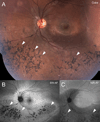
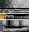
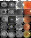
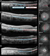
Results: Central AF rings were visible in both short-wavelength autofluorescence and NIR-AF images. Bone spicule pigmentation was nonreflective in NIR reflectance, hypoautofluorescent with short-wavelength autofluorescence and NIR-AF imaging, and presented as intraretinal hyperreflective foci in spectral domain optical coherence tomography images. In areas beyond the AF ring outer border, the photoreceptor ellipsoid zone band was absent in spectral domain optical coherence tomography and the visibility of choroidal vessels in short-wavelength autofluorescence, NIR-AF, and NIR reflectance images was indicative of reduced RPE pigmentation. Choroidal visibility was most pronounced in the zone approaching peripheral areas of bone spicule pigmentation; here RPE/Bruch membrane thinning became apparent in spectral domain optical coherence tomography.
Conclusion: These findings are consistent with a process by which RPE cells vacate their monolayer and migrate into inner retina in response to photoreceptor cell degeneration. The remaining RPE spread undergo thinning and consequently become less pigmented. An explanation for the absence of NIR-AF melanin signal in relation to bone spicule pigmentation is not forthcoming.
Figures


Similar articles
-
Intraretinal Correlates of Reticular Pseudodrusen Revealed by Autofluorescence and En Face OCT.Invest Ophthalmol Vis Sci. 2017 Sep 1;58(11):4769-4777. doi: 10.1167/iovs.17-22338. Invest Ophthalmol Vis Sci. 2017. PMID: 28973322 Free PMC article.
-
Photoreceptor cells as a source of fundus autofluorescence in recessive Stargardt disease.J Neurosci Res. 2019 Jan;97(1):98-106. doi: 10.1002/jnr.24252. Epub 2018 Apr 27. J Neurosci Res. 2019. PMID: 29701254 Free PMC article.
-
Correlations among near-infrared and short-wavelength autofluorescence and spectral-domain optical coherence tomography in recessive Stargardt disease.Invest Ophthalmol Vis Sci. 2014 Oct 23;55(12):8134-43. doi: 10.1167/iovs.14-14848. Invest Ophthalmol Vis Sci. 2014. PMID: 25342616 Free PMC article.
-
[Pathophysiology of macular diseases--morphology and function].Nippon Ganka Gakkai Zasshi. 2011 Mar;115(3):238-74; discussion 275. Nippon Ganka Gakkai Zasshi. 2011. PMID: 21476310 Review. Japanese.
-
Diagnostic imaging in patients with retinitis pigmentosa.J Med Invest. 2012;59(1-2):1-11. doi: 10.2152/jmi.59.1. J Med Invest. 2012. PMID: 22449988 Review.
Cited by
-
A Distinct Phenotype of Eyes Shut Homolog (EYS)-Retinitis Pigmentosa Is Associated With Variants Near the C-Terminus.Am J Ophthalmol. 2018 Jun;190:99-112. doi: 10.1016/j.ajo.2018.03.008. Epub 2018 Mar 14. Am J Ophthalmol. 2018. PMID: 29550188 Free PMC article.
-
Assessment of inner retinal oxygen metrics and thickness in a mouse model of inherited retinal degeneration.Exp Eye Res. 2021 Apr;205:108480. doi: 10.1016/j.exer.2021.108480. Epub 2021 Feb 2. Exp Eye Res. 2021. PMID: 33539865 Free PMC article.
-
Primary versus Secondary Elevations in Fundus Autofluorescence.Int J Mol Sci. 2023 Aug 2;24(15):12327. doi: 10.3390/ijms241512327. Int J Mol Sci. 2023. PMID: 37569703 Free PMC article. Review.
-
Significance of Hyperreflective Foci as an Optical Coherence Tomography Biomarker in Retinal Diseases: Characterization and Clinical Implications.J Ophthalmol. 2021 Dec 17;2021:6096017. doi: 10.1155/2021/6096017. eCollection 2021. J Ophthalmol. 2021. PMID: 34956669 Free PMC article. Review.
-
Acupuncture for retinitis pigmentosa: a comprehensive review.Int Ophthalmol. 2025 Jul 15;45(1):292. doi: 10.1007/s10792-025-03656-6. Int Ophthalmol. 2025. PMID: 40663303 Review.
References
-
- Bunker CH, Berson EL, Bromley WC, et al. Prevalence of retinitis pigmentosa in Maine. Am J Ophthalmol. 1984;97:357–365. - PubMed
-
- Hartong DT, Berson EL, Dryja TP. Retinitis pigmentosa. Lancet. 2006;368:1795–1809. - PubMed
-
- Battaglia Parodi M, La Spina C, Triolo G, et al. Correlation of SD-OCT findings and visual function in patients with retinitis pigmentosa. Graefes Arch Clin Exp Ophthalmol. 2015 - PubMed
-
- Delori FC, Keilhauer C, Sparrow JR, Staurenghi G. Origin of fundus autofluorescence. In: Holz FG, Schmitz-Valckenberg S, Spaide RF, Bird AC, editors. Atlas of Fundus Autofluorescence Imaging. Berlin Heidelberg: Springer-Verlag; 2007. pp. 17–29.
Publication types
MeSH terms
Substances
Grants and funding
LinkOut - more resources
Full Text Sources
Other Literature Sources
Miscellaneous

