

Decreased Peak Expiratory Flow Associated with Muscle Fiber-Type Switching in Spinal and Bulbar Muscular Atrophy
- PMID: 28005993
- PMCID: PMC5179045
- DOI: 10.1371/journal.pone.0168846
Decreased Peak Expiratory Flow Associated with Muscle Fiber-Type Switching in Spinal and Bulbar Muscular Atrophy
Abstract
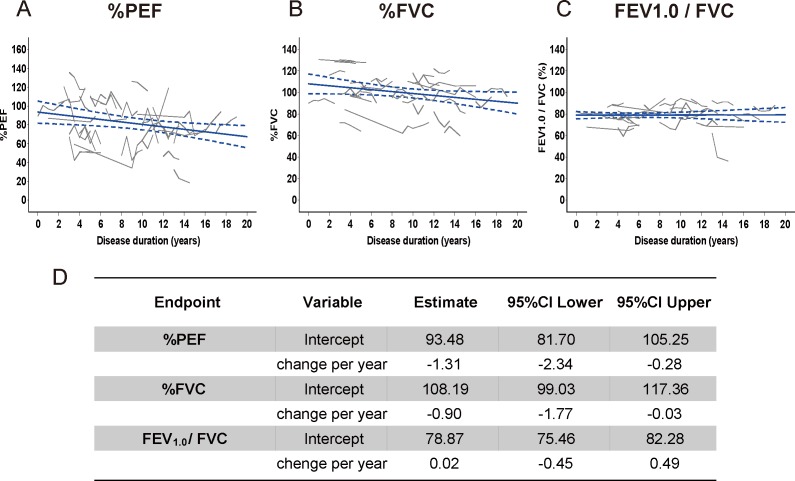
The aim of this study was to characterize the respiratory function profile of subjects with spinal and bulbar muscular atrophy (SBMA), and to explore the underlying pathological mechanism by comparing the clinical and biochemical indices of this disease with those of amyotrophic lateral sclerosis (ALS). We enrolled male subjects with SBMA (n = 40) and ALS (n = 25) along with 15 healthy control subjects, and assessed their respiratory function, motor function, and muscle strength. Predicted values of peak expiratory flow (%PEF) and forced vital capacity were decreased in subjects with SBMA compared with controls. In SBMA, both values were strongly correlated with the trunk subscores of the motor function tests and showed deterioration relative to disease duration. Compared with activities of daily living (ADL)-matched ALS subjects, %PEF, tongue pressure, and grip power were substantially decreased in subjects with SBMA. Both immunofluorescence and RT-PCR demonstrated a selective decrease in the expression levels of the genes encoding the myosin heavy chains specific to fast-twitch fibers in SBMA subjects. The mRNA levels of peroxisome proliferator-activated receptor gamma coactivator 1-alpha and peroxisome proliferator-activated receptor delta were up-regulated in SBMA compared with ALS and controls. In conclusion, %PEF is a disease-specific respiratory marker for the severity and progression of SBMA. Explosive muscle strength, including %PEF, was selectively affected in subjects with SBMA and was associated with activation of the mitochondrial biogenesis-related molecular pathway in skeletal muscles.
Conflict of interest statement
The authors have declared that no competing interests exist.
Figures


Similar articles
-
Glycolytic-to-oxidative fiber-type switch and mTOR signaling activation are early-onset features of SBMA muscle modified by high-fat diet.Acta Neuropathol. 2016 Jul;132(1):127-44. doi: 10.1007/s00401-016-1550-4. Epub 2016 Mar 12. Acta Neuropathol. 2016. PMID: 26971100 Free PMC article.
-
Increased mitophagy in the skeletal muscle of spinal and bulbar muscular atrophy patients.Hum Mol Genet. 2017 Mar 15;26(6):1087-1103. doi: 10.1093/hmg/ddx019. Hum Mol Genet. 2017. PMID: 28087734 Free PMC article.
-
Prominent fatigue in spinal muscular atrophy and spinal and bulbar muscular atrophy: evidence of activity-dependent conduction block.Clin Neurophysiol. 2013 Sep;124(9):1893-8. doi: 10.1016/j.clinph.2012.12.053. Epub 2013 Apr 30. Clin Neurophysiol. 2013. PMID: 23643309 Clinical Trial.
-
Cell-Clearing Systems Bridging Repeat Expansion Proteotoxicity and Neuromuscular Junction Alterations in ALS and SBMA.Int J Mol Sci. 2020 Jun 4;21(11):4021. doi: 10.3390/ijms21114021. Int J Mol Sci. 2020. PMID: 32512809 Free PMC article. Review.
-
Pathogenesis and therapy of spinal and bulbar muscular atrophy (SBMA).Prog Neurobiol. 2012 Dec;99(3):246-56. doi: 10.1016/j.pneurobio.2012.05.007. Epub 2012 May 15. Prog Neurobiol. 2012. PMID: 22609045 Review.
Cited by
-
Neuromuscular junction pathology is correlated with differential motor unit vulnerability in spinal and bulbar muscular atrophy.Acta Neuropathol Commun. 2022 Jul 5;10(1):97. doi: 10.1186/s40478-022-01402-y. Acta Neuropathol Commun. 2022. PMID: 35791011 Free PMC article.
-
Beyond motor neurons: expanding the clinical spectrum in Kennedy's disease.J Neurol Neurosurg Psychiatry. 2018 Aug;89(8):808-812. doi: 10.1136/jnnp-2017-316961. Epub 2018 Jan 20. J Neurol Neurosurg Psychiatry. 2018. PMID: 29353237 Free PMC article. Review.
-
Molecular Mechanisms and Therapeutics for SBMA/Kennedy's Disease.Neurotherapeutics. 2019 Oct;16(4):928-947. doi: 10.1007/s13311-019-00790-9. Neurotherapeutics. 2019. PMID: 31686397 Free PMC article. Review.
-
Simple and efficient differentiation of human iPSCs into contractible skeletal muscles for muscular disease modeling.Sci Rep. 2023 May 25;13(1):8146. doi: 10.1038/s41598-023-34445-9. Sci Rep. 2023. PMID: 37231024 Free PMC article.
-
Japanese monkeys rapidly noticed snake-scale cladded salamanders, similar to detecting snakes.Sci Rep. 2024 Nov 10;14(1):27458. doi: 10.1038/s41598-024-78595-w. Sci Rep. 2024. PMID: 39523392 Free PMC article.
References
-
- Kennedy WR, Alter M, Sung JH. Progressive proximal spinal and bulbar muscular atrophy of late onset. A sex-linked recessive trait. Neurology. 1968;18:671–680. - PubMed
MeSH terms
Substances
Grants and funding
LinkOut - more resources
Full Text Sources
Other Literature Sources
Medical
Miscellaneous
