Case Reports
. 2016 Dec 22;11(12):e0168973.
doi: 10.1371/journal.pone.0168973. eCollection 2016.

Peripheral Organs of Dengue Fatal Cases Present Strong Pro-Inflammatory Response with Participation of IFN-Gamma-, TNF-Alpha- and RANTES-Producing Cells

Affiliations
Case Reports

Peripheral Organs of Dengue Fatal Cases Present Strong Pro-Inflammatory Response with Participation of IFN-Gamma-, TNF-Alpha- and RANTES-Producing Cells

Tiago F Póvoa et al. PLoS One. .

Erratum in

Abstract

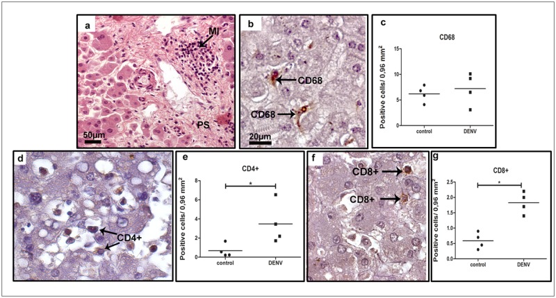
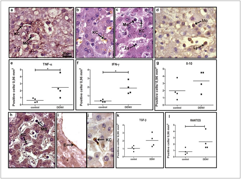
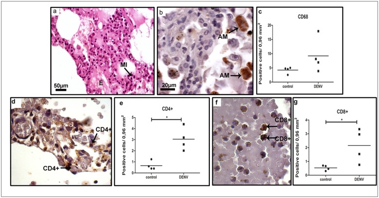
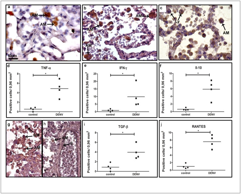
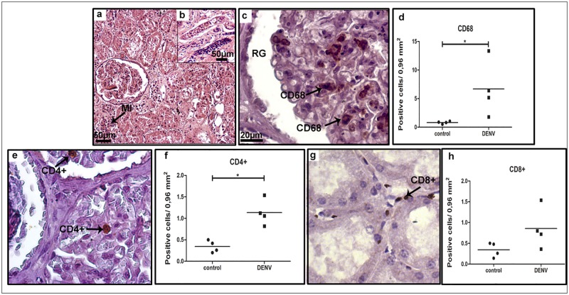
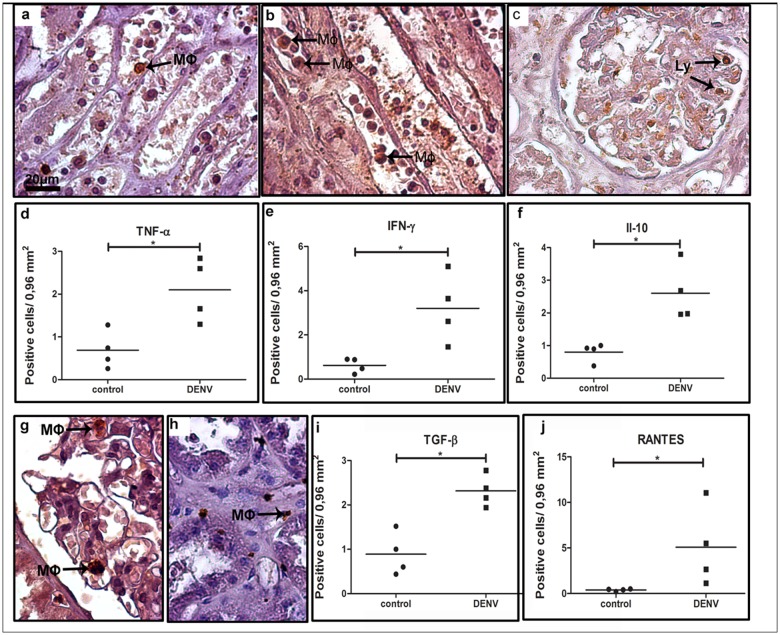
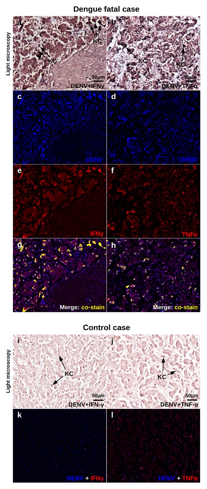
Dengue disease is an acute viral illness caused by dengue virus (DENV) that can progress to hemorrhagic stages leading to about 20000 deaths every year worldwide. Despite many clinical investigations regarding dengue, the immunopathogenic process by which infected patients evolve to the severe forms is not fully understood. Apart from differences in virulence and the antibody cross reactivity that can potentially augment virus replication, imbalanced cellular immunity is also seen as a major concern in the establishment of severe dengue. In this context, the investigation of cellular immunity and its products in dengue fatal cases may provide valuable data to help revealing dengue immunopathogenesis. Here, based in four dengue fatal cases infected by the serotype 3 in Brazil, different peripheral organs (livers, lungs and kidneys) were studied to evaluate the presence of cell infiltrates and the patterns of local cytokine response. The overall scenario of the studied cases revealed a considerable systemic involvement of infection with mononuclear cells targeted to all of the evaluated organs, as measured by immunohistochemistry (IHC). Quantification of cytokine-expressing cells in peripheral tissues was also performed to characterize the ongoing inflammatory process by the severe stage of the disease. Increased levels of IFN-γ- and TNF-α-expressing cells in liver, lung and kidney samples of post-mortem subjects evidenced a strong pro-inflammatory induction in these tissues. The presence of increased RANTES-producing cell numbers in all analyzed organs suggested a possible link between the clinical status and altered vascular permeability. Co-staining of DENV RNA and IFN-γ or TNF-α using in situ hibridization and IHC confirmed the virus-specific trigger of the pro-inflammatory response. Taken together, this work provided additional evidences that corroborated with the traditional theories regarding the "cytokine storm" and the occurrence of uneven cellular immunity in response to DENV as major reasons for progress to severe disease.


Conflict of interest statement

The authors have declared that no competing interests exist.

Figures

Fig 1
Fig 1. Characterization of cell subpopulations in liver tissues from DENV-3 fatal cases.
Sections were stained with (a) H.E. or (b, d, f) incubated with specific antibodies in immunohistochemical assays. (a) Liver of a representative dengue case showing diffuse mononuclear infiltrates around the portal space; (b) detection of hyperplasic Kupffer cells (CD68+) observed mainly in sinusoidal capillaries of the dengue cases; (c) quantification of CD68+ cells in dengue cases and controls (non-dengue cases); (d) detection of CD4+ cells manly in portal space of the dengue cases; (e) quantification of CD4+ cells in controls and dengue cases; (f) CD8+ cells detected mainly in portal space; (g) quantification of CD8+ cells. MI—mononuclear cell infiltrates. Asterisks indicate differences that are statistically significant between dengue cases and control groups, (*p < 0.05). Staining controls are shown in S1 Fig.
Fig 2
Fig 2. Detection of cytokine-producing cells in liver tissues.
(a) Detection of TNF-α in macrophages and Kupffer cells in sinusoidal capillaries; (b and c) detection of INF-α in Kupffer cells and lymphocytes; (d) production of anti-inflammatory cytokines (IL-10 in monocytes and lymphocytes circulating in sinusoidal capillaries and TGFβ detected in macrophages inside the portal space; (h) TGF-β-expressing macrophages and Kupffer cells in sinusoidal capillaries; (i and j) detection of RANTES in endothelium and Kupffer cells; (e, f, g, k and l) quantification of the number of cells expressing these cytokines in the hepatic tissue. Monocytes (Mo); Macrophages (Mϕ); Kupffer cells (KC); lymphocyte (Ly) and sinusoidal endothelium cells (E). Asterisks indicate differences that are statistically significant between groups (*p < 0.05). Staining controls are shown in S1 Fig.
Fig 3
Fig 3. Characterization of mononuclear cell subpopulations in lung tissues collected from DENV-3 fatal cases.
Lung tissue sections stained with (a) H.E. or (b, d and f) incubated with specific antibodies in immunohistochemistry assays. (a) Mononuclear infiltrates located in interstitial septa; (b) macrophages (CD68+ cells) observed in alveolar capillaries; (d and f) CD4+ and CD8+ T lymphocytes respectively, found in interstitial infiltrates septa; (c, e and g) quantification of cell subpopulations in the lung tissue. Macrophage (CD68); Mononuclear infiltrate (MI); edema (E); alveolar macrophage (AM); (CD4+) lymphocytes; (CD8+) lymphocytes. Asterisks indicate differences that are statistically significant between analyzed groups (*p < 0.05). Staining controls are shown in S2 Fig.
Fig 4
Fig 4. Cytokine-producing cells profile in lung tissues of dengue fatal cases.
Immunohistochemical analysis of lung sections collected from dengue fatal cases exhibited (a) TNF-α and (b) INF-γ production in alveolar macrophages; (c) IL-10 detected in monocytes/macrophages and lymphocytes in alveolar septa; (g) TGF-β produced by numerous macrophages and lymphocytes in the septa; (h) RANTES detected in endothelium cells and several macrophages in pulmonary tissue; (d-f, i and j) quantification of cytokine-expressing cells in the lung sections. Monocyte (Mo); Alveolar macrophage (AM); Endothelium (E); Lymphocytes (Ly). Asterisks indicate differences that are statistically significant between dengue cases and controls (*p < 0.05). Staining controls are shown in S2 Fig.
Fig 5
Fig 5. Characterization of mononuclear cells found in renal sections of post-mortem dengue cases.
(a and b) A representative kidney section stained with H.E. showing diffuse mononuclear cell infiltrate; (c) detection of (CD68+) in mesangial cells in glomeruli; (e and g) circulating CD4+ lymphocytes detected inside renal glomeruli and CD8+ cells observed in the interstitial space, respectively; (d, f and h) quantification of cell subpopulations in the kidney tissue. Mesangial cells (CD68); (CD4+) lymphocytes; (CD8+) lymphocytes. RG—renal gromerulus. Asterisks indicate differences that are statistically significant between groups (*p < 0.05). Staining controls are shown in S3 Fig.
Fig 6
Fig 6. Cytokine-producing cells in renal tissues of dengue cases.
(a) TNF-α detected in monocytes located in blood vessels; (b) Production of INF-γ observed in circulating macrophages and lymphocytes located in the interstitial space; (c) IL-10 found in endothelial cells of glomerulus; (g) TGF-β production by lymphocytes present mainly inside renal glomeruli; (h) RANTES detected in macrophages and lymphocytes in the interstitial renal space; (d-f, i and j) quantification of the number of cells expressing the above cytokines. Macrophages (Mϕ); Lymphocytes (Ly). Asterisks indicate differences that are statistically significant between analyzed groups (*p < 0.05). Staining controls are shown in S3 Fig.
Fig 7
Fig 7. Co-expression of DENV and pro-inflammatory cytokines in the liver.
Liver samples of dengue fatal case 2 were processed for in situ hybridization and IHC procedures. DENV was detected by a probe that aneals to a conserved sequence within the viral RNA negative strand. IFN-γ and TNF-α were assessed by immunohistochemistry assay. Probe-target complexes were revealed by alkaline phosphatase activity and cytokines were identified by standard DAB reactions. The chromogenic signals were converted to fluorescent-based signals using a computer based Nuance system. Representative images are shown: (a, b) Light microscopy of sinusiodal areas of dengue case stained for DENV RNA and IFN-γ or TNF-α, respectively. (c, d) Expression of DENV RNA (blue) in samples stained with IFN-γ or TNF-α, respectively. (e, f) Expression of IFN-γ or TNF-α (red) in in samples stained with DENV RNA, respectively. (g, h) Merged signals of DENV RNA and IFN-γ or TNF-α where co-expression is exhibited in yellow, respectively. (i, j) Light microscopy of sinusiodal areas of a non-dengue case stained for DENV RNA and IFN-γ or TNF-α, respectively. (k, l) Comtrol samples showing DENV and IFN-γ or TNF-α fluorescent signals, respectively. KC—Kupffer cells.

Similar articles

Cited by

References

    1. WHO: Global strategy for dengue prevention and control 2012-2020. World Health Organization; 2012. Available from: www.who.int
    1. WHO: Dengue: Guidelines for diagnosis, treatment, prevention and control. World Health Organization; 2009. Available from: www.who.int - PubMed
    1. Bhatt S, Gething PW, Brady OJ, Messina JP, Farlow AW, Moyes CL, et al. The global distribution and burden of dengue. Nature. 2013;496:504–507. 10.1038/nature12060 - DOI - PMC - PubMed
    1. Halstead SB, Cohen SN. Dengue hemorrhagic fever at 60 Years: early evolution of concepts of causation and treatment. Microbiol Mol Biol Rev. 2015;79:281–291. 10.1128/MMBR.00009-15 - DOI - PMC - PubMed
    1. Guzman MG, Halstead SB, Artsob H, Buchy P, Farrar J, Gubler DJ, et al. Dengue: a continuing global threat. Nat Rev Microbiol. 2010;8:S7–16. 10.1038/nrmicro2460 - DOI - PMC - PubMed

Publication types

MeSH terms