. 2017 Sep 1;27(7):433-451.
doi: 10.1089/ars.2016.6895. Epub 2017 Feb 7.

Augmentation of S-Nitrosoglutathione Controls Cigarette Smoke-Induced Inflammatory-Oxidative Stress and Chronic Obstructive Pulmonary Disease-Emphysema Pathogenesis by Restoring Cystic Fibrosis Transmembrane Conductance Regulator Function

Affiliations

Augmentation of S-Nitrosoglutathione Controls Cigarette Smoke-Induced Inflammatory-Oxidative Stress and Chronic Obstructive Pulmonary Disease-Emphysema Pathogenesis by Restoring Cystic Fibrosis Transmembrane Conductance Regulator Function

Manish Bodas et al. Antioxid Redox Signal. .

Abstract

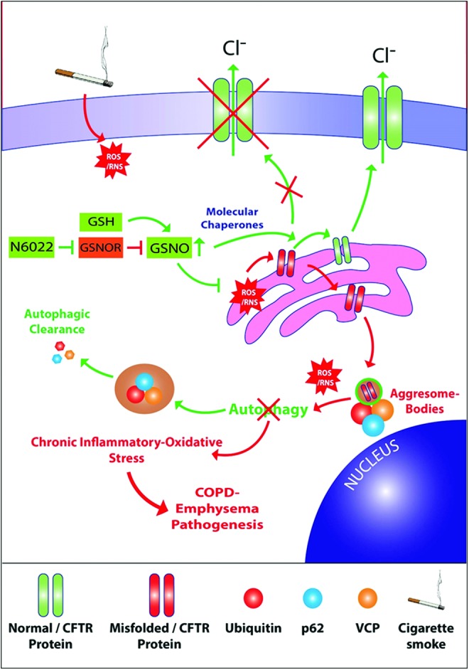
Aims: Cigarette smoke (CS)-mediated acquired cystic fibrosis transmembrane conductance regulator (CFTR)-dysfunction, autophagy-impairment, and resulting inflammatory-oxidative/nitrosative stress leads to chronic obstructive pulmonary disease (COPD)-emphysema pathogenesis. Moreover, nitric oxide (NO) signaling regulates lung function decline, and low serum NO levels that correlates with COPD severity. Hence, we aim to evaluate here the effects and mechanism(s) of S-nitrosoglutathione (GSNO) augmentation in regulating inflammatory-oxidative stress and COPD-emphysema pathogenesis.

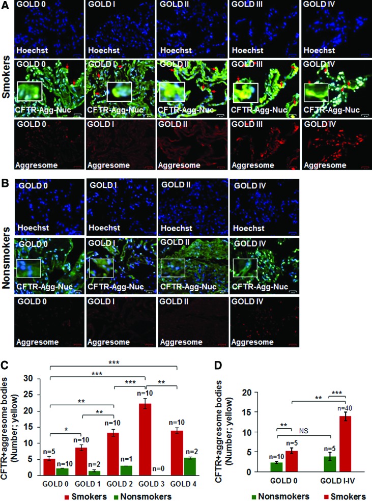
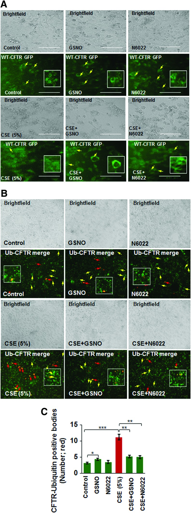
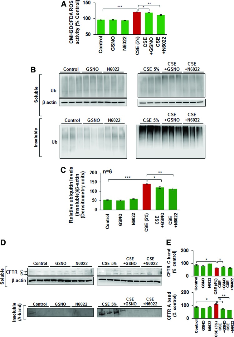
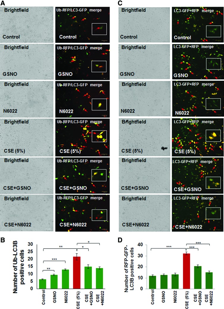
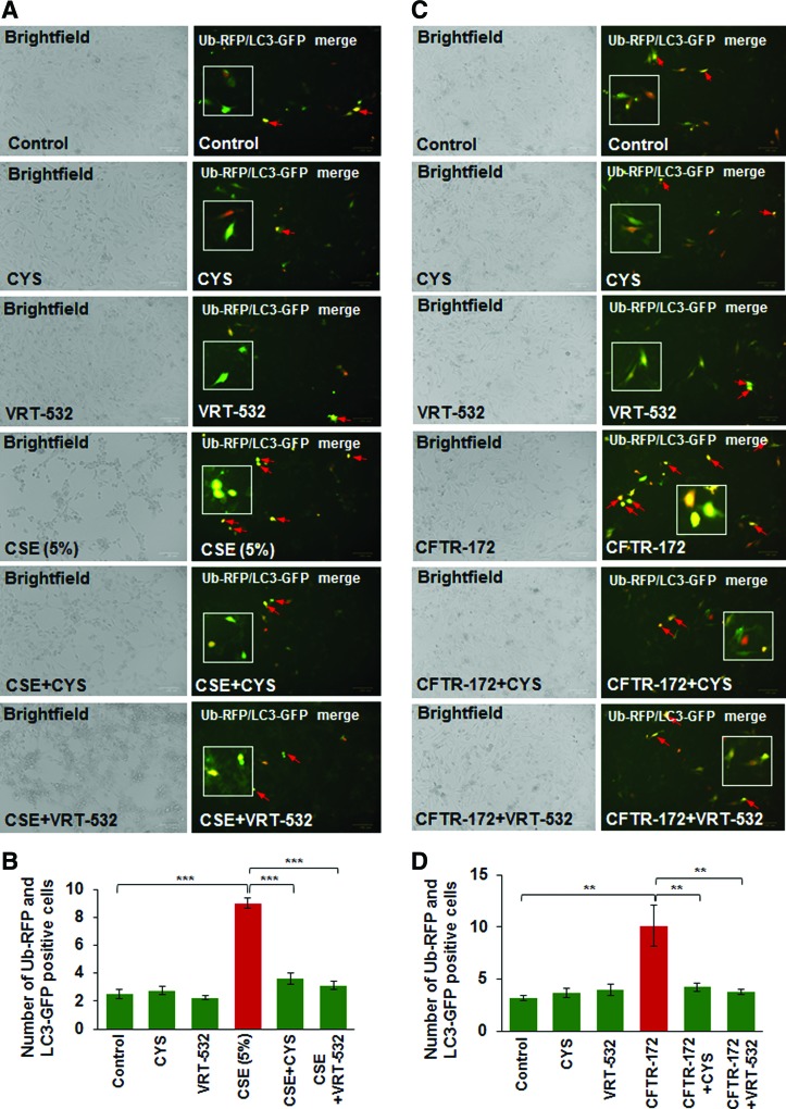
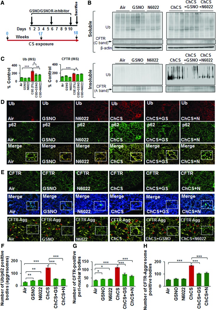
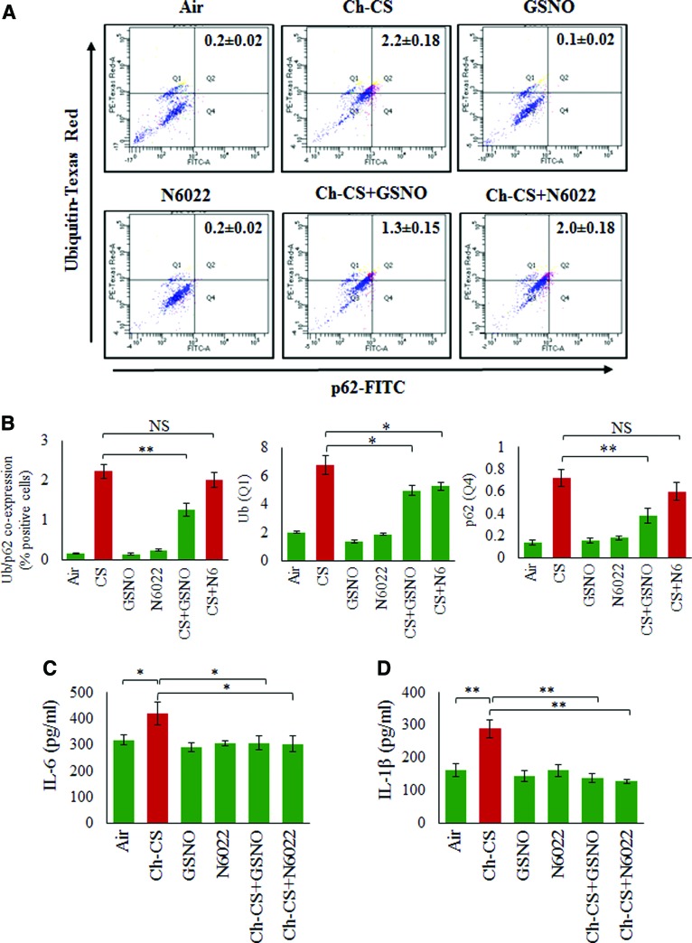
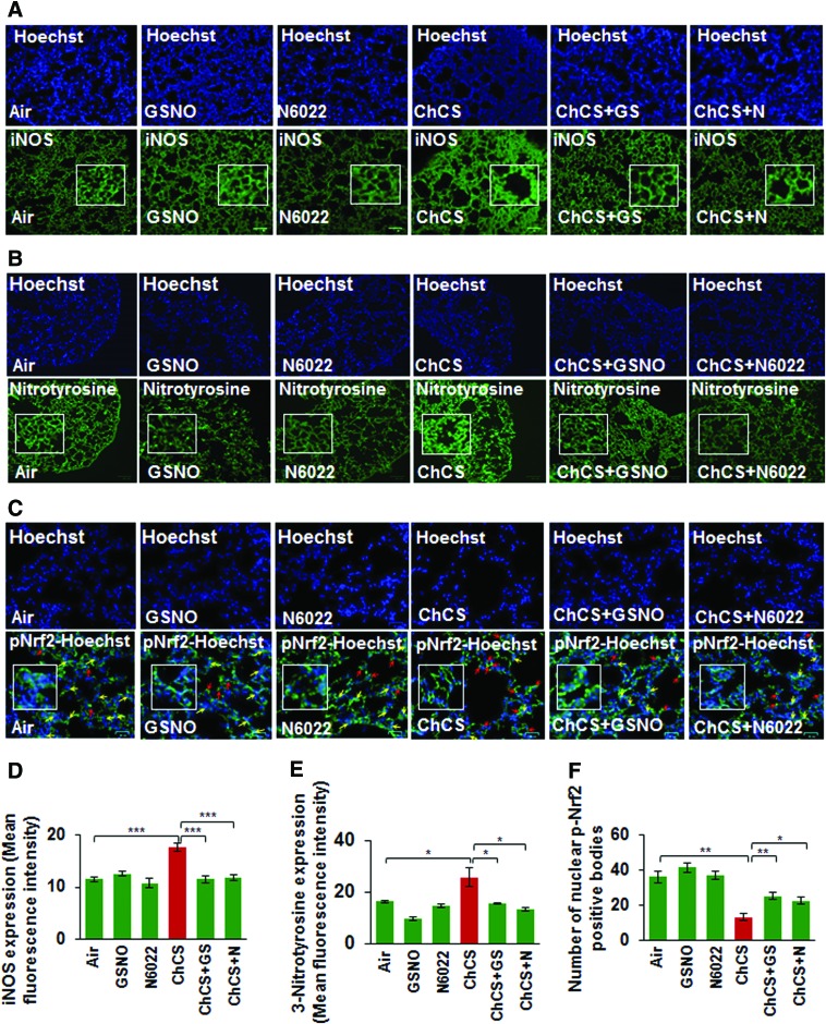
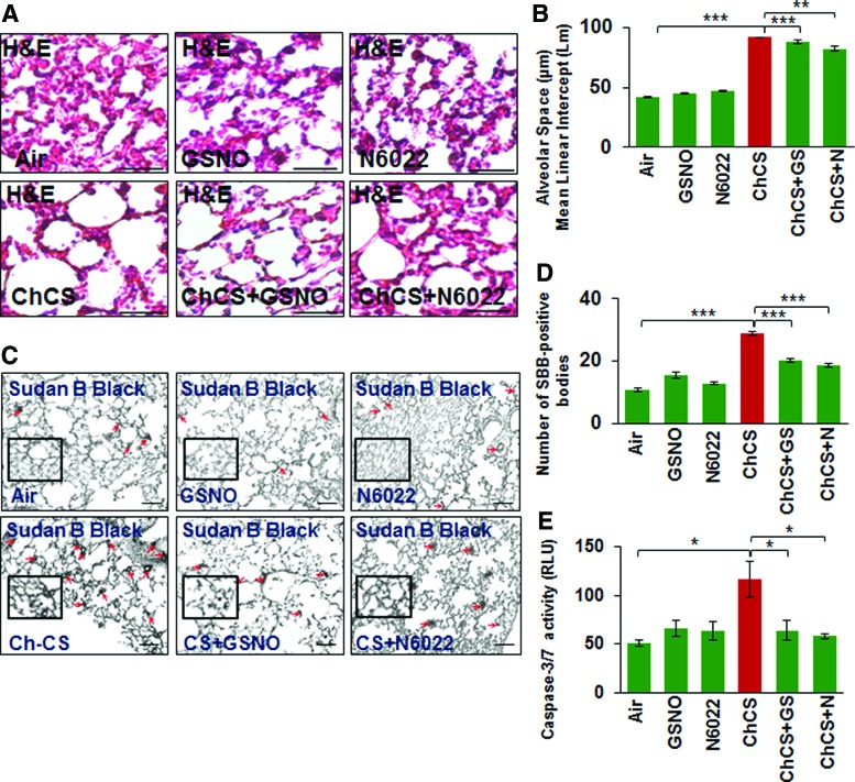
Results: Our data shows that cystic fibrosis transmembrane conductance regulator (CFTR) colocalizes with aggresome bodies in the lungs of COPD subjects with increasing emphysema severity (Global Initiative for Chronic Obstructive Lung Disease [GOLD] I - IV) compared to nonemphysema controls (GOLD 0). We further demonstrate that treatment with GSNO or S-nitrosoglutathione reductase (GSNOR)-inhibitor (N6022) significantly inhibits cigarette smoke extract (CSE; 5%)-induced decrease in membrane CFTR expression by rescuing it from ubiquitin (Ub)-positive aggresome bodies (p < 0.05). Moreover, GSNO restoration significantly (p < 0.05) decreases CSE-induced reactive oxygen species (ROS) activation and autophagy impairment (decreased accumulation of ubiquitinated proteins in the insoluble protein fractions and restoration of autophagy flux). In addition, GSNO augmentation inhibits protein misfolding as CSE-induced colocalization of ubiquitinated proteins and LC3B (in autophagy bodies) is significantly reduced by GSNO/N6022 treatment. We verified using the preclinical COPD-emphysema murine model that chronic CS (Ch-CS)-induced inflammation (interleukin [IL]-6/IL-1β levels), aggresome formation (perinuclear coexpression/colocalization of ubiquitinated proteins [Ub] and p62 [impaired autophagy marker], and CFTR), oxidative/nitrosative stress (p-Nrf2, inducible nitric oxide synthase [iNOS], and 3-nitrotyrosine expression), apoptosis (caspase-3/7 activity), and alveolar airspace enlargement (Lm) are significantly (p < 0.05) alleviated by augmenting airway GSNO levels. As a proof of concept, we demonstrate that GSNO augmentation suppresses Ch-CS-induced perinuclear CFTR protein accumulation (p < 0.05), which restores both acquired CFTR dysfunction and autophagy impairment, seen in COPD-emphysema subjects.

Innovation: GSNO augmentation alleviates CS-induced acquired CFTR dysfunction and resulting autophagy impairment.

Conclusion: Overall, we found that augmenting GSNO levels controls COPD-emphysema pathogenesis by reducing CS-induced acquired CFTR dysfunction and resulting autophagy impairment and chronic inflammatory-oxidative stress. Antioxid. Redox Signal. 27, 433-451.

Keywords: CFTR; COPD; GSNO; GSNOR; NO; emphysema.


Conflict of interest statement

No competing financial interests exist.

Figures

<b>FIG. 1.</b>
FIG. 1.
Increased colocalization of CFTR with aggresome bodies in human lung tissues with increasing emphysema severity. (A, B) The paraffin-embedded longitudinal lung tissue sections from nonemphysema (GOLD 0) and emphysema (GOLD I − IV), smokers and nonsmoker subjects were costained with aggresome-specific dye [red, bottom panel of (A, B)], CFTR (green), and Hoechst dye (nucleus, blue). The merged images (scale bar, 25 μm) were used to count the number of CFTR-positive aggresome bodies (yellow) in the perinuclear region depicted by red arrows, and high-magnification images are shown as insets. (C) The data indicate a significant statistical correlation between increased CFTR aggresome colocalization with decreasing lung function (FEV-1% predicted) and increasing severity of emphysema (GOLD I − IV) compared to nonemphysema (GOLD 0) control subjects [mean ± SEM, *p < 0.05, **p < 0.01, ***p < 0.001, r = −0.88; “n” numbers shown in (C)]. (D) The graph indicates a significant increase in CFTR aggresome colocalization in smokers (GOLD 0 and GOLD I − IV) compared to nonsmokers (GOLD 0 or GOLD I − IV). Also, smokers with greater emphysema severity (from GOLD I − IV) demonstrate a higher increase in CFTR aggresome colocalization compared to GOLD 0 smoker subjects, verifying that aggresome localization of CFTR is directly correlated to COPD-emphysema pathogenesis. Scale bar, 25 μM, *p < 0.05, **p < 0.01, ***p < 0.001. CFTR, cystic fibrosis transmembrane conductance regulator; COPD, chronic obstructive pulmonary disease; GOLD, Global Initiative for Chronic Obstructive Lung Disease; SEM, standard error of the mean. To see this illustration in color, the reader is referred to the web version of this article at www.liebertpub.com/ars
<b>FIG. 2.</b>
FIG. 2.
CSE-induced decrease in membrane CFTR and its aggresome accumulation is restored by augmenting GSNO levels. (A) Beas2b cells were transiently transfected with WT-CFTR GFP plasmid (24 h) and treated with 5% CSE and/or GSNO/N6022 (10 μM) for 12 h. The expression of WT-CFTR was analyzed by fluorescence microscopy, while the cell morphology and numbers are depicted using brightfield images of the same areas. The data shows that CSE-induced decrease in membrane CFTR expression is reinstated by GSNO augmentation (yellow arrows and insets, scale bar: 100 μm). (B, C) The WT-CFTR GFP and Ub-RFP plasmids were used to transiently transfect Beas2b cells for 24 h and cells were treated with 5% CSE and/or GSNO/N6022 (10 μM) for 12 h. Fluorescence and brightfield images were captured and the number of cells positive for both Ub (red) and WT-CFTR (green) were counted (yellow, red arrows), indicative of WT-CFTR accumulation in aggresomes. The data indicate that GSNO augmentation significantly reduces the CSE-induced increase in aggresome accumulation of WT-CFTR (red arrows) and enhances membrane WT-CFTR expression (yellow arrows) that was diminished by CSE treatment (mean ± SEM; *p < 0.05, **p < 0.01, ***p < 0.001, scale bar: 100 μm). The inset shows an enlarged selected area to better visualize the membrane expression of WT-CFTR and its colocalization with Ub. CSE, cigarette smoke extract; GFP, green fluorescent protein; GSNO, S-nitrosoglutathione; RFP, red fluorescent protein; Ub, ubiquitin; WT-CFTR, wild-type cystic fibrosis transmembrane conductance regulator. To see this illustration in color, the reader is referred to the web version of this article at www.liebertpub.com/ars
<b>FIG. 3.</b>
FIG. 3.
Restoring GSNO levels reduces CSE-induced ROS activation and autophagy impairment. (A) Beas2b cells were treated with 5% CSE and/or GSNO/N6022 (10 μM) for 12 h and CM-DCFDA ROS indicator dye was added for last 30 min to quantify changes in ROS activity. The data shows that CSE-induced ROS activation is significantly alleviated by treatment with GSNO or N6022 (mean ± SEM, n = 3, *p < 0.05, **p < 0.01, ***p < 0.001). (B, C) Western blot showing changes in accumulation of ubiquitinated proteins in the soluble and insoluble protein lysates isolated from Beas2b cells treated with 5% CSE and/or GSNO/N6022 (10 μM) for 12 h. The data indicate that CSE-mediated increase in accumulation of ubiquitinated proteins in the insoluble protein fractions (aggresomes) is significantly decreased by treatment with GSNO or N6022. Data represent mean ± SEM of six replicates (*p < 0.05, **p < 0.01, ***p < 0.001). β-actin was used as the loading control. (D, E) Beas2b cells were transiently transfected with WT-CFTR and treated with 5% CSE and/or GSNO/N6022 (10 μM) for 12 h. Western blot showing that augmenting GSNO elevates the CSE-induced decrease in mature CFTR (C-band), while significantly rescuing the CFTR protein accumulation in the aggresomes (A-band). Data analysis is shown as mean ± SEM of three replicates (*p < 0.05, **p < 0.01). These results demonstrate that augmentation of GSNO levels, either directly using GSNO or by inhibiting GSNOR can control CSE-induced oxidative stress and autophagy impairment by rescuing CFTR protein from aggresomes. GSNOR, S-nitrosoglutathione reductase; ROS, reactive oxygen species. To see this illustration in color, the reader is referred to the web version of this article at www.liebertpub.com/ars
<b>FIG. 4.</b>
FIG. 4.
GSNO or GSNOR inhibitor (N6022) treatment restores CSE-impaired autophagy. (A) Beas2b cells were transiently cotransfected with Ub-RFP and LC3B-GFP, the autophagy protein light chain-3 plasmids. After 24 h of transfection, cells were treated with CSE (5%), GSNO, and/or N6022 (10 μM) for 12 h and fluorescence images were captured. The images were used to count the number of cells positive for ubiquitinated protein accumulation (red), LC3B-containing puncta-bodies (green) and/or their colocalization (yellow, insets). Scale bar, 100 μm. (B) Data analysis is shown as mean ± SEM of three replicates (*p < 0.05, **p < 0.01, ***p < 0.001). The results demonstrate that enhancing GSNO levels by GSNO or N6022 treatment controls CSE-induced aggresome formation (Ub-LC3B positive, yellow) potentially via inhibition of ROS activity. (C) Beas2b cells were incubated with BacMam reagent (with Premo Autophagy Tandem Sensor RFP-GFP-LC3B) for 16 h followed by treatment with CSE (5%), GSNO, and/or N6022 (10 μM) for 12 h followed by fluorescence microscopy-based analysis to quantify changes in autophagy flux. The data are shown as mean ± SEM of eight replicates (***p < 0.001) and indicate that CSE-impaired autophagy flux (formation of GFP/RFP-positive autophagosomes, yellow, insets) can be restored by augmenting GSNO levels, using GSNO or N6022 treatment. To see this illustration in color, the reader is referred to the web version of this article at www.liebertpub.com/ars
<b>FIG. 5.</b>
FIG. 5.
Modulating CFTR membrane expression/function restores autophagy. (A) Beas2b cells were transiently cotransfected with Ub-RFP and LC3B-GFP, the autophagy protein light chain-3 plasmids. Twenty-four hours after transfection, the cells were treated with CSE (5%), CYS (250 μM), and/or VRT-532 (10 μM) for 12 h and fluorescence images were captured. These images were used to count the number of cells positive for ubiquitinated protein accumulation (red), LC3B-containing puncta-bodies (green), and/or their colocalization (yellow, insets). Scale bar, 100 μm. (B) Data analysis is shown as mean ± SEM of eight replicates (***p < 0.001) and indicate that CSE-induced aggresome formation can be controlled by treatment with either CYS, an antioxidant drug with autophagy-inducing properties, or a CFTR corrector- potentiator drug, VRT-532. (C) The Beas2b cells were transiently cotransfected with Ub-RFP and LC3B-GFP as described above in (A), and treated with CFTR-172 (10 μM), CYS (250 μM), and/or VRT-532 (10 μM) for 12 h followed by fluorescence microscopy to detect Ub-LC3B-positive puncta-bodies (yellow, insets, scale bar, 100 μm). The analysis of the data shown in (D) (mean ± SEM of eight replicates, **p < 0.01) implies that inhibition of CFTR function leads to aggresome formation that can be reduced by treatment with either CYS, an antioxidant drug with autophagy-inducing properties, or a CFTR corrector-potentiator drug, VRT-532. CYS, cysteamine. To see this illustration in color, the reader is referred to the web version of this article at www.liebertpub.com/ars
<b>FIG. 6.</b>
FIG. 6.
Augmenting intracellular GSNO levels controls Ch-CS-induced autophagy impairment and aggresome formation. (A) Scale depicting the timeline for Ch-CS exposure (18 weeks) and drug treatments (days 1, 5, and 9 before termination) and termination of experiment. (B) Immunoblots showing the levels of ubiquitinated proteins (Ub) and CFTR in the soluble and insoluble protein fractions isolated from murine lung lysates collected from C57BL/6 mice (n = 4) exposed to RA or Ch-CS (18 weeks, 5 h/day, 5 days/week) and/or treated with vehicle control (PBS), GSNO, and/or N6022 [4 mg/kg body weight, i.t., three total doses with a gap of 3 days as shown in (A)]. The changes in expression of protein were normalized to β-actin (loading control) for each experimental group. (C) The data shows that augmenting GSNO intracellular levels significantly controls Ch-CS-induced accumulation of ubiquitinated proteins (Ub) and CFTR in the insoluble protein fractions (aggresomes). The data represent mean ± SEM of three replicates (*p < 0.05, **p < 0.01, ***p < 0.001). (D, E) Immunostaining of longitudinal lung sections isolated from C57BL/6 mice exposed to RA or Ch-CS exposure (18 weeks, 5 h/day, 5 days/week) and/or treated with vehicle control (PBS), GSNO, and/or N6022 (4 mg/kg body weight, i.t., three total doses with a gap of 3 days) shows that induction of cellular GSNO levels significantly diminishes the colocalization of ubiquitinated proteins (Ub) and impaired autophagy marker, p62 (yellow, insets, F), and also substantially decreases CS-induced perinuclear (G, CFTR nucleus costaining, insets) aggresome localization of CFTR (CFTR aggresome costaining, red arrows, insets, H). The data imply that restoring GSNO levels rescues CFTR protein (yellow arrows) from perinuclear aggresome bodies as a potential mechanism to control CFTR-dependent autophagy and inflammation in murine lungs of CS-induced COPD-emphysema experimental group (n = 4, mean ± SEM, *p < 0.5, **p < 0.01, ***p < 0.001, scale bar, 100 μm). Ch-CS, chronic cigarette smoke; CS, cigarette smoke; i.t., intratracheal; PBS, phosphate-buffered saline; RA, room air. To see this illustration in color, the reader is referred to the web version of this article at www.liebertpub.com/ars
<b>FIG. 7.</b>
FIG. 7.
Chronic CS-induced aggresome formation and inflammation are mitigated by restoring GSNO levels. (A) Flow cytometry analysis of BALF cells isolated from C57BL/6 mice exposed to RA or Ch-CS exposure (18 weeks, 5 h/day, 5 days/week) and/or treated with vehicle control (PBS), GSNO, and/or N6022 (4 mg/kg body weight, i.t., “three” total doses with a gap of 3 days). (B) The data shows that Ch-CS-mediated increase in expression of ubiquitinated proteins (Ub) is significantly reduced by GSNO and GSNOR inhibitor (N6022) treatment, although impaired autophagy marker (p62) is inhibited only by GSNO. Moreover, CS-induced coexpression of Ub-p62 is also significantly inhibited by GSNO but the effect of GSNOR inhibition is not significant. (C, D) ELISA-based analysis of BALF from mice lungs as described in (A) shows that an increase in GSNO (by GSNO or N6022 treatment) significantly decreases the Ch-CS-induced inflammatory cytokine, IL-6 and IL-1β, levels. All graphs represent average of four replicates, mean ± SEM, *p < 0.05, **p < 0.01. The data suggests that restoring GSNO levels mitigates Ch-CS-induced autophagy impairment and inflammation highlighting its ability to control chronic inflammatory response in COPD-emphysema. BALF, bronchoalveolar lavage fluid; ELISA, enzyme-linked immunosorbent assay; IL, interleukin. To see this illustration in color, the reader is referred to the web version of this article at www.liebertpub.com/ars
<b>FIG. 8.</b>
FIG. 8.
Augmentation of GSNO controls Ch-CS-induced nitrosative/oxidative stress. (A, B) Immunostaining of longitudinal lung sections from C57BL/6 mice exposed to RA or Ch-CS exposure (18 weeks, 5 h/day, 5 days/week) and/or treated with vehicle control (PBS), GSNO, or N6022 (4 mg/kg body weight, i.t., “three” total doses with a gap of 3 days) demonstrates that Ch-CS-mediated increase in iNOS and 3-nitrotyrosine (nitrosative/oxidative stress markers) expression (high-magnification images shown as insets) is significantly diminished by restoring intracellular GSNO levels by GSNO or GSNOR inhibitor (N6022). (C) The longitudinal lung sections described in (A) were immunostained with p-Nrf2 (antioxidant response marker) and the data indicate that Ch-CS-induced decrease in nuclear localization of p-Nrf2 (yellow arrows, insets) is significantly restored by GSNO or GSNOR inhibitor (N6022)-mediated augmentation of GSNO levels. The data also demonstrate that p-Nrf2 accumulates in the perinuclear spaces (red arrows) in Ch-CS-exposed mice lungs, which can be rescued by GSNO augmentation. (D–F) Data analysis represents average of four replicates shown as mean ± SEM [*p < 0.05, **p < 0.01, ***p < 0.001, scale bar: 100 μm in (A, B), 25 μm in (C)]. These findings suggest a potential therapeutic advantage of elevating lung GSNO levels in Ch-CS-mediated COPD-emphysema. iNOS, inducible nitric oxide synthase. To see this illustration in color, the reader is referred to the web version of this article at www.liebertpub.com/ars
<b>FIG. 9.</b>
FIG. 9.
Augmenting GSNO physiological levels ameliorates structural and cellular changes related to COPD-emphysema pathogenesis. (A) The H&E staining of the longitudinal lung sections from C57BL/6 mice exposed to RA or Ch-CS exposure (18 weeks, 5 h/day, 5 days/week) and/or treated with vehicle control (PBS), GSNO, or N6022 (4 mg/kg body weight, i.t., “three” total doses with a gap of 3 days) demonstrates that restoration of intracellular GSNO significantly decreases the Ch-CS-induced alveolar airspace enlargement (Lm) indicative of emphysema. (B) Data represent mean ± SEM of four replicates (**p < 0.01, ***p < 0.001, scale bar, 50 μm). (C, D) The mice lung sections as described above in (A) were stained with senescence marker dye, Sudan Black B. The data (mean ± SEM, n = 4, scale bar, 100 μm) verifies that restoring intracellular GSNO levels, by either GSNO or GSNOR inhibitor, can significantly (***p < 0.001) control alveolar cell senescence (red arrows, insets), a typical pathological feature of COPD-emphysema. (E) The caspase-3/7 activity in the total protein lysates isolated from murine lungs of these experimental groups [described in (A)] demonstrates that GSNO augmentation diminishes Ch-CS-induced caspase-3/7 activity demonstrating its ability to control apoptosis and resulting emphysema pathogenesis. H&E, hematoxylin and eosin. To see this illustration in color, the reader is referred to the web version of this article at www.liebertpub.com/ars
<b>FIG. 10.</b>
FIG. 10.
Schematic showing mechanism for correction of acquired CFTR dysfunction in COPD-emphysema by GSNO augmentation. CS exposure leads to ROS- and RNS-mediated protein misfolding, resulting in perinuclear accumulation of CFTR and ubiquitinated proteins. Thus, CS-ROS/RNS-mediated CFTR dysfunction impairs autophagy, resulting in induction of chronic inflammatory–oxidative stress followed by development and progression of severe emphysema in COPD subjects. Moreover, diminished GSNO/NO levels, due to CS-induced peroxynitrite, a highly reactive molecule, result in low bioavailability of NO. Thus, augmentation of GSNO allows rescue of functional CFTR on the plasma membrane, in addition to restoration of autophagy that can control chronic inflammatory–oxidative stress responses. Moreover, restoring normal GSNO levels, either directly by administration of GSNO or via treatment with GSNOR inhibitor (N6022), controls CS-induced acquired CFTR dysfunction, autophagy impairment, and resulting inflammatory–oxidative stress. In summary, GSNO augmentation strategy provides substantial therapeutic advantage in ameliorating CS-mediated COPD-emphysema pathogenesis by regulating mechanisms involved in initiation and progression of lung disease. NO, nitric oxide; RNS, reactive nitrogen species. To see this illustration in color, the reader is referred to the web version of this article at www.liebertpub.com/ars

Similar articles

Cited by

References

    1. Aydin M, Altintas N, Cem Mutlu L, Bilir B, Oran M, Tulubas F, Topcu B, Tayfur I, Kucukyalcin V, Kaplan G, and Gurel A. Asymmetric dimethylarginine contributes to airway nitric oxide deficiency in patients with COPD. Clin Respir J 2015, [Epub ahead of print]; DOI: 10.1111/crj.12337 - DOI - PubMed
    1. Barnes PJ. New concepts in chronic obstructive pulmonary disease. Annu Rev Med 54: 113–129, 2003 - PubMed
    1. Barnes PJ. The cytokine network in asthma and chronic obstructive pulmonary disease. J Clin Invest 118: 3546–3556, 2008 - PMC - PubMed
    1. Barnes PJ. Inflammatory mechanisms in patients with chronic obstructive pulmonary disease. J Allergy Clin Immunol 138: 16–27, 2016 - PubMed
    1. Barnes PJ, Shapiro SD, and Pauwels RA. Chronic obstructive pulmonary disease: Molecular and cellular mechanisms. Eur Respir J 22: 672–688, 2003 - PubMed

MeSH terms

LinkOut - more resources