Review
. 2017 May;27(5):365-378.
doi: 10.1016/j.tcb.2016.11.009. Epub 2016 Dec 19.

RGMs: Structural Insights, Molecular Regulation, and Downstream Signaling

Affiliations
Review

RGMs: Structural Insights, Molecular Regulation, and Downstream Signaling

Christian Siebold et al. Trends Cell Biol. 2017 May.

Abstract

Although originally discovered as neuronal growth cone-collapsing factors, repulsive guidance molecules (RGMs) are now known as key players in many fundamental processes, such as cell migration, differentiation, iron homeostasis, and apoptosis, during the development and homeostasis of many tissues and organs, including the nervous, skeletal, and immune systems. Furthermore, three RGMs (RGMa, RGMb/DRAGON, and RGMc/hemojuvelin) have been linked to the pathogenesis of various disorders ranging from multiple sclerosis (MS) to cancer and juvenile hemochromatosis (JHH). While the molecular details of these (patho)biological effects and signaling modes have long remained unknown, recent studies unveil several exciting and novel aspects of RGM processing, ligand-receptor interactions, and downstream signaling. In this review, we highlight recent advances in the mechanisms-of-action and function of RGM proteins.

Keywords: BMP; axon guidance; immune system; iron metabolism; neogenin; proteolytic cleavage.


Figures

Figure 1
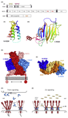
Figure 1. Molecular Determinants of Repulsive Guidance Molecules (RGMs) and Their Interactions with Neogenin (NEO1) and Bone Morphogenetic Protein (BMP) Ligands.
(A) (i) Schematic representation and domain organization of RGMs, Neogenin, and BMPs. Both the N-terminal domain (ii) [N-RGM; Protein Data Bank (PDB) ID 4UI1] and C-terminal domain (iii) (C-RGM; PDB ID 4BQ6) form distinct domains stabilized by several intramolecular disulfide bonds. Structures are shown in cartoon in rainbow coloring (blue, N terminus; red, C terminus). Note that RGD motifs are potential integrin-binding sites, but that no binding of RGMs to integrins has been reported so far. (B) The RGM–Neogenin complex (PDB ID 4BQ6). Two C-RGM molecules (blue) act as a molecular staple bringing together two Neogenin receptors (red). (C) The N-RGM–BMP complex (PDB ID 4UI1). The disulfide-linked BMP2 dimer binds two molecules of N-RGM in its wing region. The yellow asterisk indicates the position of the ‘RGD’ motif. (D) Model for RGM-mediated signaling in trans. The RGM ectodomain can be shed by proteolytic or phospholipase activity (open triangle). RGM binding to preclustered Neogenin results in stabilization and dimerization of the Neogenin ectodomain, subsequently activating downstream signaling (gray lightning bolt). The gray box highlights the RGM–Neogenin signaling hub observed in the crystal structure. (E) Model of RGM-mediated signaling in cis. RGMs can act as a physical protein bridge bringing together Neogenin and the BMP ligand, resulting in clustering. Abbreviations: FN, fibronectin; GDPH, autoproteolysis cleavage motif; GPI, glycosylphosphatidylinositol anchor; ICD, intracellular domain; IG, immunoglobulin; L, flexible linker; RGD, Arg-Gly-Asp; SP, signal peptide; TM, transmembrane; vWFD, von Willebrand Factor type D.
Figure 2
Figure 2
RGMs act as co-receptors for bone morphogenetic proteins (BMPs) and have been proposed to act as a structural bridge between BMPs and Neogenin. A recently proposed model suggests that RGMs induce endocytosis of the BMP receptor complex, thereby activating canonical SMAD signaling. Interactions between RGMs and BMP signaling have been implicated in iron homeostasis and endochondral bone development. Binding of RGM to Neogenin inhibits interactions between Lrig2 and Neogenin, allowing ectodomain shedding by A disintegrin and metalloproteinase domain-containing protein 17 (ADAM17) leading to signal termination. In general, RGM–Neogenin binding leads to the activation of RhoA through Unc5 and LARG, and inactivation of Ras through focal adhesion kinase (FAK) and p120 RasGAP to induce growth cone collapse. However, signaling is dependent on the proteolytic processing of RGMs, since C-RGM triggers RhoA-dependent signaling, while the effects of N-RGM rely on shedding of the Neogenin intracellular domain by γ-secretase and LMO4. The Neogenin intracellular domain has been proposed to move into the nucleus, possible together with LMO4, and regulate gene transcription. In epithelial cells, Neogenin binds and localizes the WAVE regulatory complex (WRC), leading to actin nucleation by Arp2/3, which also requires activation by Rac1 and adherence junction stability. The extent to which these signaling pathways are specific to select cell types or cellular functions remains to be determined. Key Figure Signaling Mechanisms Downstream of Repulsive Guidance Molecules (RGMs)
Figure 3
Figure 3. Proteolytic Processing of Repulsive Guidance Molecules (RGMa) Generates N- and C-RGMa Fragments that Regulate Distinct Aspects of Retinotectal Path Finding in vivo.
(A) Autocatalytic processing (arrowhead) and proteolysis by subtilisin kexin isozyme-1 (SKI-1) and furin generates C- and N-RGMa fragments. (B) Ectopic expression of C- and N-RGMa peptides in the chick optic tectum results in distinct axon-targeting defects. Normally, two gradients of Neogenin (blue) in the eye and RGMa (in the tectum) allow correct anterior–posterior targeting of retinal axons. In control experiments, all axons from retinal ganglion cells in the eye terminate before the terminal front (TF) in the tectum and arborize in layers a–f of the stratum griseum et fibrosum superficiale (SGFS). Ectopic expression of C-RGMa results in axonal overshooting beyond the TF, with aberrant retinal axons remaining restricted to the superficial stratum opticum (SO) layer. By contrast, ectopic expression of N-RGMa induces overshooting beyond the TF and into deeper layers (beyond SGFS layer g). Abbreviations: N, nasal; T, temporal.

References

    1. Kolodkin AL, Pasterkamp RJ. SnapShot: axon guidance II. Cell. 2013;153:722.e1. - PubMed
    1. Severyn CJ, et al. Molecular biology, genetics and biochemistry of the repulsive guidance molecule family. Biochem J. 2009;422:393–403. - PMC - PubMed
    1. Rajagopalan S, et al. Neogenin mediates the action of repulsive guidance molecule. Nat Cell Biol. 2004;6:756–762. - PubMed
    1. Matsunaga E, et al. RGM and its receptor neogenin regulate neuronal survival. Nat Cell Biol. 2004;6:749–755. - PubMed
    1. Babitt JL, et al. Bone morphogenetic protein signaling by hemojuvelin regulates hepcidin expression. Nat Genet. 2006;38:531–539. - PubMed

Publication types