

Recreating the Cardiac Microenvironment in Pluripotent Stem Cell Models of Human Physiology and Disease
- PMID: 28007424
- PMCID: PMC5403626
- DOI: 10.1016/j.tcb.2016.11.010
Recreating the Cardiac Microenvironment in Pluripotent Stem Cell Models of Human Physiology and Disease
Abstract
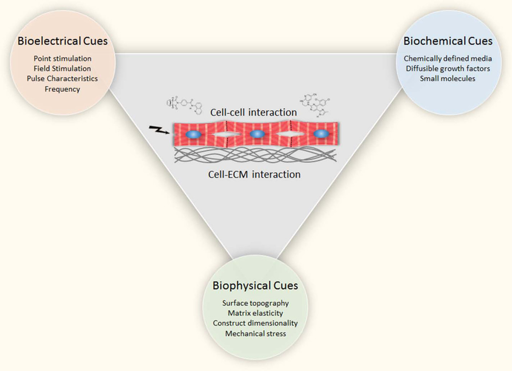
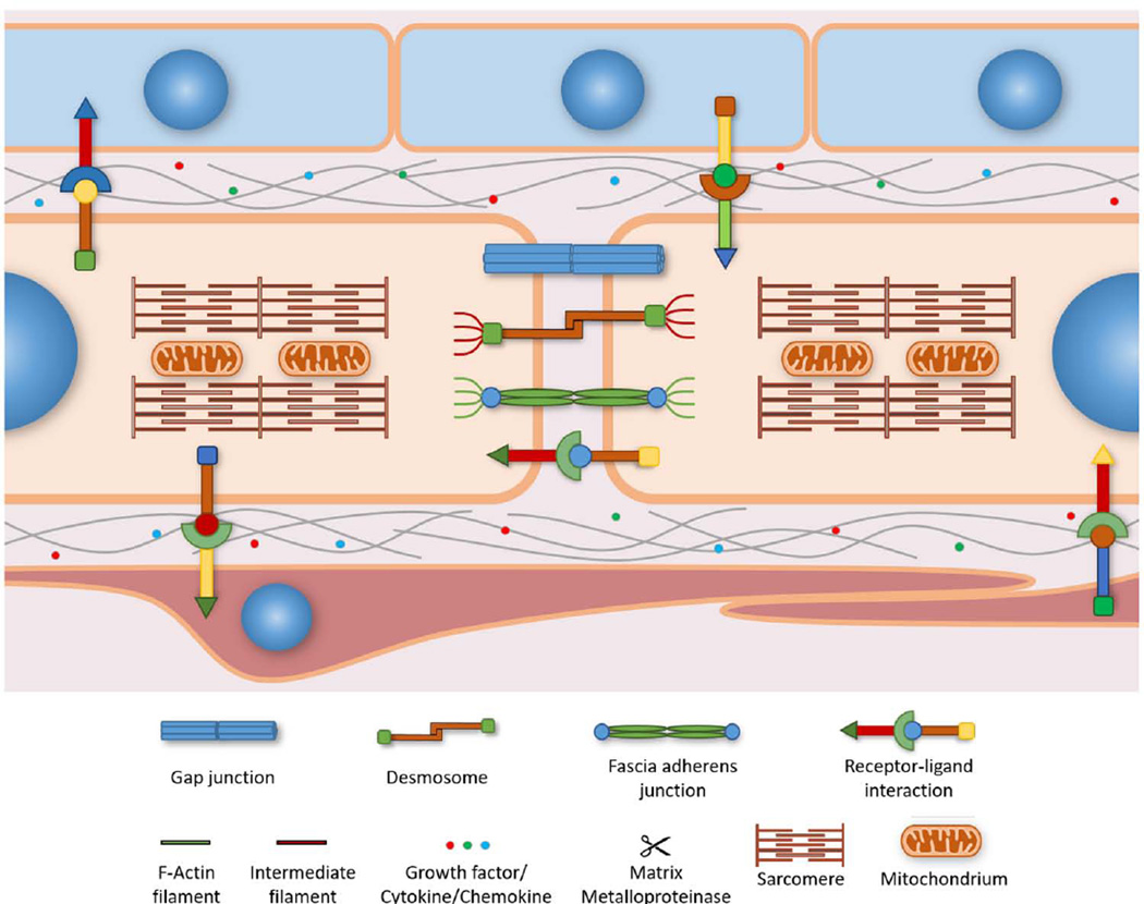
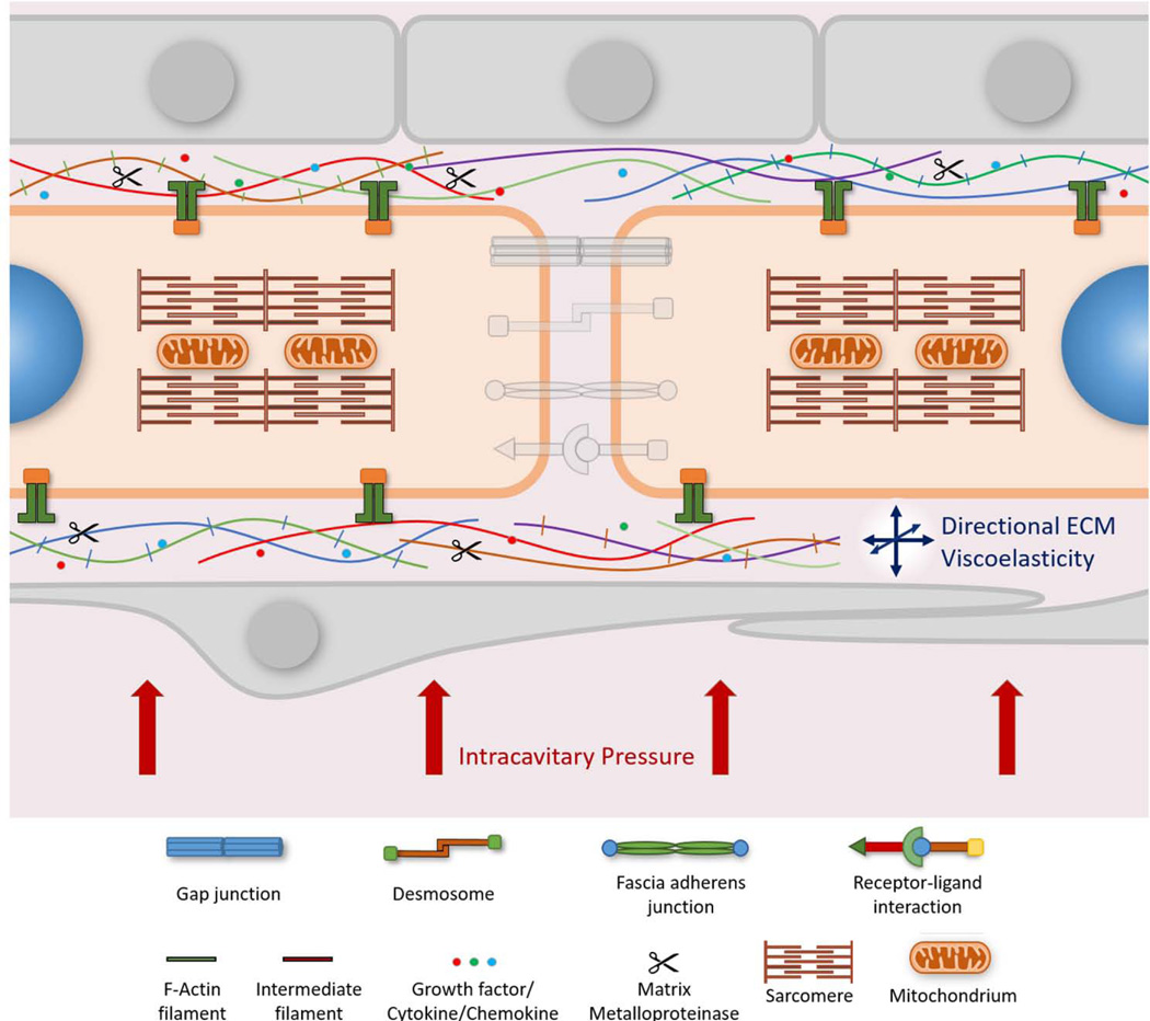
The advent of human pluripotent stem cell (hPSC) biology has opened unprecedented opportunities for the use of tissue engineering to generate human cardiac tissue for in vitro study. Engineering cardiac constructs that recapitulate human development and disease requires faithful recreation of the cardiac niche in vitro. Here we discuss recent progress in translating the in vivo cardiac microenvironment into PSC models of the human heart. We review three key physiologic features required to recreate the cardiac niche and facilitate normal cardiac differentiation and maturation: the biochemical, biophysical, and bioelectrical signaling cues. Finally, we discuss key barriers that must be overcome to fulfill the promise of stem cell biology in preclinical applications and ultimately in clinical practice.
Keywords: cardiac myocytes; cardiac niche; cellular microenvironment; disease modeling; pluripotent stem cells; tissue engineering.
Copyright © 2016 Elsevier Ltd. All rights reserved.
Figures
Similar articles
-
Human Pluripotent Stem Cell Mechanobiology: Manipulating the Biophysical Microenvironment for Regenerative Medicine and Tissue Engineering Applications.Stem Cells. 2015 Nov;33(11):3187-96. doi: 10.1002/stem.2105. Epub 2015 Jul 29. Stem Cells. 2015. PMID: 26189759 Review.
-
On human pluripotent stem cell control: The rise of 3D bioengineering and mechanobiology.Biomaterials. 2015 Jun;52:26-43. doi: 10.1016/j.biomaterials.2015.01.078. Epub 2015 Feb 21. Biomaterials. 2015. PMID: 25818411 Free PMC article. Review.
-
Physical developmental cues for the maturation of human pluripotent stem cell-derived cardiomyocytes.Stem Cell Res Ther. 2014 Oct 20;5(5):117. doi: 10.1186/scrt507. Stem Cell Res Ther. 2014. PMID: 25688759 Free PMC article. Review.
-
Human pluripotent stem cells: Prospects and challenges as a source of cardiomyocytes for in vitro modeling and cell-based cardiac repair.Adv Drug Deliv Rev. 2016 Jan 15;96:3-17. doi: 10.1016/j.addr.2015.05.004. Epub 2015 May 14. Adv Drug Deliv Rev. 2016. PMID: 25980938 Free PMC article. Review.
-
Design and formulation of functional pluripotent stem cell-derived cardiac microtissues.Proc Natl Acad Sci U S A. 2013 Dec 3;110(49):E4698-707. doi: 10.1073/pnas.1311120110. Epub 2013 Nov 19. Proc Natl Acad Sci U S A. 2013. PMID: 24255110 Free PMC article.
Cited by
-
Emerging technologies for cardiac tissue engineering and artificial hearts.Smart Med. 2023 Feb 16;2(1):e20220040. doi: 10.1002/SMMD.20220040. eCollection 2023 Feb. Smart Med. 2023. PMID: 39188557 Free PMC article.
-
YAP repression of the WNT3 gene controls hESC differentiation along the cardiac mesoderm lineage.Genes Dev. 2017 Nov 15;31(22):2250-2263. doi: 10.1101/gad.307512.117. Epub 2017 Dec 21. Genes Dev. 2017. PMID: 29269485 Free PMC article.
-
Remodeling the Human Adult Stem Cell Niche for Regenerative Medicine Applications.Stem Cells Int. 2017;2017:6406025. doi: 10.1155/2017/6406025. Epub 2017 Sep 27. Stem Cells Int. 2017. PMID: 29090011 Free PMC article. Review.
-
Glucocorticoids and programming of the microenvironment in heart.J Endocrinol. 2019 Jul 1;242(1):T121-T133. doi: 10.1530/JOE-18-0672. J Endocrinol. 2019. PMID: 31018174 Free PMC article. Review.
-
Recent advances in lineage differentiation from stem cells: hurdles and opportunities?F1000Res. 2018 Feb 23;7:220. doi: 10.12688/f1000research.12596.1. eCollection 2018. F1000Res. 2018. PMID: 29552337 Free PMC article. Review.
References
Publication types
MeSH terms
Grants and funding
LinkOut - more resources
Full Text Sources
Other Literature Sources
