Review
. 2017 Mar 20:605:20-31.
doi: 10.1016/j.gene.2016.12.014. Epub 2016 Dec 19.

Structure, biochemistry, and biology of PAK kinases

Affiliations
Review

Structure, biochemistry, and biology of PAK kinases

Rakesh Kumar et al. Gene. .

Abstract

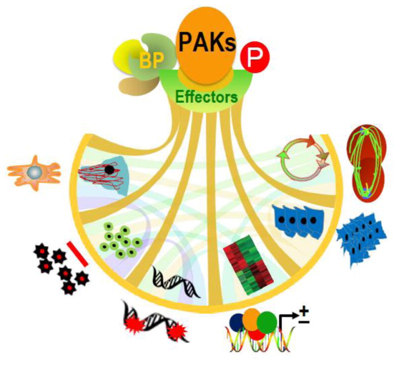
PAKs, p21-activated kinases, play central roles and act as converging junctions for discrete signals elicited on the cell surface and for a number of intracellular signaling cascades. PAKs phosphorylate a vast number of substrates and act by remodeling cytoskeleton, employing scaffolding, and relocating to distinct subcellular compartments. PAKs affect wide range of processes that are crucial to the cell from regulation of cell motility, survival, redox, metabolism, cell cycle, proliferation, transformation, stress, inflammation, to gene expression. Understandably, their dysregulation disrupts cellular homeostasis and severely impacts key cell functions, and many of those are implicated in a number of human diseases including cancers, neurological disorders, and cardiac disorders. Here we provide an overview of the members of the PAK family and their current status. We give special emphasis to PAK1 and PAK4, the prototypes of groups I and II, for their profound roles in cancer, the nervous system, and the heart. We also highlight other family members. We provide our perspective on the current advancements, their growing importance as strategic therapeutic targets, and our vision on the future of PAKs.

Keywords: Cancer; Cytoskeleton remodeling; Heart; Nervous system; PAK; p21-Activated kinase.


Conflict of interest statement

The authors have no conflicts of interest to declare.

Figures

Figure 1
Figure 1
PAK stimulation by upstream signals contributes to phenotypic signaling through binding partners (BP) and phosphorylation of effector substrates. Representative PAK-regulated cancerous phenotypes include – cytoskeleton remodeling, cell motility, inhibition of apoptosis, DNA damage response, gene expression, transformation and invasion, deregulated cell cycle progression and metastasis (from left to the right).
Figure 2
Figure 2
Structural domains of group I and group II PAKs. For PAK1, detail organization of polypeptide chain was shown. Numerals indicate residue numbers at the boundaries of various subdivisions. The N-terminal auto-regulatory region (70–149aa) and the kinase domain (272–523aa) are shown. The detailed substructure of the auto-regulatory region including overlapped PBD (p21-binding domain, in blue) and AID (auto inhibitory domain, in green) is shown in the expanded upper part of the PAK1 diagram. | indicates proline-rich region and indicates the interaction motif for PIX. For other PAKs, PBD, kinase domain, and proline-rich region are shown for each family member.
Figure 3
Figure 3
Models of PAK activation. P21-activated kinases (PAKs) have a conserved carboxyl-terminal serine/threonine kinase domain with a single phosphorylation (P) site and an amino-terminal regulatory domain. The regulatory domain of group I PAKs (PAK1–3) contain PBD (p21-binding domain) and overlapped AID (auto-inhibitory domain), which is structurally distinct from that of group II PAKs (PAK4–6). There are two suggested models for PAK4 activation.
Figure 4
Figure 4
Structure and partial transcripts of human PAK genes. The diagram/table and transcripts of PAK1~6 genes are based on the ENSEMBL database (PAK1 ENSG00000149269, PAK2 ENSG00000180370, PAK3 ENSG00000077264, PAK4 ENSG00000130669, PAK5 ENSG00000101349, PAK6 ENSG00000137843). The blues are alternatively spliced coding exons and the white ones are UTRs. The tables are based on the known protein coding transcripts, not including noncoding RNAs.
Figure 5
Figure 5
Representative substrates of group I and II PAKs and their subcellular localization.

References

    1. Abo A, et al. PAK4, a novel effector for Cdc42Hs, is implicated in the reorganization of the actin cytoskeleton and in the formation of filopodia. EMBO J. 1998;17:6527–6540. - PMC - PubMed
    1. Acconcia F, et al. Phosphorylation-dependent regulation of nuclear localization and functions of integrin-linked kinase. Proc Natl Acad Sci U S A. 2007;104:6782–6787. - PMC - PubMed
    1. Adam L, et al. Heregulin regulates cytoskeletal reorganization and cell migration through the p21-activated kinase-1 via phosphatidylinositol-3 kinase. J Biol Chem. 1998;273:28238–28246. - PubMed
    1. Advani SJ, et al. Kinase-independent role for CRAF-driving tumour radioresistance via CHK2. Nat Commun. 2015;6:8154. - PMC - PubMed
    1. Ahmed T, et al. A PAK4-LIMK1 pathway drives prostate cancer cell migration downstream of HGF. Cell Signal. 2008;20:1320–1328. - PubMed

MeSH terms

LinkOut - more resources