. 2017 Mar 1;215(5):703-712.
doi: 10.1093/infdis/jiw632.

Synergistic Interaction Between Phage Therapy and Antibiotics Clears Pseudomonas Aeruginosa Infection in Endocarditis and Reduces Virulence

Affiliations

Synergistic Interaction Between Phage Therapy and Antibiotics Clears Pseudomonas Aeruginosa Infection in Endocarditis and Reduces Virulence

Frank Oechslin et al. J Infect Dis. .

Abstract

Background: Increasing antibiotic resistance warrants therapeutic alternatives. Here we investigated the efficacy of bacteriophage-therapy (phage) alone or combined with antibiotics against experimental endocarditis (EE) due to Pseudomonas aeruginosa, an archetype of difficult-to-treat infection.

Methods: In vitro fibrin clots and rats with aortic EE were treated with an antipseudomonas phage cocktail alone or combined with ciprofloxacin. Phage pharmacology, therapeutic efficacy, and resistance were determined.

Results: In vitro, single-dose phage therapy killed 7 log colony-forming units (CFUs)/g of fibrin clots in 6 hours. Phage-resistant mutants regrew after 24 hours but were prevented by combination with ciprofloxacin (2.5 × minimum inhibitory concentration). In vivo, single-dose phage therapy killed 2.5 log CFUs/g of vegetations in 6 hours (P < .001 vs untreated controls) and was comparable with ciprofloxacin monotherapy. Moreover, phage/ciprofloxacin combinations were highly synergistic, killing >6 log CFUs/g of vegetations in 6 hours and successfully treating 64% (n = 7/11) of rats. Phage-resistant mutants emerged in vitro but not in vivo, most likely because resistant mutations affected bacterial surface determinants important for infectivity (eg, the pilT and galU genes involved in pilus motility and LPS formation).

Conclusions: Single-dose phage therapy was active against P. aeruginosa EE and highly synergistic with ciprofloxacin. Phage-resistant mutants had impaired infectivity. Phage-therapy alone or combined with antibiotics merits further clinical consideration.

Keywords: Pseudomonas aeruginosa; bacteriophage; endocarditis; phage therapy; resistance; antibiotic.


Figures

Figure 1.
Figure 1.
Activity of phage cocktail PP1131 in in vitro fibrin clots. Clots were produced from rat plasma and infected with 108 colony-forming units (CFUs)/mL of either phage-resistant Pseudomonas aeruginosa strain PA7 (A) or phage-susceptible strain CHA (B). Clots were left untreated (solid lines) or exposed to 108 PFUs/mL of PP1131 for 24 hours at 37°C (dashed lines). Phage titers in noninfected clots (solid lines) and in PA7-infected or CHA-infected clots (dashed lines) were determined 6 hours and 24 hours after exposure to the phage cocktail. Clots infected with strain PA7 lysed in spite of phage treatment (C), whereas those infected with phage-susceptible CHA and treated with phages remained intact (D). P values were determined using the Mann–Whitney test.
Figure 2.
Figure 2.
Bactericidal synergism between phages and selected antibiotics. Bacterial killing by phage-antibiotic combinations was tested by using 108 PFUs/mL of PP1131 with 2.5-times the minimum inhibitory concentration (MIC) of ciprofloxacin (A) or meropenem (B) (MIC of 0.19 µg/mL and 0.125 µg/mL, respectively). Each value represents the mean ± SD of 4–16 independent clots. Abbreviation: ABT, antibiotic.
Figure 3.
Figure 3.
Pharmacokinetics/pharmacodynamics (PK/PD) and therapeutic efficacy of phage cocktail PP1131 and ciprofloxacin in rats with experimental endocarditis (EE) due to Pseudomonas aeruginosa CHA. A, Pharmacokinetics in the absence of infection of phage cocktail PP1131 in rat plasma after either a single intravenous bolus injection (1 mL of 1010 PFUs/mL in 1 minute, dashed line, Bolus) or the same amount of phages administered through continuous infusion (0.1 mL/h of 1010 PFUs/mL over 24 hours, solid line: continuous infusion). Each value represents the mean ± SD from 8–10 individual animals. B, Bacterial loads of infected vegetations after 6 hours of phage treatment administered through continuous infusion or in single bolus and treatment with ciprofloxacin alone or combined with phages. Each dot represents the vegetation of a single animal. The mode of injection and type of treatment are indicated at the top of columns (C.I.: continuous infusion; φ: phages). Statistical results are indicated at the bottom of columns (†: control vs all types of treatment, P < .0005; ‡: combination vs control and all other types of treatments, P < .0001; results were compared by the Mann–Whitney test). C, Phage titers in vegetations measured in uninfected rats treated with bolus phage injection (control) or infected rats treated with the same regimens as in Figure 2B (***, P < .0001 using the Mann–Whitney test). D, Correlation between the decrease in vegetation bacterial loads and the vegetation phage titers resulting from in situ phage amplification (continuous infusion and bolus injection pooled together; correlation value [r] = −0.66; Pearson 2-tailed correlation test: P = .003). Abbreviation: cip, ciprofloxacin.
Figure 4.
Figure 4.
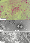
Light microscopy and transmission electron microscopy of rat vegetations after 6 hours of phage therapy. A, Semithin section (200 nm) of control vegetation infected with Pseudomonas aeruginosa CHA stained using the modified Braun Brenn staining protocol [37]. Arrows indicate bacteria, stained in red. B, Transmission electron microscopy of ultrathin sections (50 nm) of control vegetation infected with P. aeruginosa CHA positive-stained using uranium acetate and lead citrate. Arrows indicate bacteria. C, Example of a negative staining of a myoviridae phage present in the cocktail; other phages morphotypes including podoviridae were also present. D, Transmission electron microscopy of vegetation infected by P. aeruginosa CHA and treated with the phage cocktail. Arrows indicate phage capsids inside lysed bacteria. Abbreviation: RBC, red blood cell.
Figure 5.
Figure 5.
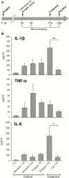
Cytokine quantification in rat plasma during experimental endocarditis. A, Protocol of blood sampling for cytokine quantification. B, Levels of interleukin 1β (IL-1β), interleukin 6 (IL-6), and tumor necrosis factor α (TNF-α) measured after 6 hours of phage or antibiotherapy (treatment). Controls included rats 24 hours after surgery (inoculum, T0), untreated but infected rats for 18 hours or 24 hours (bacteria T18 and T24), and uninfected rats receiving phage for 6 hours (Phages T6). Each value represents the mean ± SEM from 4–10 individual animals (*, P = .03; **, P = .005 using the Mann–Whitney test). Abbreviation: cip, ciprofloxacin.
Figure 6.
Figure 6.
Infectivity of phage-resistant Pseudomonas aeruginosa mutants in rats with catheter-induced vegetations. A, Two phage resistant P. aeruginosa mutants, 24/2 and 19/2, were isolated in vitro from fibrin clots and showed different phage resistance patterns. Lysis zones at the site of the phage deposits indicated phage-sensitivity. The absence of lysis indicated phage resistance. B, The isolates’ infectivity was tested in rats with catheter-induced vegetations. Both variants showed a loss of infectivity (>40% for 24/2 and >70% for 19/2) as compared with the parent strain CHA. P values were determined using Fisher’s exact test.
Figure 7.
Figure 7.
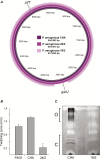
Characterization of phage-resistant Pseudomonas aeruginosa mutants. The 2 phage-resistant P. aeruginosa mutants, 24/2 and 19/2, isolated in vitro were further characterized regarding to genomic content and phenotypes. A, Variant 24/2 displayed a 15-basepair deletion in the pilT gene, and variant 19/2 displayed a 362-kb chromosomal deletion that encompassed the galU gene. B, Impaired twitching motility resulting from the pilT alteration in variant 24/2, as compared with wild-type PAO1 and parent CHA. C, Impaired LPS synthesis resulting from the galU deletion in variant 19/2 with absence of O-antigen (O) and LPS core (C), as compared with parent CHA.

Comment in

References

    1. Chanishvili N. Phage therapy—history from Twort and d’Herelle through Soviet experience to current approaches. Adv Virus Res 2012; 83:3–40. - PubMed
    1. Abedon ST. Phage therapy of pulmonary infections. Bacteriophage 2015; 5:e1020260. - PMC - PubMed
    1. Lang G, Kehr P, Mathevon H, Clavert JM, Séjourne P, Pointu J. Bacteriophage therapy of septic complications of orthopaedic surgery. Rev Chir Orthop Reparatrice Appar Mot 1979; 65:33–7. - PubMed
    1. Speck P, Smithyman A. Safety and efficacy of phage therapy via the intravenous route. FEMS Microbiol Lett 2016; 363. - PubMed
    1. Chhibber S, Kaur S, Kumari S. Therapeutic potential of bacteriophage in treating Klebsiella pneumoniae B5055-mediated lobar pneumonia in mice. J Med Microbiol 2008; 57:1508–13. - PubMed