. 2017 Feb 17;292(7):2646-2659.
doi: 10.1074/jbc.M116.771808. Epub 2016 Dec 22.

c-JUN Dimerization Protein 2 (JDP2) Is a Transcriptional Repressor of Follicle-stimulating Hormone β (FSHβ) and Is Required for Preventing Premature Reproductive Senescence in Female Mice

Affiliations

c-JUN Dimerization Protein 2 (JDP2) Is a Transcriptional Repressor of Follicle-stimulating Hormone β (FSHβ) and Is Required for Preventing Premature Reproductive Senescence in Female Mice

Carrie R Jonak et al. J Biol Chem. .

Abstract

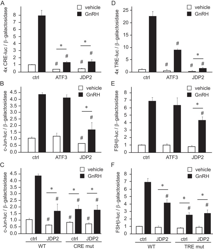
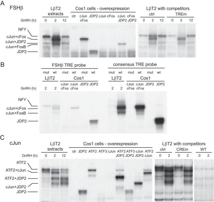
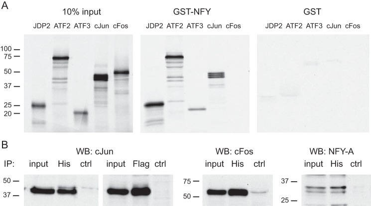
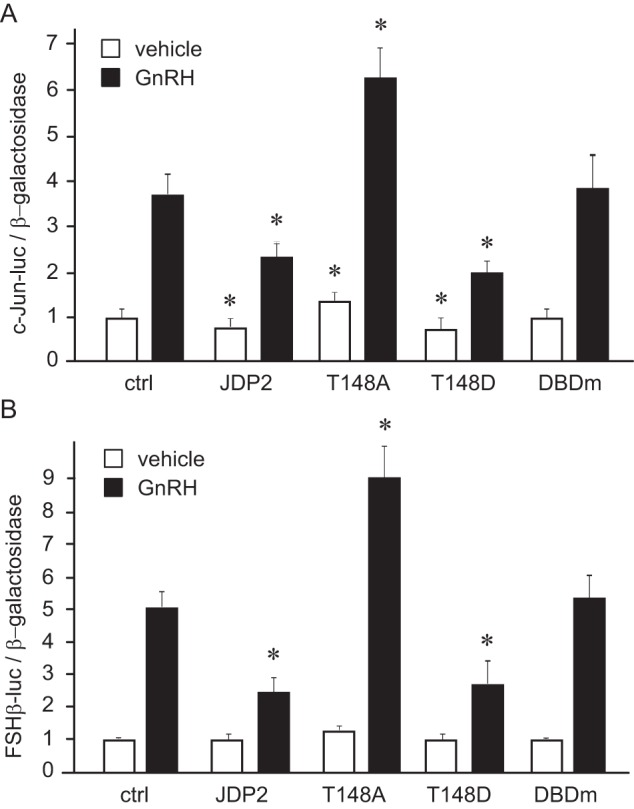
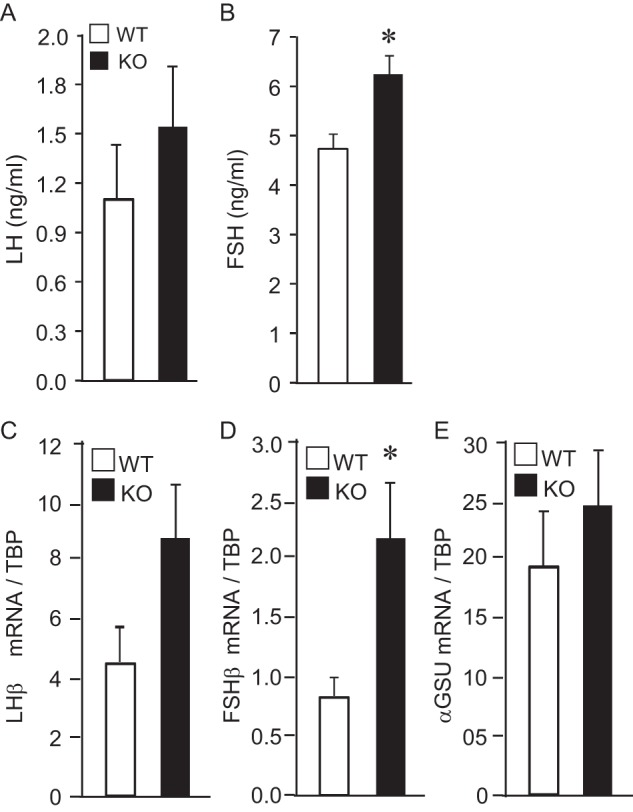
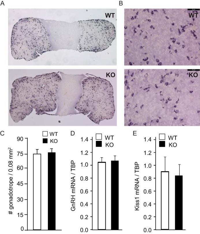
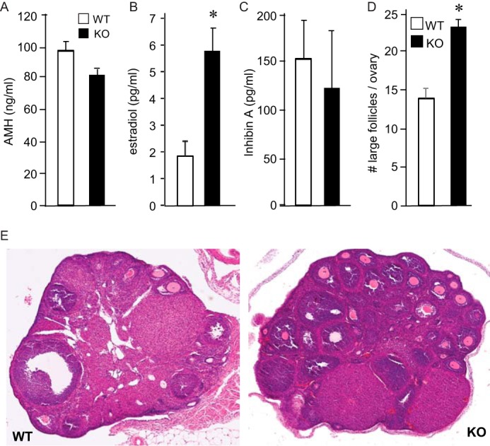
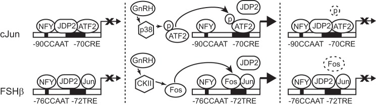
Follicle-stimulating hormone (FSH) regulates follicular growth and stimulates estrogen synthesis in the ovaries. FSH is a heterodimer consisting of an α subunit, also present in luteinizing hormone, and a unique β subunit, which is transcriptionally regulated by gonadotropin-releasing hormone 1 (GNRH). Because most FSH is constitutively secreted, tight transcriptional regulation is critical for maintaining FSH levels within a narrow physiological range. Previously, we reported that GNRH induces FSHβ (Fshb) transcription via induction of the AP-1 transcription factor, a heterodimer of c-FOS and c-JUN. Herein, we identify c-JUN-dimerization protein 2 (JDP2) as a novel repressor of GNRH-mediated Fshb induction. JDP2 exhibited high basal expression and bound the Fshb promoter at an AP-1-binding site in a complex with c-JUN. GNRH treatment induced c-FOS to replace JDP2 as a c-JUN binding partner, forming transcriptionally active AP-1. Subsequently, rapid c-FOS degradation enabled reformation of the JDP2 complex. In vivo studies revealed that JDP2 null male mice have normal reproductive function, as expected from a negative regulator of the FSH hormone. Female JDP2 null mice, however, exhibited early puberty, observed as early vaginal opening, larger litters, and early reproductive senescence. JDP2 null females had increased levels of circulating FSH and higher expression of the Fshb subunit in the pituitary, resulting in elevated serum estrogen and higher numbers of large ovarian follicles. Disruption of JDP2 function therefore appears to cause early cessation of reproductive function, a condition that has been associated with elevated FSH in women.

Keywords: AP-1 transcription factor (AP-1); c-FOS; c-JUN transcription factor; endocrinology; follicle-stimulating hormone (FSH); gene expression; gene transcription.


Conflict of interest statement

The authors declare that they have no conflicts of interest with the contents of this article

Figures

FIGURE 1.
FIGURE 1.
Time line of GNRH induction of gonadotrope genes. A, FSHβ (Fshb; solid line) and c-JUN (c-Jun; dashed line) mRNA expression was determined by qPCR following 10 nm GNRH treatment of LβT2 cells for the times indicated on the x axis and normalized to the Tbp mRNA. B, mRNA of the putative repressors, Jdp2 (solid line) and Atf3 (dashed line), was analyzed in the same samples and also normalized to Tbp mRNA. The experiment was repeated three times, and results are presented as an average ± S.E. (error bars). C, protein levels of transcriptional regulators in the whole cell lysates following GNRH treatment for the times indicated above each lane were analyzed by Western blotting.
FIGURE 2.
FIGURE 2.
JDP2 diminishes GNRH induction of c-Jun and FSHβ. A, four tandem copies of the CRE element (TGACGTCA) were linked to the minimal heterologous promoter and luciferase reporter in pGL3 backbone and transfected into LβT2 cells with the herpesvirus thymidine kinase-driven β-galactosidase gene as an internal control for transfection efficiency; additionally, cells were co-transfected with empty vector control (ctrl) or expression vector for ATF3 or JDP2. After overnight starvation, cells were treated with vehicle or 10 nm GNRH for 5 h, after which the luciferase and β-galactosidase values were obtained. B and C, c-JUN luciferase plasmid (−1000 bp) was used as a reporter. D, four copies of TRE element (TGAGTCA) served as a reporter. E and F, −1000 bp FSHβ luciferase was transfected as a reporter. *, significant change (p < 0.05) in -fold induction by GNRH, where the GNRH-treated luciferase/β-galactosidase ratio was normalized to vehicle-treated samples transfected with the same expression vector. #, significantly lower luciferase expression from the empty vector control (vehicle-treated samples transfected with overexpression vector compared with vehicle-treated empty vector, and GNRH-treated samples transfected with overexpression compared with GNRH-treated vector control). White bars, vehicle; black bars, GNRH-treated. Error bars, S.E.
FIGURE 3.
FIGURE 3.
JDP2 binds FSHβ (A and B) and c-Jun (C) promoters. Nuclear extract from either LβT2 cells treated with GNRH for the times indicated above the lanes or COS-1 cells transfected with overexpression vectors for proteins indicated above the corresponding lanes was incubated with a 30-bp radiolabeled probe that encompasses the AP-1/TRE site in the FSHβ promoter (A), a 30-bp radiolabeled probe encompassing the AP-1/TRE site in the FSHβ promoter (TTGGTCA) or its mutation (TTGaaaA) compared with AP-1/TRE consensus (TGAGTCA) or its mutation (TGAaaaA) (B), or a 30-bp radiolabeled probe encompassing the CRE site in the c-JUN promoter (C). EMSA was performed three times, and representative gels subjected to autoradiography are shown.
FIGURE 4.
FIGURE 4.
JDP2 interacts with NFY. A, GST pull-down assays demonstrate that NFY can interact directly with JDP2, ATF2, ATF3, and c-JUN, but not c-FOS. 35S-Labeled proteins, indicated above each panel, were used in the binding assay with GST-NFY fusion protein or control GST, labeled above each panel. In the input panel, 10% of the in vitro transcribed and translated labeled proteins that were used in the binding reaction were run on the gel as a control for their expression and labeling. B, cells were transfected with histidine tag-JDP2 (His) or FLAG-tagged-c-FOS, and complexes were precipitated (IP) with antibodies to His tag or FLAG tag, run on the gel, and transferred, and membranes were probed with antibodies to c-JUN, c-FOS, and NFY-A to determine proteins that interact with JDP2. WB, Western blotting.
FIGURE 5.
FIGURE 5.
Threonine 148 is necessary for JDP2 repressor activity. c-JUN luciferase (A) or FSHβ luciferase (B) reporter were transfected into LβT2 cells with empty vector control, wild-type JDP2 (JDP2), or JDP2 mutated at the threonine 148 to alanine (T148A) or to glutamic acid (T148D) or with JDP2 with a mutated DNA-binding domain (DBDm) and treated with vehicle or GNRH for 5 h. *, statistically significant difference (p < 0.05) in reporter expression with JDP2 overexpression following vehicle or GNRH treatment, compared with empty vector control following vehicle or GNRH, respectively. White bars, vehicle; black bars, GNRH-treated. Error bars, S.E.
FIGURE 6.
FIGURE 6.
Histone 3 acetylation increases following GNRH treatment. Chromatin immunoprecipitation was performed using antibodies against acetylated histone H3 and total histone H3 following LβT2 cell treatment with GNRH for the times indicated below each bar. The percentage of precipitated chromatin with each antibody at each time point was calculated using serial dilution of total input chromatin, and then acetylated H3 was normalized to total H3. *, statistically significant increase (p < 0.05) in acetylated H3 compared with the zero time point. Error bars, S.E.
FIGURE 7.
FIGURE 7.
JDP2 null females exhibit reproductive anomalies. White bars, wild type (WT); black bars, JDP2 nulls (KO); 5 mice/group. *, significant difference (p < 0.05) in analyzed reproductive parameters in the female JDP2 null animals from the wild type animals. Error bars, S.E.
FIGURE 8.
FIGURE 8.
JDP2 null females have higher levels of FSH and higher expression of Fshb. A and B, serum from six 8-week-old females per genotype was analyzed by a radioimmunoassay for LH and FSH. C–E, whole pituitary mRNA was extracted from 6 animals/group, and mRNA levels for Lhb (C), Fshb (D), and common Cga encoding α-GSU (E) were determined by qPCR. White bars, wild type; black bars, JDP2 nulls. *, significant difference in the JDP2 null animals from the wild type animals; p < 0.05. Error bars, S.E.
FIGURE 9.
FIGURE 9.
Pituitaries and gonadotrope number are unchanged in JDP2 null females. A and B, pituitaries were stained with antibodies to LH, and representative images are shown at low magnification (A) and higher magnification (B) (×40 objective; bar, 50 μm). C, gonadotropes from three different 8-week-old females per group were counted, and numbers are presented per 0.08 mm2. White bar, wild type; black bar, JDP2 nulls. D and E, hypothalamus mRNA was extracted from 6 females/genotype, and Gnrh and kisspeptin (Kiss1) mRNA were analyzed by qPCR. White bars, wild type (WT); black bars, JDP2 nulls (KO).
FIGURE 10.
FIGURE 10.
JDP2 null females exhibit elevated estrogen levels and higher number of large follicles in the ovaries. White bars, wild type; black bars, JDP2 nulls. A–C, serum AMH, estradiol, and inhibin A were analyzed in estrus females by ELISA. D, large follicles with multilayer granulosa cells were counted throughout the ovary. *, significant difference in the JDP2 null animals from the wild type animals. E, bottom panels, representative image of H&E stain of the wild-type (WT) and null (KO) ovaries. Error bars, S.E.
FIGURE 11.
FIGURE 11.
JDP2 repression of c-Jun and FSHβ gene expression. JDP2 is present in the gonadotrope at the basal level and inhibits c-JUN and FSHβ transcription, indicated with X. JDP2 binds c-JUN and FSHβ promoters at the CRE and TRE elements, respectively, and interacts with basal transcription factor NFY. JDP2 also interacts with ATF2 that binds the c-JUN promoter at the basal state alone and as a heterodimer with JDP2 (top left) and with c-JUN that is expressed at some basal level and binds the FSHβ promoter as a heterodimer with JDP2 (bottom left). GNRH treatment leads to phosphorylation and activation of ATF2 by p38 MAPK, which causes dissociation of JDP2 and induction of c-JUN transcription, indicated with an arrow (top middle). GNRH treatment also leads to induction of c-FOS via calcium calmodulin kinase II (CKII) activation, after which c-FOS displaces JDP2 as a c-JUN binding partner and activates FSHβ transcription (bottom middle). Dephosphorylation of ATF2 (top right) or degradation of c-FOS proteins due to their short half-life (bottom right) leads to recruitment of JDP2 and cessation of transcription until the next pulse of GNRH.

Similar articles

Cited by

References

    1. Ortolano G. A., Haisenleder D. J., Dalkin A. C., Iliff-Sizemore S. A., Landefeld T. D., Maurer R. A., and Marshall J. C. (1988) Follicle-stimulating hormone β subunit messenger ribonucleic acid concentrations during the rat estrous cycle. Endocrinology 123, 2946–2948 - PubMed
    1. Zmeili S. M., Papavasiliou S. S., Thorner M. O., Evans W. S., Marshall J. C., and Landefeld T. D. (1986) α and luteinizing hormone β subunit messenger ribonucleic acids during the rat estrous cycle. Endocrinology 119, 1867–1869 - PubMed
    1. Huhtaniemi I. (2006) Mutations along the pituitary-gonadal axis affecting sexual maturation: novel information from transgenic and knockout mice. Mol. Cell Endocrinol. 254, 84–90 - PubMed
    1. Huhtaniemi I., Ahtiainen P., Pakarainen T., Rulli S. B., Zhang F. P., and Poutanen M. (2006) Genetically modified mouse models in studies of luteinising hormone action. Mol. Cell Endocrinol. 252, 126–135 - PubMed
    1. Lamminen T., Jokinen P., Jiang M., Pakarinen P., Simonsen H., and Huhtaniemi I. (2005) Human FSH beta subunit gene is highly conserved. Mol. Hum. Reprod. 11, 601–605 - PubMed

Publication types

MeSH terms

Substances