. 2017 Feb 17;292(7):2679-2689.
doi: 10.1074/jbc.M116.769216. Epub 2016 Dec 22.

Interaction of p190A RhoGAP with eIF3A and Other Translation Preinitiation Factors Suggests a Role in Protein Biosynthesis

Affiliations

Interaction of p190A RhoGAP with eIF3A and Other Translation Preinitiation Factors Suggests a Role in Protein Biosynthesis

Prasanna Parasuraman et al. J Biol Chem. .

Abstract

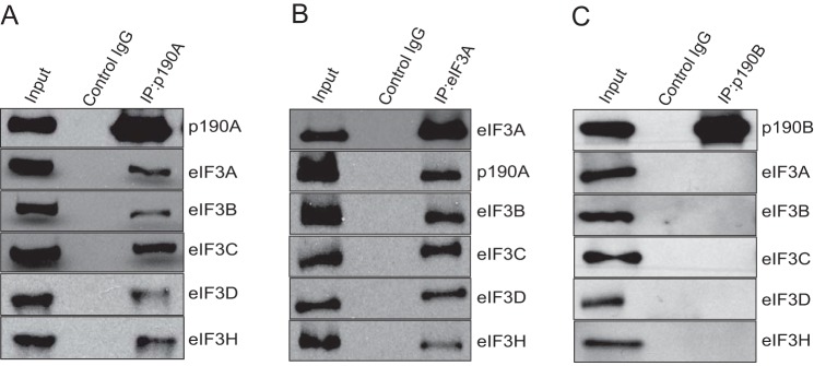
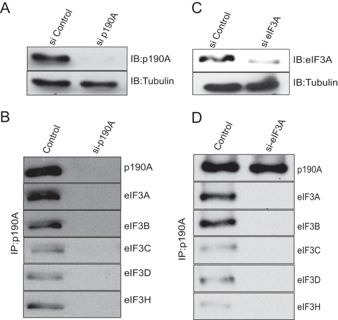
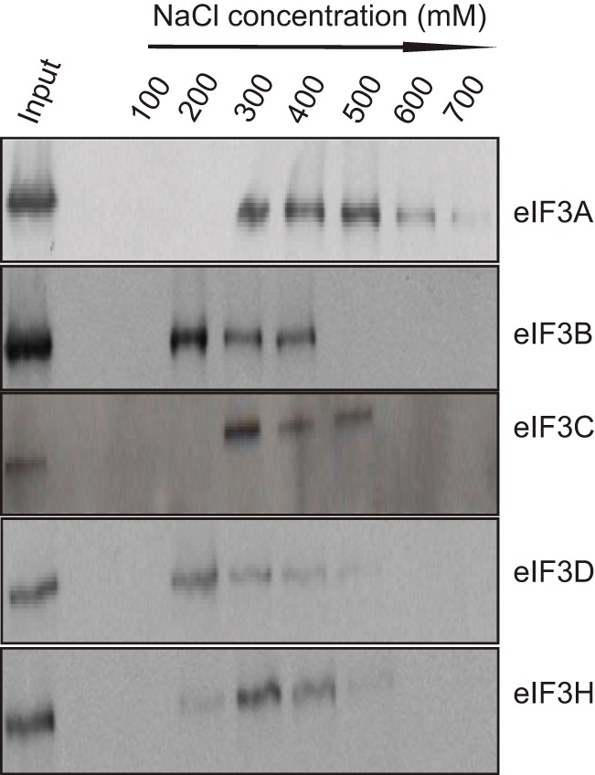
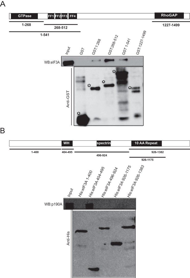
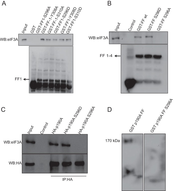
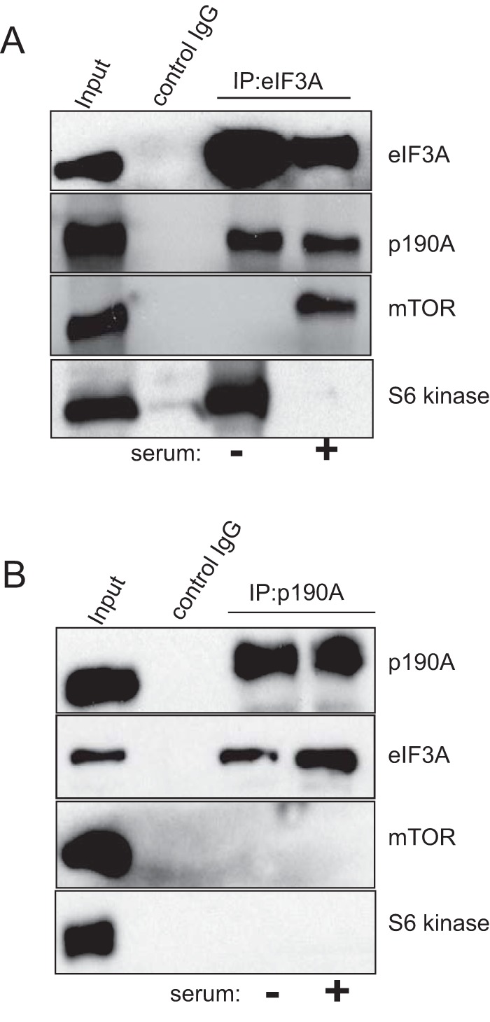
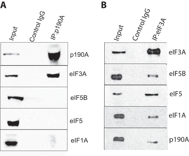
The negative regulator of Rho family GTPases, p190A RhoGAP, is one of six mammalian proteins harboring so-called FF motifs. To explore the function of these and other p190A segments, we identified interacting proteins by tandem mass spectrometry. Here we report that endogenous human p190A, but not its 50% identical p190B paralog, associates with all 13 eIF3 subunits and several other translational preinitiation factors. The interaction involves the first FF motif of p190A and the winged helix/PCI domain of eIF3A, is enhanced by serum stimulation and reduced by phosphatase treatment. The p190A/eIF3A interaction is unaffected by mutating phosphorylated p190A-Tyr308, but disrupted by a S296A mutation, targeting the only other known phosphorylated residue in the first FF domain. The p190A-eIF3 complex is distinct from eIF3 complexes containing S6K1 or mammalian target of rapamycin (mTOR), and appears to represent an incomplete preinitiation complex lacking several subunits. Based on these findings we propose that p190A may affect protein translation by controlling the assembly of functional preinitiation complexes. Whether such a role helps to explain why, unique among the large family of RhoGAPs, p190A exhibits a significantly increased mutation rate in cancer remains to be determined.

Keywords: GTPase activating protein (GAP); Rho (Rho GTPase); cell biology; mass spectrometry (MS); protein biosynthesis; protein complexes; protein domains; translation ini; translation initiation factor; translational initiation.


Conflict of interest statement

The authors declare that they have no conflicts of interest with the contents of this article

Figures

FIGURE 1.
FIGURE 1.
Subcellular localization of p190A RhoGAP and affinity purification of associated proteins. A, in HeLa cell fractions prepared by differential centrifugation, most p190A was found in the detergent-soluble membrane fraction. β2-Adrenergic receptor (β2-AR) and HDAC1 antibodies were used to monitor fraction purity. Equal amounts of protein were loaded in each lane (B). Silver-stained gel, showing that multiple proteins eluted from an anti-p190A affinity matrix did not interact with a non-immune IgG affinity matrix. The samples represent growth factor-stimulated cells. Essentially identical results were obtained when an extract of serum-starved HeLa cells was similarly analyzed (not shown).
FIGURE 2.
FIGURE 2.
eIF3 subunits co-precipitate with p190A, and vice versa. A, five endogenous HeLa cell eIF3 subunits co-immunoprecipitate with endogenous p190A. B, p190A, eIF3B, eIF3C, eIF3F, and eIF3H co-precipitate with eIF3A. C, endogenous p190B precipitated from HeLa cells does not detectably associate with the five tested eIF3 subunits.
FIGURE 3.
FIGURE 3.
Co-precipitation of p190A and eIF3 subunits is prevented by knockdown of eIF3A or p190A. HeLa cells were transfected with a siRNA control, or with p190A (panels A and B) or eIF3A (panels C and D) siRNA Smartpools. After transfection cells were cultured for 72 h prior to lysis and processing for immunoprecipitation. Panels A and C show the levels of p190A or eIF3A knockdown achieved. Panels B and D show that p190A or eIF3A knockdown prevents the co-precipitation of the proteins indicated to the right. The control lanes in panels B and D show p190A co-precipitation of the indicated eIF3 subunits from untreated HeLa cells.
FIGURE 4.
FIGURE 4.
Among five tested eIF3 subunits, the p190A-eIF3A co-precipitation is most resistant to increased ionic strength. HeLa cell extracts were prepared by lysing cells in 50 mm NaCl containing RIPA buffer. p190A immunoprecipitates collected on Protein A-Sepharose beads were washed with 0.2 ml of RIPA buffer of the indicated ionic strengths, after which 50 μl of the washes were subjected to SDS-PAGE and immunoblotting using the indicated antibodies.
FIGURE 5.
FIGURE 5.
Mapping of the p190A and eIF3A interacting segments. The indicated segments of p190A (diagram in panel A) and eIF3A (panel B) were produced as GST fusion proteins. The central region of p190A could not be made as a soluble fusion protein and has not been analyzed. The anti-GST or anti-His tag blots in panels A and B show the sizes and relative amounts of GST proteins used in the pull downs. The eIF3A Western blot (WB) in panel A show that eIF3A interacts weakly with the p190A GTPase domain (residues 1–268), and more strongly with a fusion protein representing all four FF domains (residues 268–512). Interaction is also observed with a fusion protein representing p190A residues 1–512. The p190A Western blot in panel B shows that only the winged helix (WH) segment of the eIF3A PCI domain interacts with p190A in this assay.
FIGURE 6.
FIGURE 6.
Further characterization of the p190A/eIF3A interaction. A, similar amounts of eIF3A are pulled down by wild-type and nucleotide binding defective GST-p190A1–512 fusion proteins. The anti-GST blot shows that similar amounts of fusion protein were used in the pull downs. B, GST pulldown showing that eIF3A specifically interacts with a fusion protein representing the first p190A FF domain. The protein segments represented by the four indicated GST fusion proteins are documented under “Experimental Procedures.” C, the amount of eIF3A that co-precipitated with N terminally HA-tagged full-length p190A (lane 3) is reduced by prior λ-phosphatase treatment (lane 4). Addition of a phosphatase inhibitor partially prevented this reduction (lane 5). Lane 1 shows input proteins; lane 2 is empty. D, quantification of the amount of eIF3A co-precipitating with HA-p190A. Shown is the average band intensity of three biological replicates. Error bars indicate S.D. Column numbers refer to the conditions specified in panel C.
FIGURE 7.
FIGURE 7.
Missense mutations of first FF domain residues differentially affect eIF3A interactions. A, Ser296 and Tyr308 are the only known phosphorylated residues in the first FF domain. Phosphomimicking S/D or Y/D and non-phosphorylatable S/A or Y/A missense mutations of Ser296 or Tyr308, as well as of an adjacent Tyr310 residue were generated in the context of the GST-FF1 fusion protein and used to pull down eIF3A. The S296A mutant fusion protein was the only one that did not interact with eIF3A. B, the S296A missense mutation in the context of the GST-FF1–4 (residues 268–512) fusion protein also prevented the pull down of eIF3A. C, eIF3A co-precipitates with full-length wild-type and S296D p190A, but not with S296A p190. N terminally HA-tagged p190A vectors were transfected in HeLa cells, and proteins were precipitated using an anti-HA monoclonal antibody. D, blots of size-fractionated HeLa cell lysates were sequentially probed with wild-type or S296A mutant GST-FF1 fusion proteins, and with an anti-GST antibody. The band labeled eIF3A detected by the wild-type but not by the mutant probe is of the size expected for eIF3A. WB, Western blot.
FIGURE 8.
FIGURE 8.
p190A, mTOR, and S6 kinase form distinct complexes with eIF3A. The input lanes in both panels show blots of cell lysates probed with the antisera indicated on the right. The lanes labeled “control IgG” show that eIF3A, p190A, mTOR, or S6 kinase are not precipitated by a non-immune antibody. Panel A shows that p190A co-IPs with eIF3A in both serum-starved and -stimulated cells, whereas mTOR and S6 kinase show reciprocal interactions with eIF3A, as previously reported. Panel B shows that unlike eIF3A, mTOR and S6 kinase do not co-precipitate with p190A in either serum-starved or -stimulated cells.
FIGURE 9.
FIGURE 9.
Not all preinitiation complex subunits co-precipitate with p190A. A, eiF3A co-precipitates with p190A, but eIF5, eIF5B, and eIF1A do not. B, all indicated proteins co-precipitate with eIF3A.

Similar articles

Cited by

References

    1. Jaffe A. B., and Hall A. (2005) Rho GTPases: biochemistry and biology. Annu. Rev. Cell Dev. Biol. 21, 247–269 - PubMed
    1. Hall A. (2012) Rho family GTPases. Biochem. Soc. Trans. 40, 1378–1382 - PubMed
    1. Bernards A. (2003) GAPs galore! a survey of putative Ras superfamily GTPase activating proteins in man and Drosophila. Biochim. Biophys. Acta 1603, 47–82 - PubMed
    1. Ellis C., Moran M., McCormick F., and Pawson T. (1990) Phosphorylation of GAP and GAP-associated proteins by transforming and mitogenic tyrosine kinases. Nature 343, 377–381 - PubMed
    1. Moran M. F., Polakis P., McCormick F., Pawson T., and Ellis C. (1991) Protein-tyrosine kinases regulate the phosphorylation, protein interactions, subcellular distribution, and activity of p21ras GTPase-activating protein. Mol. Cell. Biol. 11, 1804–1812 - PMC - PubMed

Publication types

MeSH terms

Substances

LinkOut - more resources