Frontline Science: Myeloid-derived suppressor cells (MDSCs) facilitate maternal-fetal tolerance in mice
- PMID: 28007981
- PMCID: PMC5380379
- DOI: 10.1189/jlb.1HI1016-306RR
Frontline Science: Myeloid-derived suppressor cells (MDSCs) facilitate maternal-fetal tolerance in mice
Abstract
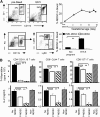
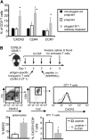
During successful pregnancy, a woman is immunologically tolerant of her genetically and antigenically disparate fetus, a state known as maternal-fetal tolerance. How this state is maintained has puzzled investigators for more than half a century. Diverse, immune and nonimmune mechanisms have been proposed; however, these mechanisms appear to be unrelated and to act independently. A population of immune suppressive cells called myeloid-derived suppressor cells (MDSCs) accumulates in pregnant mice and women. Given the profound immune suppressive function of MDSCs, it has been suggested that this cell population may facilitate successful pregnancy by contributing to maternal-fetal tolerance. We now report that myeloid cells with the characteristics of MDSCs not only accumulate in the circulation and uterus of female mice following mating but also suppress T cell activation and function in pregnant mice. Depletion of cells with the phenotype and function of MDSCs from gestation d 0.5 through d 7.5 resulted in implantation failure, increased T cell activation, and increased T cell infiltration into the uterus, whereas induction of MDSCs restored successful pregnancy and reduced T cell activation. MDSC-mediated suppression during pregnancy was accompanied by the down-regulation of L-selectin on naïve T cells and a reduced ability of naïve T cells to enter lymph nodes and become activated. Because MDSCs regulate many of the immune and nonimmune mechanisms previously attributed to maternal-fetal tolerance, MDSCs may be a unifying mechanism promoting maternal-fetal tolerance, and their induction may facilitate successful pregnancy in women who spontaneously abort or miscarry because of dysfunctional maternal-fetal tolerance.
Keywords: G-CSF; T cell activation; immune suppression; implantation; myeloid cells; pregnancy.
© Society for Leukocyte Biology.
Figures


Comment in
-
Editorial: The rebirth of myeloid-derived suppressor cells: from adversary in cancer to ally in reproductive health.J Leukoc Biol. 2017 May;101(5):1079. doi: 10.1189/jlb.1CE1216-525R. J Leukoc Biol. 2017. PMID: 28468987 No abstract available.
Similar articles
-
Granulocytic myeloid-derived suppressor cells maintain feto-maternal tolerance by inducing Foxp3 expression in CD4+CD25-T cells by activation of the TGF-β/β-catenin pathway.Mol Hum Reprod. 2016 Jul;22(7):499-511. doi: 10.1093/molehr/gaw026. Epub 2016 Mar 25. Mol Hum Reprod. 2016. PMID: 27016139
-
Bushen Antai recipe alleviates embryo absorption by enhancing immune tolerance and angiogenesis at the maternal-fetal interface via mobilizing MDSCs in abortion-prone mice.Phytomedicine. 2024 Jan;123:155164. doi: 10.1016/j.phymed.2023.155164. Epub 2023 Oct 25. Phytomedicine. 2024. PMID: 37952407
-
17β-Oestradiol enhances the expansion and activation of myeloid-derived suppressor cells via signal transducer and activator of transcription (STAT)-3 signalling in human pregnancy.Clin Exp Immunol. 2016 Jul;185(1):86-97. doi: 10.1111/cei.12790. Epub 2016 Apr 13. Clin Exp Immunol. 2016. PMID: 26969967 Free PMC article.
-
MDSCs in pregnancy: Critical players for a balanced immune system at the feto-maternal interface.Cell Immunol. 2019 Dec;346:103990. doi: 10.1016/j.cellimm.2019.103990. Epub 2019 Sep 24. Cell Immunol. 2019. PMID: 31703912 Review.
-
Myeloidderived suppressor cells: Escorts at the maternal-fetal interface.Front Immunol. 2023 Feb 2;14:1080391. doi: 10.3389/fimmu.2023.1080391. eCollection 2023. Front Immunol. 2023. PMID: 36817414 Free PMC article. Review.
Cited by
-
LILRB4 regulates the function of decidual MDSCs via the SHP-2/STAT6 pathway during Toxoplasma gondii infection.Parasit Vectors. 2023 Jul 17;16(1):237. doi: 10.1186/s13071-023-05856-4. Parasit Vectors. 2023. PMID: 37461040 Free PMC article.
-
Granulocytic Myeloid-Derived Suppressor Cells (GR-MDSC) in Breast Milk (BM); GR-MDSC Accumulate in Human BM and Modulate T-Cell and Monocyte Function.Front Immunol. 2018 May 17;9:1098. doi: 10.3389/fimmu.2018.01098. eCollection 2018. Front Immunol. 2018. PMID: 29868036 Free PMC article.
-
Myeloid-Derived Suppressor Cells and Their Potential Application in Transplantation.Transplantation. 2018 Mar;102(3):359-367. doi: 10.1097/TP.0000000000002022. Transplantation. 2018. PMID: 29189485 Free PMC article. Review.
-
Myeloid-derived suppressor cells: Cancer, autoimmune diseases, and more.Oncotarget. 2022 Nov 17;13:1273-1285. doi: 10.18632/oncotarget.28303. Oncotarget. 2022. PMID: 36395389 Free PMC article. Review.
-
The Safe Baculovirus-Based PrM/E DNA Vaccine Protected Fetuses Against Zika Virus in A129 Mice.Vaccines (Basel). 2021 Apr 30;9(5):438. doi: 10.3390/vaccines9050438. Vaccines (Basel). 2021. PMID: 33946611 Free PMC article.
References
-
- Medawar P. B. (1961) Immunological tolerance. Science 133, 303–306. - PubMed
-
- Erlebacher A. (2013) Mechanisms of T cell tolerance towards the allogeneic fetus. Nat. Rev. Immunol. 13, 23–33. - PubMed
-
- Saito S., Sasaki Y., Sakai M. (2005) CD4(+)CD25high regulatory T cells in human pregnancy. J. Reprod. Immunol. 65, 111–120. - PubMed
-
- Jasper M. J., Tremellen K. P., Robertson S. A. (2006) Primary unexplained infertility is associated with reduced expression of the T-regulatory cell transcription factor Foxp3 in endometrial tissue. Mol. Hum. Reprod. 12, 301–308. - PubMed
Publication types
MeSH terms
Grants and funding
LinkOut - more resources
Full Text Sources
Other Literature Sources

