. 2017 Feb;140(2):370-386.
doi: 10.1093/brain/aww295. Epub 2016 Dec 21.

Phospholipase A2-activating protein is associated with a novel form of leukoencephalopathy

Affiliations

Phospholipase A2-activating protein is associated with a novel form of leukoencephalopathy

Tzipora C Falik Zaccai et al. Brain. 2017 Feb.

Abstract

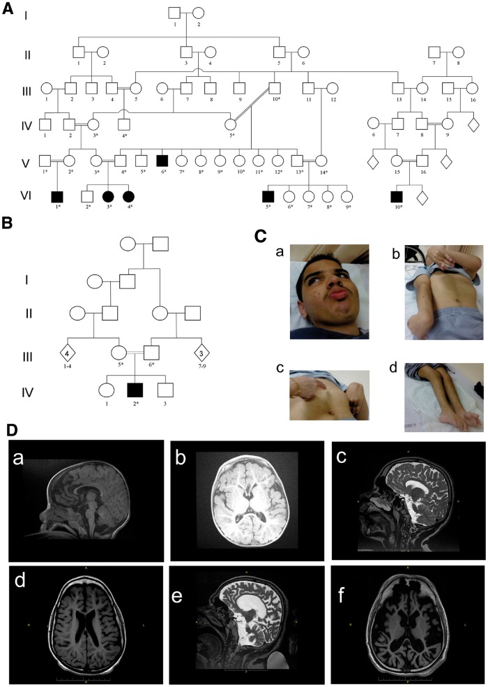
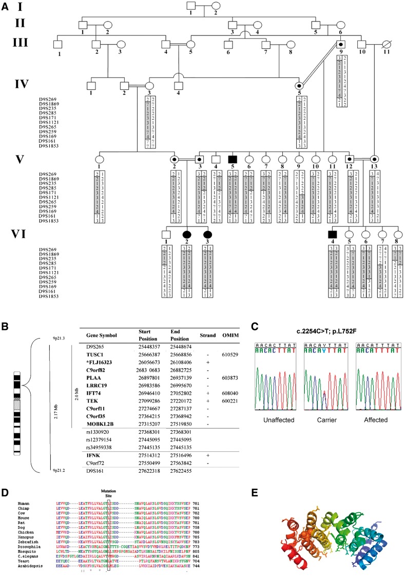
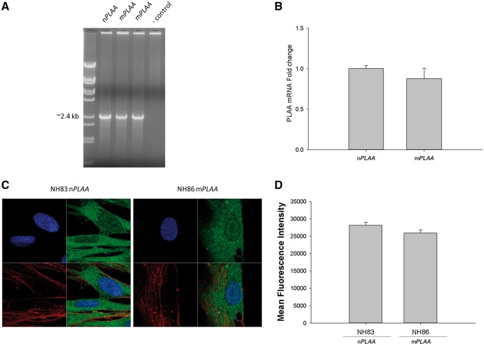
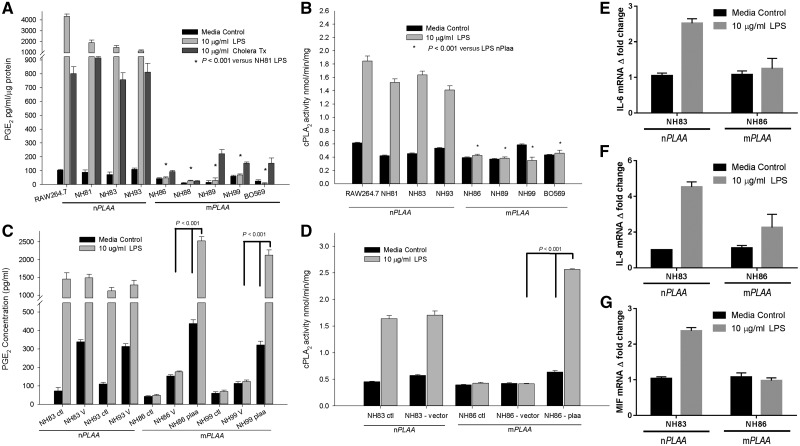
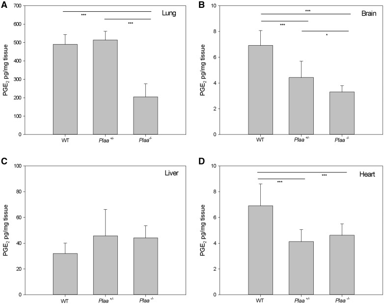
Leukoencephalopathies are a group of white matter disorders related to abnormal formation, maintenance, and turnover of myelin in the central nervous system. These disorders of the brain are categorized according to neuroradiological and pathophysiological criteria. Herein, we have identified a unique form of leukoencephalopathy in seven patients presenting at ages 2 to 4 months with progressive microcephaly, spastic quadriparesis, and global developmental delay. Clinical, metabolic, and imaging characterization of seven patients followed by homozygosity mapping and linkage analysis were performed. Next generation sequencing, bioinformatics, and segregation analyses followed, to determine a loss of function sequence variation in the phospholipase A2-activating protein encoding gene (PLAA). Expression and functional studies of the encoded protein were performed and included measurement of prostaglandin E2 and cytosolic phospholipase A2 activity in membrane fractions of fibroblasts derived from patients and healthy controls. Plaa-null mice were generated and prostaglandin E2 levels were measured in different tissues. The novel phenotype of our patients segregated with a homozygous loss-of-function sequence variant, causing the substitution of leucine at position 752 to phenylalanine, in PLAA, which causes disruption of the protein's ability to induce prostaglandin E2 and cytosolic phospholipase A2 synthesis in patients' fibroblasts. Plaa-null mice were perinatal lethal with reduced brain levels of prostaglandin E2 The non-functional phospholipase A2-activating protein and the associated neurological phenotype, reported herein for the first time, join other complex phospholipid defects that cause leukoencephalopathies in humans, emphasizing the importance of this axis in white matter development and maintenance.

Keywords: autosomal recessive; complex phospholipid defects; phospholipase A2-activating protein (PLAA); progressive leukoencephalopathy; startle response.


Figures

Figure 1
Figure 1
Pedigree of the investigated families. (A) Family I: six affected individuals (filled symbols). (B) Family II: containing another affected individual. A high rate of consanguinity and an autosomal-recessive pattern of inheritance are evident. (C) Photographs of Patient VI5 (Pedigree A) illustrating: coarse facial features (a) pectus carinatum, dystonic posturing, rigidity/freezing and shortening of tendons (b and c), and rocker bottom feet (d). (D) Patients’ brain MRI. [D(a and b)] T1 brain MRI of Patient IV2 (Family II), at 1 year of age, shows white matter atrophy. Corpus callosum is complete but thin. (c) (T2 MRI) and d (T1 MRI). Brain MRI of Patient VI3 (Family I), at 14 years, shows moderate white matter atrophy and severe corpus callosum thinning. (e) T2 MRI and (f) T1 MRI. Brain MRI of Patient V4 (Family I), at age 32 years, shows severe general atrophy. The cortex is usually preserved but very thin, corpus callosum is complete but also very thin. The basal ganglia appear normal.
Figure 2
Figure 2
Identification of the gene and protein associated with leukoencephalopathy in the studied patients. (A) Haplotypes for each family member were constructed for 11 microsatellite markers spanning the neurodegenerative interval. Markers analysed are given on the left, according to their physical order. Haplotypes are represented by bars, with the disease-associated haplotype shaded in grey. Reduction in the affected linked region to 1.9 Mb was due to healthy individuals VI6 and IV4 who bear fraction of the affected haplotype in a homozygous manner. (B) Physical location of genes and predicted transcripts in the chromosome 9 linked interval. Asterisk denotes genes not approved by the HUGO Gene Nomenclature Committee (HGNC). ‘Strand’ refers to transcription orientation. Bold names indicate gene analysed by direct sequencing. Physical location obtained from UCSC Human Genome Browser Gateway (hg19 assembly). (C) Analysis of the c.2254C > T mutation in exon 14 of PLAA. Sequence analysis is shown for an unaffected individual, an obligatory carrier, and an affected individual. (D) Sequence alignment of human PLAA to orthologues in the mutation area. The leucine at position 752 (boxed) in this protein is highly conserved throughout evolution. (E) Effect of L752F (Leu→Phe) substitution on PLAA structure. Shown is a ribbon diagram of the PUL domain of PLAA (PDB ID 3EBB) which adopts a banana like shaped Armadillo domain. The conserved residues of PLAA (Homo sapiens, Mus musculus, Rattus norvegicus, Xenopus laevis, and S. cerevisiae) are displayed in stick-representation and form the putative binding site of PLAA. Mutation of Leu752 shown in ball-representation disrupts the rigid leucine network that tightly holds together the Armadillo domain.
Figure 3
Figure 3
mRNA levels for PLAA and confocal microscopy of fibroblasts for the presence of PLAA protein. (A) Presence of full-length transcript for PLAA from fibroblasts of affected patients and the control subject based on PCR. (B) RT-qPCR for the detection of PLAA transcript from fibroblasts of a patient versus the healthy control normalized to four house-keeping genes coding for human 18 S RNA, GAPDH, POLB, and L19 ribosomal protein. Arithmetic means ± SD from three biological replicates performed in triplicate are shown. (C) Fibroblasts (nPLAA or mPLAA) were counterstained with DAPI (blue) for the nucleus and with fluorophore conjugated phalloidin (red) for actin. Cells were fixed, subjected to immunofluorescence staining for PLAA (green), and observed by confocal microscopy. (D) Mean fluorescence intensity of regions of interest corresponding to the cytoplasm and nucleus of imaged cells (ImageJ processing software, NIH). Figure represents results from three sets of images and error bars represent SD.
Figure 4
Figure 4
PGE2 levels and cPLA2 activity are low in patients' fibroblasts, and could be rescued. (A) Levels of PGE2 in cell culture media after 24 h of stimulation with LPS or cholera toxin (CT). Levels of PGE2 were normalized against protein concentrations in the supernatants. All cells were primary human fibroblasts except RAW 264.7 cells, which are murine macrophage like cells and used as a positive control. (B) Activity of cPLA2 in the membrane fractions of fibroblasts and RAW 264.7 macrophages. Cells were stimulated with or without LPS for 24 h before harvesting and purification of the membrane fractions. The cPLA2 activity was normalized to amount of proteins added to the assay. (C) PGE2 levels in the cell culture media after transfection with CMV promoter-based pIRES2-DsRed2 plasmid containing the native PLAA gene and a fluorescent marker of transfection. Cells were treated as follows: ctl = no transfection; V = transfection with empty vector; PLAA = transfection with plasmid vector containing the wild-type or native PLAA. (D) cPLA2 activity from membrane fractions of fibroblasts after transfection with a plasmid containing the nPLAA in a CMV promoter-based vector system and a fluorescent marker of transfection. (E–G) Fold-changes in transcripts for IL6, IL8, and MIF based on RT-qPCR. Arithmetic means ± SD from three independent experiments performed in triplicate are plotted and the data analysed using one-way ANOVA with Tukey post hoc correction.
Figure 5
Figure 5
PGE2 levels in embryonic mouse tissues. Wild-type, Plaa+/−, and Plaa−/− embryos were sacrificed at embryonic Day 18.5 and organs were isolated and prostaglandin levels determined for the lung (A), brain (B), liver (C), and heart (D). Data represented arithmetic means ± SD from tissues representing three wild-type, three Plaa+/−, and four Plaa−/− embryos and obtained from three independent littermates. Significance was determined by one-way ANOVA with Tukey post hoc correction. *P < 0.05 ***P < 0.001.
Figure 6
Figure 6
Histopathology of embryonic mouse tissues (5 µm) at embryonic Day 18.5. Lungs (A), brain cerebral cortex (B), and skin (C) were haematoxylin and eosin stained and analysed in a blinded fashion. Tissues representing two wild-type, two Plaa+/−, and four Plaa−/− embryos were analysed. Multiple fields for each tissue were visualized and typical representations are shown with scale bars of 100 µm (magnification ×100; top rows in A–C) and 50 µm (maginification ×200; bottom rows in A and B). Arrows in the haematoxylin and eosin stained slides indicate examples of mature neurons, while the arrowheads indicate dark round cells as examples of immature neurons or oligodendroglia. Plaa−/− mouse embryos showed an increasing number of less matured and undifferentaited neurons.
Figure 7
Figure 7
Histopathology and myelin staining of brain cerebral cortex. Sections of the brain cerebral cortex stained either with haematoxylin and eosin (5 µm section) or Luxol® fast blue (10 µm section) for myelin staining. (A) Wild-type mouse embryo; (B) Plaa+/− mouse embryo; and (C) Plaa−/− mouse embryo. Multiple fields for each tissue were visualized and typical representations are shown with a scale bar of 20 µm (magnification ×400). Arrows in the haematoxylin and eosin stained slides indicated examples of mature neurons, while the arrowheads pointed towards dark round cells as examples of immature neurons or oligodendroglia. Plaa−/− mouse embryos showed an increasing number of less matured and undifferentaited neurons. Arrows in all of the Luxol® fast blue stained slides indicated nerve processes with possible minimal early myelin (blue or turquoise colour).

Similar articles

Cited by

References

    1. Akella A, Deshpande SB. Pulmonary surfactants and their role in pathophysiology of lung disorders. Indian J Exp Biol 2013; 51: 5–22. - PubMed
    1. Atrouni S, Daraze A, Tamraz J, Cassia A, Caillaud C, Megarbane A. Leukodystrophy associated with oligodontia in a large inbred family: fortuitous association or new entity? Am J Med Genet A 2003; 118A: 76–81. - PubMed
    1. Baugh EH, Lyskov S, Weitzner BD, Gray JJ. Real-time PyMOL visualization for Rosetta and PyRosetta. PLoS One 2011; 6: e21931. - PMC - PubMed
    1. Bazan NG, Colangelo V, Lukiw WJ. Prostaglandins and other lipid mediators in Alzheimer's disease. Prostaglandins Other Lipid Mediat 2002; 68–9: 197–210. - PubMed
    1. Bomont P, Cavalier L, Blondeau F, Ben Hamida C, Belal S, Tazir M, et al. The gene encoding gigaxonin, a new member of the cytoskeletal BTB/kelch repeat family, is mutated in giant axonal neuropathy. Nat Genet 2000; 26: 370–4. - PubMed

Publication types

MeSH terms