

Choroidal microvascular proliferation secondary to diabetes mellitus
- PMID: 28008140
- PMCID: PMC5356776
- DOI: 10.18632/oncotarget.14020
Choroidal microvascular proliferation secondary to diabetes mellitus
Abstract
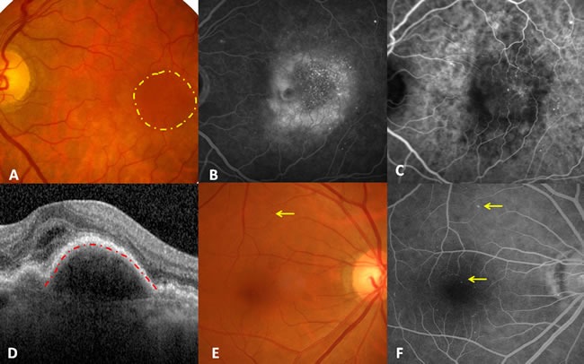
Diabetes is a common endocrine disorder associated with peripheral microvascular diseases such as proliferative retinal microangiopathy (or diabetic retinopathy), which may lead to blindness. Unfortunately, diabetic microvascular abnormalities in the choroid are underestimated in clinical practice. Recent literature has revealed that the severity of diabetic retinopathy is aggravated by choroidopathy resulting from hyperglycemia. Here, we introduce a case of diabetic retinopathy with choroidal neovascularization membrane but without signs of retinal microvascular proliferation or drusen. We investigated the pathogenesis of choroidal microvascular proliferation secondary to diabetes. We postulate that choroidal neovascularization is an intraocular microvascular complication of diabetes mellitus. Intravitreal anti-vascular endothelial growth factor therapy may be a treatment option for microvascular proliferation in both retina and choroids.
Keywords: Pathology Section; choroidal neovasculation; fluorescein angiography; indocyanine green angiography; microangiopathy; proliferative diabetic choroidopathy.
Conflict of interest statement
No potential conflicts of interest relevant to this article were reported.
Figures
References
-
- Congdon NG, Friedman DS, Lietman T. Important causes of visual impairment in the world today. JAMA. 2010(290):2057–2060. - PubMed
-
- Vassilev ZP, Ruigomez A, Soriano-Gabarro M, Garcia Rodriguez LA. Diabetes, cardiovascular morbidity, and risk of age-related macular degeneration in a primary care population. Invest Ophthalmol Vis Sci. 2015(56):1585–1592. - PubMed
-
- Cho BJ, Heo JW, Shin JP, Ahn J, Kim TW, Chung H. Epidemiological association between systemic diseases and age-related macular degeneration: the Korea National Health and Nutrition Examination Survey 2008-2011. Invest Ophthalmol Vis Sci. 2014(55):4430–4437. - PubMed
-
- Topouzis F, Anastasopoulos E, Augood C, Bentham GC, Chakravarthy U, de Jong PT, Rahu M, Seland J, Soubrane G, Tomazzoli L, Vingerling JR, Vioque J, Young IS, Fletcher AE. Association of diabetes with age-related macular degeneration in the EUREYE study. Br J Ophthalmol. 2009(93):1037–1041. - PubMed
Publication types
MeSH terms
LinkOut - more resources
Full Text Sources
Other Literature Sources
Medical
