. 2016 Dec;78(4):507-516.
doi: 10.18999/nagjms.78.4.507.

What do we know about pulmonary blastoma?: review of literature and clinical case report

Affiliations

What do we know about pulmonary blastoma?: review of literature and clinical case report

Dorota Brodowska-Kania et al. Nagoya J Med Sci. 2016 Dec.

Abstract

Pulmonary blastoma (PB) is a rare form of lung tumour and is accountable for 0.25-0.5% of primary pulmonary malignancies. Initially pulmonary blastoma was divided into three subtypes: biphasic pulmonary blastoma (BPB) consisting of an epithelial and mesenchymal component, well differentiated fetal adenocarcinoma (WDFA) built of well differentiated epithelium and a mesenchymal component and malignant pleuropulmonary blastoma (PPB). Prognosis in this type of cancer is really poor. We present a current review of literature and a clinical case report. Treatment of PB is very difficult. Data and recommendations about the treatment of pulmonary blastoma are still available therefore we should use only observations and clinical case reports.

Keywords: blastoma pulmonary; chronic kidney disease; lung cancer.


Figures
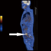
Fig. 1
Fig. 1
PET-CT, sagittal plane, patient after implantation of the aortic graft. Arrow marks the focus of intensively increased glucose uptake in the retroperitoneal space, on the border of the abdominal aorta and the left iliac artery – limited inflammatory infiltration of the graft.
Fig. 2A
Fig. 2A
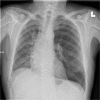
Chest X-ray, posterior-anterior view – persistent peribronchial thickening in the lower lobes.
Fig. 2B
Fig. 2B
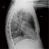
Chest X-ray, latero-lateral view. Oval lesions in area Th4/Th5
Fig. 3
Fig. 3
CT scan shows tumour in the right lung measuring 52×30 mm. No enlarged mediastinal lymph nodes.
Fig. 4
Fig. 4
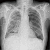
Chest X-ray, posterior-anterior view after the surgical removal of the intermediate lobe of the right lung. Drain in the right pleural cavity. The postoperative chest radiograph revealed no pneumothorax.
Fig. 5A
Fig. 5A
Pulmonary Blastoma. Hematoxylin and eosin stain (H&E). Biphasic tumor composed of bronchioling epithelial tubules or cords set in an undifferentiated stroma. On top of the image stroma with chondroid differentitation. (magnification 100×)
Fig. 5B
Fig. 5B
Pulmonary Blastoma. Cytokeratin AE 1/3 stain. (magnification 100×)
Fig. 5C
Fig. 5C
Pulmonary Blastoma. Vimentin stain. The mesenhymal components and pseudocapsula. (magnification 100×)
Fig. 5D
Fig. 5D
Pulmonary Blastoma. Trichrom Masson stain. (magnification 100×)
Fig. 6
Fig. 6
Image of PET-CT in the sagittal plane on the same patient after surgery. Arrow, marked focus moderately increased metabolism 18F-FDG in the retroperitoneal space – residual reactive changes. (Changes with a history catheter sepsis). No outbreaks of increased metabolic fluorodeoxyglucose. No evidence of cancer of increased FDG metabolism.

References

    1. Smyth R, Fabre A, Dodd J, Bartosik W, Gallagher C, McKone E. Pulmonary blastoma: a case report and review of the literature. BMC Res Notes, 2014; 13: 294. - PMC - PubMed
    1. Barnett N, Barnard W. Some unusual thoracic tumors. Br J Surg, 1945; 32: 441–457.
    1. Jethava A, Dasanu C. Adult biphasic pulmonary blastoma. Conn Med, 2013; 71: 19–22. - PubMed
    1. Spencer H. Pulmonary blastomas. J Pathol Bacteriol, 1961; 82: 161–165.
    1. Travis W, Brambilla E, Muller-Hermelink H, Harris C. World Health Organization classification of tumors: pathology and genetics of tumors of the lung, pleura, thymus and heart. IARC Press: Lyon; 2004. - PubMed

LinkOut - more resources