. 2016 Dec 15:11:6809-6824.
doi: 10.2147/IJN.S123442. eCollection 2016.

Rescuing cholinergic neurons from apoptotic degeneration by targeting of serotonin modulator-and apolipoprotein E-conjugated liposomes to the hippocampus

Affiliations

Rescuing cholinergic neurons from apoptotic degeneration by targeting of serotonin modulator-and apolipoprotein E-conjugated liposomes to the hippocampus

Yung-Chih Kuo et al. Int J Nanomedicine. .

Abstract

β-Amyloid (Aβ)-targeting liposomes (LIP) with surface serotonin modulator (SM) and apolipoprotein E (ApoE) were utilized to facilitate the delivery of nerve growth factor (NGF) across the blood-brain barrier (BBB) for neuroprotection in the hippocampus. The therapeutic efficacy of SM- and ApoE-grafted LIP carrying NGF (NGF-SM-ApoE-LIP) was assessed by an in vitro Alzheimer's disease (AD) model of degenerated SK-N-MC cells and an in vivo AD model of Aβ-insulted Wistar rats. The experimental evidences revealed that the modified SM and ApoE on the surface of LIP increased the permeation of NGF across the BBB without serious damage to structural integrity of tight junction. When compared with free NGF, NGF-SM-ApoE-LIP upregulated the expression of phosphorylated neurotrophic tyrosine kinase receptor type 1 on cholinergic neurons and significantly improved their survival. In addition, NGF-SM-ApoE-LIP could reduce the secretion of acetylcholinesterase and malondialdehyde and rescue hippocampal neurons from apoptosis in rat brains. The synergistic effect of SM and ApoE is promising in the induction of NGF to inhibit the neurotoxicity of Aβ and NGF-SM-ApoE-LIP can be a potent antiapoptotic pharmacotherapy for clinical care of patients with AD.

Keywords: Alzheimer’s disease; apolipoprotein E; blood–brain barrier; liposome; nerve growth factor; serotonin modulator.


Conflict of interest statement

The authors report no conflicts of interest in this work.

Figures

Figure 1
Figure 1
Physicochemical characterization of NGF-SM-ApoE-LIP. Notes: (A) Average particle diameter and zeta potential; CSM =40 μg/mL; CApoE =40 μg/mL; (○) average particle diameter; n=3; (□) zeta potential; n=3; inset: corresponding SEM images at rCL =10%, rCL =20%, rCL =30%, (B) TEM images; rCL =30%; (a) NGF-ApoE-LIP; CApoE =40 μg/mL; (b) NGF-SM-LIP; CSM =40 μg/mL; (c) NGF-SM-ApoE-LIP; CSM =40 μg/mL; CApoE =40 μg/mL, (C) grafting efficiency; (○) grafting efficiency of ApoE on NGF-LIP; CSM =0 μg/mL; n=3; (□) grafting efficiency of SM on NGF-ApoE-LIP; CApoE =40 μg/mL; n=3, (D) XPS spectra; (a) NGF-LIP; rCL =0%; (b) NGF-LIP; rCL =30%; (c) NGF-ApoE-LIP; rCL =30%; CApoE =40 μg/mL; (d) NGF-SM-ApoE-LIP; rCL =30%; CSM =40 μg/mL; CApoE =40 μg/mL. Abbreviations: CSM, concentration of serotonin modulator; CApoE, concentration of apolipoprotein E; rCL, weight percentage of cardiolipin in bilayer (%).
Figure 2
Figure 2
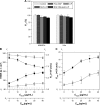
Effect of NGF-SM-ApoE-LIP on the BBB permeation. Notes: rCL =30%. (A) Viability of HBMECs and HAs; CSM =40 μg/mL; CApoE =40 μg/mL; n=3, (B) TEER and permeability coefficient of PI across HBMEC/HA; (○) TEER; NGF-ApoE-LIP; (X1 and Y1 axes); (●) TEER; NGF-SM-ApoE-LIP; CApoE =40 μg/mL; X2 and Y1 axes; (- - -): intact TEER; Y1 axis; n=3, (□) permeability coefficient of PI; NGF-ApoE-LIP; X1 and Y2 axes; (■) permeability coefficient of PI; NGF-SM-ApoE-LIP; CApoE =40 μg/mL; X2 and Y2 axes; (– – –): intact permeability coefficient of PI; Y2 axis; n=3, (C) permeability coefficient of NGF across HBMEC/HA; (○) NGF-ApoE-LIP; (●) NGF-SM-ApoE-LIP; CApoE =40 μg/mL; n=3, (D) fluorescent NGF-SM-ApoE-LIP internalized by HBMECs; CApoE =40 μg/mL; (a) NGF-LIP; (b) NGF-ApoE-LIP; (c) NGF-SM-ApoE-LIP; CSM =20 μg/mL; (d) NGF-SM-ApoE-LIP; CSM =40 μg/mL; (a1, b1, c1, d1), (a2, b2, c2, d2), (a3, b3, c3, d3), and (a4, b4, c4, d4) are red (against LDLR antibody in [a] and [b] and against both LDLR and SR antibody in [c] and [d]), green (LIP carrier), blue (DAPI for nucleus), and merged channels, respectively. a1, a2, and a3 are obtained from excitation at 555 nm, 490 nm, and 350 nm, respectively. a4 is a merged image from a1–a3. The difference between a1–a4 is the same for b, c, and d. Abbreviations: rCL, weight percentage of cardiolipin in bilayer (%); CSM, concentration of serotonin modulator; CApoE, concentration of apolipoprotein E; HBMEC, human brain-microvascular endothe lial cell; HA, human astrocyte; BBB, blood–brain barrier; TEER, transendothelial electrical resistance; LIP, liposome; SM, serotonin modulator; NGF, nerve growth factor.
Figure 2
Figure 2
Effect of NGF-SM-ApoE-LIP on the BBB permeation. Notes: rCL =30%. (A) Viability of HBMECs and HAs; CSM =40 μg/mL; CApoE =40 μg/mL; n=3, (B) TEER and permeability coefficient of PI across HBMEC/HA; (○) TEER; NGF-ApoE-LIP; (X1 and Y1 axes); (●) TEER; NGF-SM-ApoE-LIP; CApoE =40 μg/mL; X2 and Y1 axes; (- - -): intact TEER; Y1 axis; n=3, (□) permeability coefficient of PI; NGF-ApoE-LIP; X1 and Y2 axes; (■) permeability coefficient of PI; NGF-SM-ApoE-LIP; CApoE =40 μg/mL; X2 and Y2 axes; (– – –): intact permeability coefficient of PI; Y2 axis; n=3, (C) permeability coefficient of NGF across HBMEC/HA; (○) NGF-ApoE-LIP; (●) NGF-SM-ApoE-LIP; CApoE =40 μg/mL; n=3, (D) fluorescent NGF-SM-ApoE-LIP internalized by HBMECs; CApoE =40 μg/mL; (a) NGF-LIP; (b) NGF-ApoE-LIP; (c) NGF-SM-ApoE-LIP; CSM =20 μg/mL; (d) NGF-SM-ApoE-LIP; CSM =40 μg/mL; (a1, b1, c1, d1), (a2, b2, c2, d2), (a3, b3, c3, d3), and (a4, b4, c4, d4) are red (against LDLR antibody in [a] and [b] and against both LDLR and SR antibody in [c] and [d]), green (LIP carrier), blue (DAPI for nucleus), and merged channels, respectively. a1, a2, and a3 are obtained from excitation at 555 nm, 490 nm, and 350 nm, respectively. a4 is a merged image from a1–a3. The difference between a1–a4 is the same for b, c, and d. Abbreviations: rCL, weight percentage of cardiolipin in bilayer (%); CSM, concentration of serotonin modulator; CApoE, concentration of apolipoprotein E; HBMEC, human brain-microvascular endothe lial cell; HA, human astrocyte; BBB, blood–brain barrier; TEER, transendothelial electrical resistance; LIP, liposome; SM, serotonin modulator; NGF, nerve growth factor.
Figure 3
Figure 3
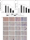
Effect of NGF-SM-ApoE-LIP on the survival of neurons treated with Aβ1–42. CSM =40 μg/mL; CApoE =40 μg/mL. Notes: (A) Viability of SK-N-MC cells; rCL =30%; *P<0.05; **P<0.01; n=3, (B) p-TrkA level of SK-N-MC cells; rCL =30%; (a): Western blot; (b): Rp, *P<0.05; n=3, (C) immunochemical staining images of NGF-SM-ApoE-LIP interacting with SK-N-MC cells; (a) NGF-LIP; rCL =0%; (b) NGF-LIP; rCL =30%; (c) NGF-SM-LIP; rCL =30%; (d) NGF-SM-ApoE-LIP; rCL =30%; (a1, b1, c1, d1), (a2, b2, c2, d2), (a3, b3, c3, d3), and (a4, b4, c4, d4) are red (against Aβ1–42 antibody), green (LIP carrier), blue (DAPI for nucleus), and merged channels, respectively. a1, a2, and a3 are obtained from excitation at 555 nm, 490 nm, and 350 nm, respectively. a4 is a merged image from a1–a3. The difference between a1–a4 is the same for b, c, and d. Abbreviations: CSM, concentration of serotonin modulator; CApoE, concentration of apolipoprotein E; rCL, weight percentage of cardiolipin in bilayer (%); Rp, protein ratio of p-TrkA to GAPDH; LIP, liposome; SM, serotonin modulator; NGF, nerve growth factor.
Figure 3
Figure 3
Effect of NGF-SM-ApoE-LIP on the survival of neurons treated with Aβ1–42. CSM =40 μg/mL; CApoE =40 μg/mL. Notes: (A) Viability of SK-N-MC cells; rCL =30%; *P<0.05; **P<0.01; n=3, (B) p-TrkA level of SK-N-MC cells; rCL =30%; (a): Western blot; (b): Rp, *P<0.05; n=3, (C) immunochemical staining images of NGF-SM-ApoE-LIP interacting with SK-N-MC cells; (a) NGF-LIP; rCL =0%; (b) NGF-LIP; rCL =30%; (c) NGF-SM-LIP; rCL =30%; (d) NGF-SM-ApoE-LIP; rCL =30%; (a1, b1, c1, d1), (a2, b2, c2, d2), (a3, b3, c3, d3), and (a4, b4, c4, d4) are red (against Aβ1–42 antibody), green (LIP carrier), blue (DAPI for nucleus), and merged channels, respectively. a1, a2, and a3 are obtained from excitation at 555 nm, 490 nm, and 350 nm, respectively. a4 is a merged image from a1–a3. The difference between a1–a4 is the same for b, c, and d. Abbreviations: CSM, concentration of serotonin modulator; CApoE, concentration of apolipoprotein E; rCL, weight percentage of cardiolipin in bilayer (%); Rp, protein ratio of p-TrkA to GAPDH; LIP, liposome; SM, serotonin modulator; NGF, nerve growth factor.
Figure 4
Figure 4
Biochemical analysis and histochemical staining of the brain of AD rats treated with NGF-SM-ApoE-LIP. rCL =30%; CSM =40 μg/mL; CApoE =40 μg/mL. Notes: (A) MDA level of the brain CA1 tissue after treating with LIP nanocarriers; *P<0.05; #P>0.05; **P<0.01; n=6, (B) AChE activity of the brain CA1 tissue after treating with LIP nanocarriers; *P<0.05; **P<0.01; n=6, (C) Nissl staining of the brain CA1 tissue after treating with LIP nanocarriers; (a) control; (b) Aβ1–42; (c) Aβ1–42 + NGF; (d) Aβ1–42 + NGF-LIP; (e) Aβ1–42 + NGF-SM-ApoE-LIP; (a1, b1, c1, d1, e1) 40×; (a2, b2, c2, d2, e2) 100×; (a3, b3, c3, d3, e3) 200×; (a4, b4, c4, d4, e4) 400×. a1, a2, a3, and a4 were obtained from magnification at 40×, 100×, 200×, and 400×, respectively. The difference between a1–a4 is the same for b, c, d, and e. Abbreviations: rCL, weight percentage of cardiolipin in bilayer (%); CSM, concentration of serotonin modulator; CApoE, concentration of apolipoprotein E; LIP, liposome; SM, serotonin modulator; NGF, nerve growth factor; MDA, malondialdehyde.

Similar articles

Cited by

References

    1. Goedert M, Spillantini MG. A century of Alzheimer’s disease. Science. 2006;314:777–781. - PubMed
    1. Alzheimer’s Association 2016 Alzheimer’s disease facts and figures. Alzheimers Dement. 2016;12:1–79. - PubMed
    1. Obara Y, Yamauchi A, Takehara S, Niemoto W, Takahashi M, Stork PJ. ERK5 activity is required for nerve growth factor-induced neurite outgrowth and stabilization of tyrosine hydroxylase in PC12 cells. J Biol Chem. 2009;284:64–73. - PMC - PubMed
    1. Sofroniew MV, Howe CL, Mobley WC. Nerve growth factor signaling, neuroprotection, and neural repair. Annu Rev Neurosci. 2001;24:1217–1281. - PubMed
    1. Bottger D, Ullrich C, Humpel C. Monocytes deliver bioactive nerve growth factor through a brain capillary endothelial cell-monolayer in vitro and counteract degeneration of cholinergic neurons. Brain Res. 2010;1312:108–119. - PubMed

MeSH terms

LinkOut - more resources