. 2016 Dec 13:9:7503-7513.
doi: 10.2147/OTT.S115268. eCollection 2016.

Early detection of poor outcome in patients with metastatic colorectal cancer: tumor kinetics evaluated by circulating tumor cells

Affiliations

Early detection of poor outcome in patients with metastatic colorectal cancer: tumor kinetics evaluated by circulating tumor cells

Virgílio Souza E Silva et al. Onco Targets Ther. .

Abstract

Background: Colorectal cancer (CRC) is the third most prevalent cancer worldwide. New prognostic markers are needed to identify patients with poorer prognosis, and circulating tumor cells (CTCs) seem to be promising to accomplish this.

Patients and methods: A prospective study was conducted by blood collection from patients with metastatic CRC (mCRC), three times, every 2 months in conjunction with image examinations for evaluation of therapeutic response. CTC isolation and counting were performed by Isolation by Size of Epithelial Tumor Cells (ISET).

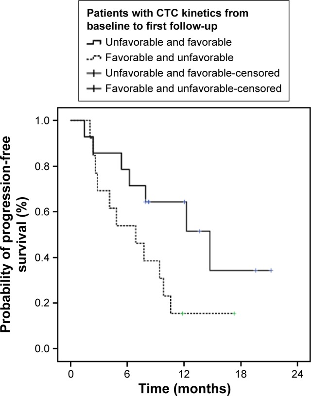
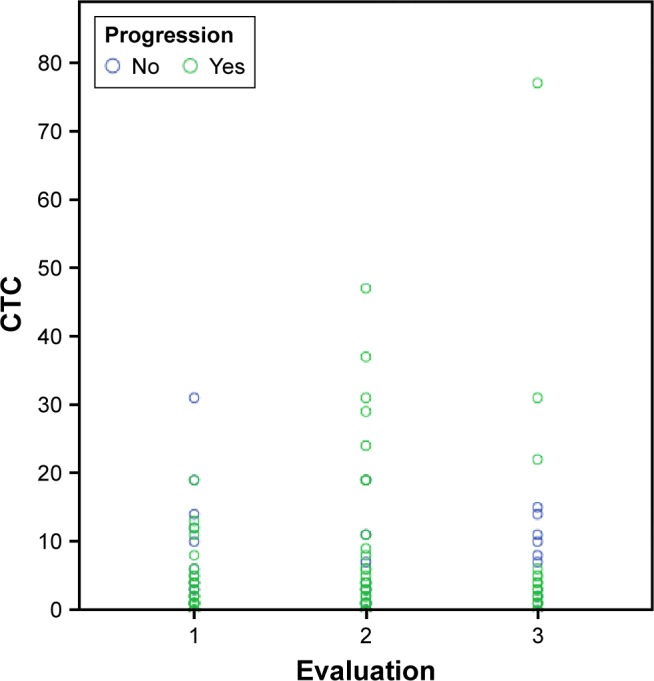
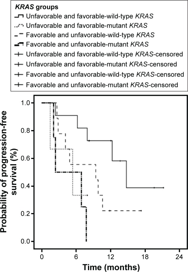
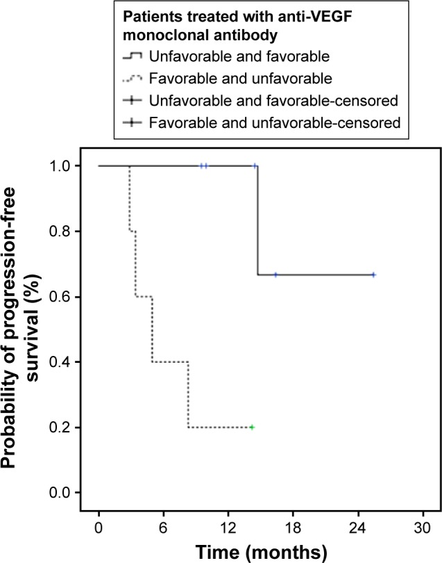
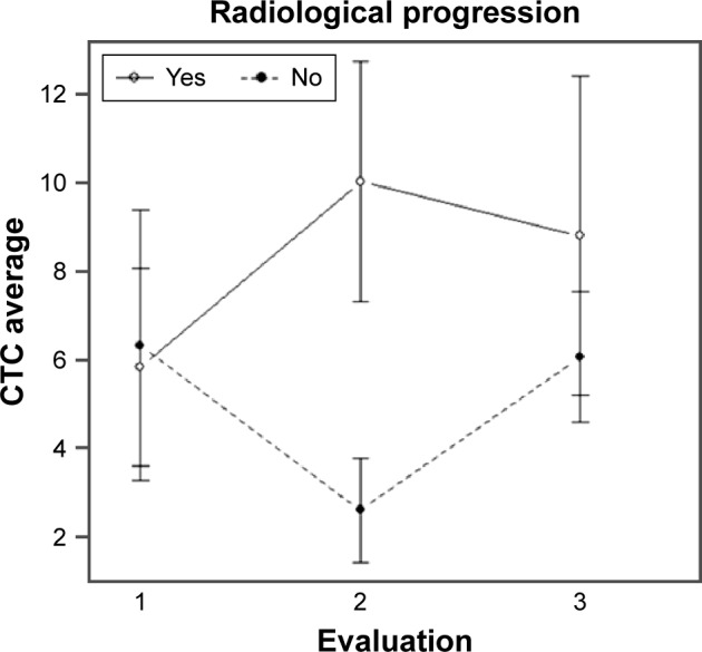
Results: A total of 54 patients with mCRC with a mean age of 57.3 years (31-82 years) were included. Among all patients, 60% (n=32) were carriers of wild-type KRAS (WT KRAS) tumors and 90% of them (n=29) were exposed to monoclonal antibodies along with systemic treatment. Evaluating CTC kinetics, when we compared the baseline (pretreatment) CTC level (CTC1) with the level at first follow-up (CTC2), we observed that CTC1-positive patients (CTCs above the median), who became negative (CTCs below the median) had a favorable evolution (n=14), with a median progression-free survival (PFS) of 14.7 months. This was higher than that for patients with an unfavorable evolution (CTC1- that became CTC2+; n=13, 6.9 months; P=0.06). Patients with WT KRAS with favorable kinetics had higher PFS (14.7 months) in comparison to those with WT KRAS with unfavorable kinetics (9.4 months; P=0.02). Moreover, patients whose imaging studies showed radiological progression had an increased quantification of CTCs at CTC2 compared to those without progression (P=0.04).

Conclusion: This study made possible the presentation of ISET as a feasible tool for evaluating CTC kinetics in patients with mCRC, which can be promising in their clinical evaluation.

Keywords: ISET; circulating tumor cells; kinetics; metastatic colorectal cancer.


Conflict of interest statement

The authors report no conflicts of interest in this work.

Figures

Figure 1
Figure 1
Cells isolated by ISET. Notes: (A) CTC visualized by hematoxylin–eosin. (B) Immunostaining of leukocyte with CD45 and counterstaining with DAB. Asterisk indicates a CTC; thin arrows represent pores of ISET membrane; and the thick arrow shows a leukocyte. Both images were taken at ×600 magnification using a light microscope (Research System Microscope BX61 – Olympus, Tokyo, Japan) coupled to a digital camera (SC100 – Olympus, Tokyo, Japan). Abbreviations: CTC, circulating tumor cell; DAB, 3,3′-daminobenzidine; ISET, isolation by size of epithelial tumors.
Figure 2
Figure 2
Progression-free survival evaluation in patients with CTC kinetic alteration on the first two analyses (CTC1 and CTC2). Notes: Continuous line: positive patients with favorable kinetics and prognosis (CTC1+ and CTC2−). Dotted line: patients with unfavorable kinetics and prognosis (CTC1− and CTC2+; P=0.06). Abbreviation: CTC, circulating tumor cell.
Figure 3
Figure 3
Progression-free survival evaluation in months, in patients without kinetic alteration of CTCs (CTC1+ and CTC2+) and (CTC1− and CTC2−). Notes: Continuous line: patients with positive CTC maintenance. Dotted line: patients with negative CTC maintenance (P=0.60). Abbreviation: CTC, circulating tumor cell.
Figure 4
Figure 4
Radiological evolution and CTC counting. Notes: Patient 1: (A1): image of hepatic lesion evidence (arrows) obtained before the collection of CTC1 (May 2013); (B1): image (September 2013) of hepatic lesion evidence (arrows) in progression when image A1 compared with image after the collection of CTC2; (C1): image of hepatic lesion evidence (arrows) in progression when compared with image B1 obtained after collection of CTC3 (November 2013). Patient 2: (A2): image of lymph node involvement evidence (arrows) obtained before the collection of CTC1 (May 2013); (B2): image (September 2013) of evidence of progression of lymph node disease (arrows) compared with illustration A2. Abbreviation: CTC, circulating tumor cell.
Figure 5
Figure 5
Dot plot figure. Notes: Each circle represents a patient analyzed three times: baseline (CTC1), first follow-up (CTC2), and second follow-up (CTC3). Blue circles refer to the absence of disease progression and the green ones to the presence. Abbreviation: CTC, circulating tumor cell.
Figure 6
Figure 6
Progression-free survival based on CTC kinetics and the status of the KRAS mutation. Notes: Continuous line: patients with wild type KRAS and favorable kinetics (CTC1+ and CTC2−; 14.7 months). Dotted line: patients with mutant KRAS and favorable kinetics (CTC1+ and CTC2−; 5.3 months). Dashed line: patients with wild-type and unfavorable kinetics (CTC1− and CTC2+; 9.4 months). Dashed-dotted line: patients with mutant KRAS and unfavorable kinetics (CTC1− and CTC2+; 2.4 months) (P=0.02). Abbreviation: CTC, circulating tumor cell.
Figure 7
Figure 7
Progression-free survival based on CTC kinetics and the treatment with anti-VEGF monoclonal antibody. Notes: Continuous line: patients treated with anti-VEGF with favorable kinetics (CTC1+ and CTC2−). Dotted line: patients treated with anti-VEGF with unfavorable kinetics (CTC1− and CTC2+; P=0.007). Abbreviation: CTC, circulating tumor cell.
Figure 8
Figure 8
Correlation between CTC quantification and radiological progression. Notes: Continuous line: patients with radiological progression and increased CTC quantification in the second collection (CTC2). Dotted line: patients without radiographic progression who did not have higher CTC in the second collection (CTC2; P=0.04). Abbreviation: CTC, circulating tumor cell.

References

    1. Ferlay J, Soerjomataram I, Ervik M, et al. GLOBOCAN 2012 v1.0, Cancer Incidence and Mortality Worldwide: IARC CancerBase No. 11. Lyon, France: International Agency for Research on Cancer; 2013.
    1. Lee WS, Yun SH, Chun HK, et al. Pulmonary resection for metastases from colorectal cancer: prognostic factors and survival. Int J Colorectal Dis. 2007;22(6):699–704. - PubMed
    1. Van Cutsem E, Nordlinger B, Adam R, et al. European Colorectal Metastases Treatment Group Towards a pan-European consensus on the treatment of patients with colorectal liver metastases. Eur J Cancer. 2006;42(14):2212–2221. - PubMed
    1. Yoo PS, Lopez-Soler RI, Longo WE, Cha CH. Liver resection for metastatic colorectal cancer in the age of neoadjuvant chemotherapy and bevacizumab. Clin Colorectal Cancer. 2006;6(3):202–207. - PubMed
    1. Chang GJ. Challenge of primary tumor management in patients with stage IV colorectal cancer. J Clin Oncol. 2012;30:3165–3166. - PubMed