. 2017 Mar;187(3):383-398.
doi: 10.1111/cei.12895. Epub 2016 Dec 23.

Defective expression of apoptosis-related molecules in multiple sclerosis patients is normalized early after autologous haematopoietic stem cell transplantation

Affiliations

Defective expression of apoptosis-related molecules in multiple sclerosis patients is normalized early after autologous haematopoietic stem cell transplantation

G L V de Oliveira et al. Clin Exp Immunol. 2017 Mar.

Abstract

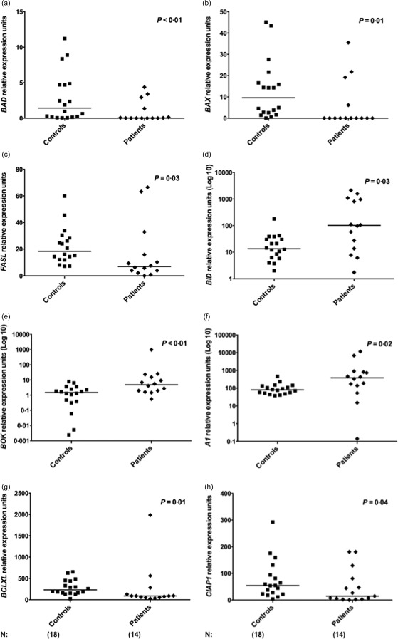
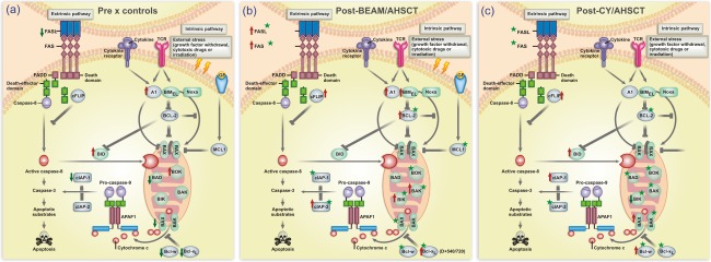
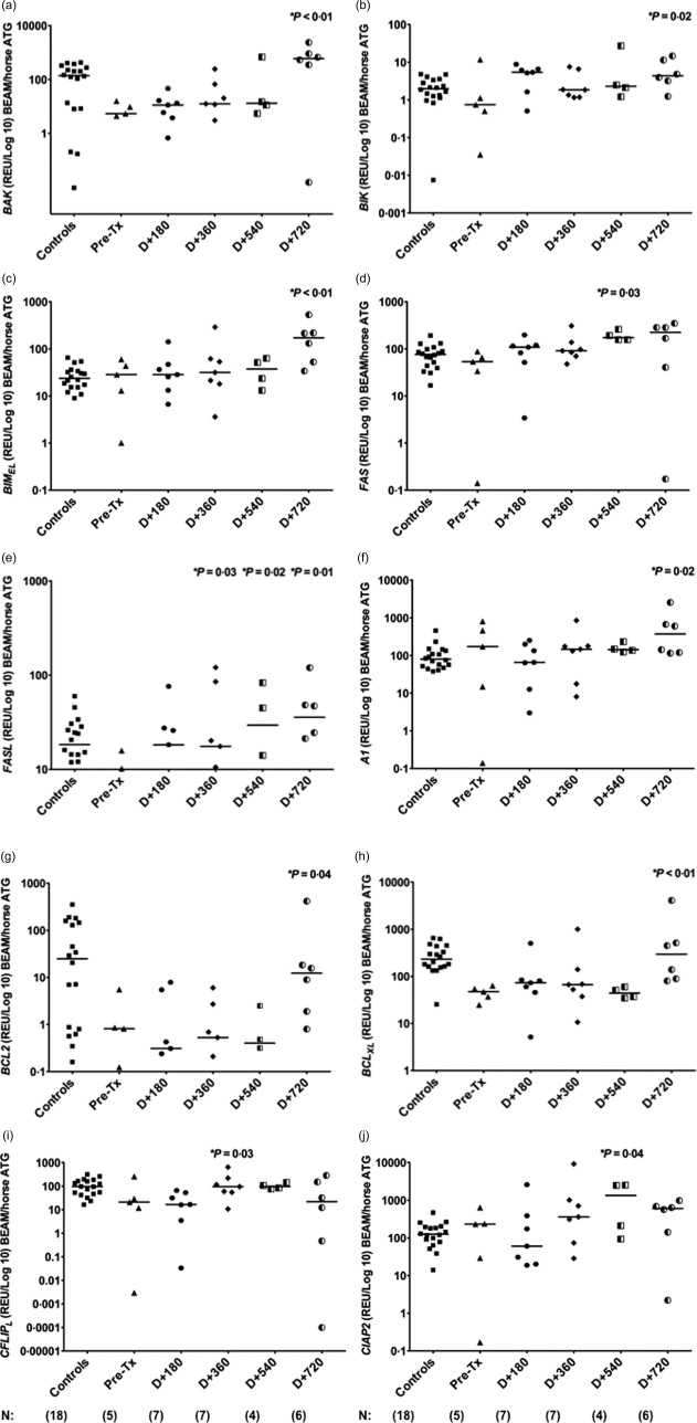
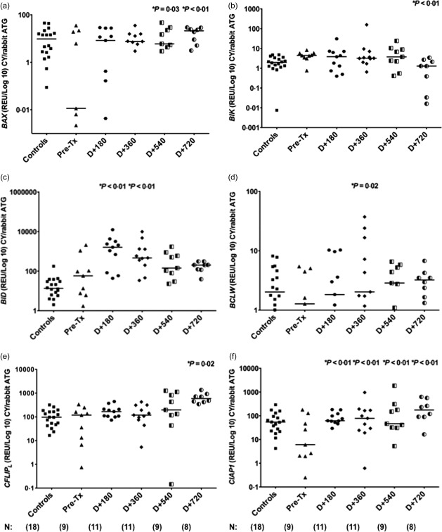
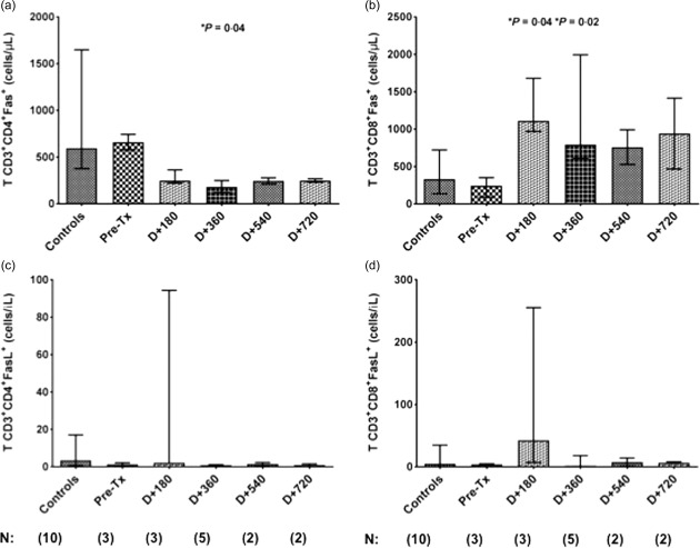
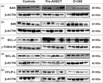
Defective apoptosis might be involved in the pathogenesis of multiple sclerosis (MS). We evaluated apoptosis-related molecules in MS patients before and after autologous haematopoietic stem cell transplantation (AHSCT) using BCNU, Etoposide, AraC and Melphalan (BEAM) or cyclophosphamide (CY)-based conditioning regimens. Patients were followed for clinical and immunological parameters for 2 years after AHSCT. At baseline, MS patients had decreased proapoptotic BAD, BAX and FASL and increased A1 gene expression when compared with healthy counterparts. In the BEAM group, BAK, BIK, BIMEL , FAS, FASL, A1, BCL2, BCLXL , CFLIPL and CIAP2 genes were up-regulated after AHSCT. With the exception of BIK, BIMEL and A1, all genes reached levels similar to controls at day + 720 post-transplantation. Furthermore, in these patients, we observed increased CD8+ Fas+ T cell frequencies after AHSCT when compared to baseline. In the CY group, we observed increased BAX, BCLW, CFLIPL and CIAP1 and decreased BIK and BID gene expressions after transplantation. At day + 720 post-AHSCT, the expression of BAX, FAS, FASL, BCL2, BCLXL and CIAP1 was similar to that of controls. Protein analyses showed increased Bcl-2 expression before transplantation. At 1 year post-AHSCT, expression of Bak, Bim, Bcl-2, Bcl-xL and cFlip-L was decreased when compared to baseline values. In summary, our findings suggest that normalization of apoptosis-related molecules is associated with the early therapeutic effects of AHSCT in MS patients. These mechanisms may be involved in the re-establishment of immune tolerance during the first 2 years post-transplantation.

Keywords: apoptosis-related molecules; autologous haematopoietic stem cell transplantation; autoreactive cells; immune tolerance; multiple sclerosis.


Figures

Figure 1
Figure 1
Deregulated expression of apoptosis‐related genes in multiple sclerosis (MS) patients. (a–e) Expression of BAD, BAX, FASL, BID and BOK pro‐apoptotic genes in peripheral blood mononuclear cells (PBMCs) from healthy controls (n = 18) and MS patients (n = 14). (f–h) Expression of A1, BCLXL and CIAP1 anti‐apoptotic genes in controls and patients. Apoptosis‐related gene expression was evaluated by real‐time polymerase chain reaction (PCR). Statistical analyses were performed by Mann–Whitney U‐test. Significance was set at P < 0·05. Horizontal bars represent median values. Symbols correspond to individual subjects.
Figure 2
Figure 2
Schematic diagram of the extrinsic and intrinsic apoptotic pathways highlighting results of the study. (a) Alterations of gene expression in peripheral blood mononuclear cells (PBMCs) from multiple sclerosis (MS) patients compared with those from healthy subjects. Red arrows indicate up‐regulated genes and green arrows indicate down‐regulated genes. (b) Abnormalities of gene expression in PBMCs from MS patients after BCNU, Etoposide, AraC and Melphalan/horse anti‐human thymocyte globulin autologous haematopoietic stem cell transplantation (BEAM/hATG AHSCT) (day + 720) compared with pre‐AHSCT values. (c) Abnormalities of gene expression in PBMCs from MS patients after cyclophosphamide (CY)/rabbit ATG (rATG) AHSCT (day + 720) compared with pre‐AHSCT values. In (b) and (c), red arrows indicate up‐regulated genes and green arrows indicate down‐regulated genes in MS patients following transplantation. Stars indicate modulated genes that reached healthy control levels at day + 720. [Colour figure can be viewed at wileyonlinelibrary.com]
Figure 3
Figure 3
Modulation of apoptosis‐related genes in multiple sclerosis (MS) patients after AHSCT with BCNU, Etoposide, AraC and Melphalan/horse anti‐human thymocyte globulin autologous haematopoietic stem cell transplantation (BEAM/hATG AHSCT) conditioning regimen. (a–e) Expression of BAK, BIK, BIMEL, FAS and FASL pro‐apoptotic genes in peripheral blood mononuclear cells (PBMCs) from healthy controls (n = 18) and MS patients (n = 5) before and at day + 180 (n = 7), day + 360 (n = 7), day + 540 (n = 4) and day + 720 (n = 6) post‐AHSCT with BEAM/hATG conditioning regimen. (f–j) Expression of A1, BCL2, BCLXL, CFLIPL and CIAP2 anti‐apoptotic genes in PBMCs from healthy controls and MS patients before and at day + 180, day + 360, day + 540 and day + 720 post‐AHSCT with BEAM/hATG conditioning regimen. Apoptosis‐related gene expression was evaluated by real‐time polymerase chain reaction (PCR). Statistical analyses were performed by linear regression model with mixed effects. For multiple comparisons, we used orthogonal contrasts post‐test. Significance was set at P < 0·05. Horizontal bars represent medians. Symbols correspond to individual subjects.
Figure 4
Figure 4
Modulation of apoptosis‐related genes in multiple sclerosis (MS) patients after autologous haematopoietic stem cell transplantation (AHSCT) with cyclophosphamide/rabbit anti‐human thymocyte globulin (CY/rATG) conditioning regimen. (a–c) Expression of BAX, BIK and BID pro‐apoptotic genes in peripheral blood mononuclear cells (PBMCs) from healthy controls (n = 18) and MS patients (n = 9) before and at day + 180 (n = 11), day + 360 (n = 11), day + 540 (n = 9) and day + 720 (n = 8) post‐AHSCT with CY/rATG conditioning regimen. (d–f) Expression of BCLW, CFLIPL and CIAP1 anti‐apoptotic genes in PBMCs from healthy controls and MS patients before and at day + 180, day + 360, day + 540 and day = 720 post‐CY/rATG conditioning regimen. Apoptosis‐related gene expression was evaluated by real‐time polymerase chain reaction (PCR). Statistical analyses were performed by linear regression model with mixed effects. For multiple comparisons we used the post‐test by orthogonal contrasts. Significance was set at P < 0·05. Horizontal bars represent medians and symbols correspond to individual subjects.
Figure 5
Figure 5
Reconstitution of CD4+Fas+ and CD8+Fas+ T cells in multiple sclerosis (MS) patients after AHSCT. (a) CD4+Fas+ T cell counts (cells/μl) in healthy controls (n = 10) and in MS patients before (n = 3) and at day (D) + 180 (n = 3), day + 360 (n = 5), day + 540 (n = 2) and day + 720 (n = 2) post‐BCNU, Etoposide, AraC and Melphalan/horse anti‐human thymocyte globulin autologous haematopoietic stem cell transplantation (BEAM/hATG AHSCT) conditioning regimen. (b) CD8+Fas+ T cell counts (cells/μl) in healthy controls and in MS patients before and at day + 180, day + 360, day + 540 and day + 720. Statistical analyses were performed by linear regression model with mixed effects. For multiple comparisons, we used the post‐test by orthogonal contrasts. Significance was set at P < 0·05. Bars represent the medians with the range.
Figure 6
Figure 6
Expression of apoptosis‐related proteins in multiple sclerosis (MS) patients after cyclophosphamide/autologous haematopoietic stem cell transplantation (CY/AHSCT). (a,b) Expression of Bak and Bim pro‐apoptotic proteins in healthy controls and MS patients before and at day + 360 post‐AHSCT with CY/rATG conditioning regimen (n = 5 for each group). Protein expression was determined by Western blotting. (c–e) Bcl‐2, Bcl‐xL and cFlip‐L anti‐apoptotic protein expression in healthy controls and MS patients before and at day + 360 post‐AHSCT with CY/rATG conditioning regimen. Statistical analyses were performed by Kruskal–Wallis test followed by Dunn's post‐test. Significance was set at P < 0·05.

Similar articles

Cited by

References

    1. Nylander A, Hafler DA. Multiple sclerosis. J Clin Invest 2012; 122:1180–8. - PMC - PubMed
    1. Dendrou CA, Fugger L, Friese MA. Immunopathology of multiple sclerosis. Nat Rev Immunol 2015; 15:545–58. - PubMed
    1. Stys PK, Zamponi GW, Van Minnen J, Geurts JJ. Will the real multiple sclerosis please stand up? Nat Rev Neurosci 2012; 13:507–14. - PubMed
    1. Hafler DA, Compston A, Sawcer S et al Risk alleles for multiple sclerosis identified by a genomewide study. N Engl J Med 2007; 357:851–62. - PubMed
    1. Ascherio A, Munger KL. Environmental risk factors for multiple sclerosis. Part I: The role of infection. Ann Neurol 2007; 61:288–99. - PubMed

Publication types

Substances