. 2017 Jan 30;109(2):81-91.
doi: 10.1002/bdra.23542.

High levels of iron supplementation prevents neural tube defects in the Fpn1ffe mouse model

Affiliations

High levels of iron supplementation prevents neural tube defects in the Fpn1ffe mouse model

Bethany A Stokes et al. Birth Defects Res. .

Abstract

Background: Periconception maternal nutrition and folate in particular are important factors influencing the incidence of neural tube defects (NTDs). Many but not all NTDs are prevented by folic acid supplementation and there is a pressing need for additional strategies to prevent these birth defects. Other micronutrients such as iron are potential candidates, yet a clear role for iron deficiency in contributing to NTDs is lacking. Our previous studies with the flatiron (ffe) mouse model of Ferroportin1 (Fpn1) deficiency suggest that iron is required for neural tube closure and forebrain development raising the possibility that iron supplementation could prevent NTDs.

Methods: We determined the effect of periconception iron and/or folic acid supplementation on the penetrance of NTDs in the Fpn1ffe mouse model. Concurrently, measurements of folate and iron were made to ensure supplementation had the intended effects.

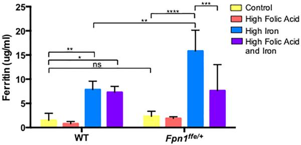
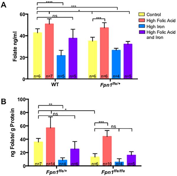
Results: High levels of iron supplementation significantly reduced the incidence of NTDs in Fpn1ffe mutants. Fpn1 deficiency resulted in reduced folate levels in both pregnant dams and embryos. Yet folic acid supplementation did not prevent NTDs in the Fpn1ffe model. Similarly, forebrain truncations were rescued with iron. Surprisingly, the high levels of iron supplementation used in this study caused folate deficiency in wild-type dams and embryos.

Conclusion: Our results demonstrate that iron supplementation can prevent NTDs and forebrain truncations in the Fpn1ffe model. Surprisingly, high levels of iron supplementation and iron overload can cause folate deficiency. If iron is essential for neural tube closure, it is possible that iron deficiency might contribute to NTDs. Birth Defects Research 109:81-91, 2017. © 2016 The Authors Birth Defects Research Published by Wiley Periodicals, Inc.

Keywords: exencephaly; folic acid supplementation; iron deficiency; neural tube defects; spina bifida.


Figures

Figure 1
Figure 1. Periconceptional supplementation with a high iron diet but not folic acid prevents NTDs in the Fpn1ffe mouse line
A-A”. A. Normal neural tube closure in a 9.5 dpc Fpn1ffe/+ embryo compared to A’ exencephaly in an Fpn1ffe/ffe mutant from a dam fed the control diet. A” normal morphology and neural tube closure in an Fpn1ffe/ffe mutant from a dam fed the high iron diet. Size bar = 1 mm. B. Frequency of NTDs in Fpn1ffe/ffe mutant embryos from dams supplemented with control (yellow bar), high folic acid (10 ppm, orange bar), high iron (0.5% carbonyl iron, blue bar) or high folic acid and iron (purple bar) diets for 4 weeks before mating. Statistical significance was determined by the Fisher's Exact test and p-values: =0.0002 ***, ≤0.0001 **** or non significant (ns). The number of samples represented in each group is indicated.
Figure 2
Figure 2. Effects of iron and folate supplementation on embryo size and developmental stage
A. Comparison of crown to rump length (A) and somite numbers (B) of embryos from Fpn1ffe/ffe mutant embryos (White Bars in A) and wildtype littermates (Colored Bars in A) dissected at 9.5 dpc from dams supplemented with control (yellow), high folic acid (10 ppm, orange), high iron (0.5% carbonyl iron, blue) or high folic acid and iron (purple) diets for 4 weeks before mating. C. Comparison of weights of Fpn1ffe/ffe mutant embryos and wildtype littermates dissected at 11.5 dpc from dams fed the various diets for 4 weeks before mating. Statistical significance was determined by unpaired t-test in A and C, and by 2- factor ANOVA with post-hoc Sidak's multiple comparisons test in B. P-values: ≤0.05 *, ≤0.01 ** or non significant (ns).
Figure 3
Figure 3. Supplementation with a high iron diet increases iron stores in wildtype and Fpn1ffe/+ dams
Serum was obtained from pregnant dams upon dissection of embryos at 9.5 or 11.5 dpc. Dams were supplemented with control (yellow bar), high folic acid (10 ppm, orange bar), high iron (0.5% carbonyl iron, blue bar) or high folic acid and iron (purple bar) diets for 4 weeks before mating. Ferritin levels were determined by ELISA and served as a proxy for stored iron levels. Maternal serum ferritin was measured in 3 samples in the wildtype (WT) group and 5 in the Fpn1ffe/+ group. Statistical significance was determined by 2-factor ANOVA with post-hoc Sidak's multiple comparisons test. P-values ≤0.05 *, ≤0.01 **, ≤0.001 ***, ≤0.0001 **** or non significant (ns).
Figure 4
Figure 4. Folate levels in dams and embryos
A. Determination of red blood cell folate levels in pregnant dams. Whole blood was obtained from pregnant wildtype (WT) or Fpn1ffe/+ dams at 9.5 or 11.5 dpc. Dams were supplemented with control (yellow bar), high folic acid (10 ppm, orange bar), high iron (0.5% carbonyl iron, blue bar) or high folic acid and iron (purple bar) diets for 4 weeks before mating. B. Determination of folate levels in 11.5 dpc wildtype (Fpn1+/+) and Fpn1ffe/ffe embryos from dams fed the various diets. The number of samples represented in each group is indicated. The Sidak's test was used to determine significance of multiple comparisons within a genotype and the Tukey test across genotypes. P-values: ≤0.05 *, ≤0.01**, ≤0.001 ***, ≤0.0001 **** or non significant (ns). The number of samples represented in each group is indicated.
Figure 5
Figure 5. Forebrain truncations in Fpn1ffe/ffe mutant embryos are rescued by supplementation with a high iron diet
A. In situ hybridization to detect Six3 expression in 9.5 dpc embryos. The forebrain was measured from the rostral point of the optic vesicle (stained by Six3) to the most rostral point of the forebrain as indicated by white line. B. Forebrain measurements in 9.5 dpc wildtype (Fpn1+/+) and Fpn1ffe/ffe embryos from dams fed control (yellow bar), high folic acid (10 ppm, orange bar), high iron (0.5% carbonyl iron, blue bar) or high folic acid and iron (purple bar) diets for 4 weeks before mating. Statistical significance was determined by the unpaired t-test. P-values: ≤0.0005 *** or non significant (ns). The number of samples represented in each group is indicated.

Similar articles

Cited by

References

    1. Acampora D, Avantaggiato V, Tuorto F, Briata P, Corte G, Simeone A. Visceral endoderm-restricted translation of Otx1 mediates recovery of Otx2 requirements for specification of anterior neural plate and normal gastrulation. Development. 1998;125:5091–5104. - PubMed
    1. Arakawa T, Ohara K, Kakizaki R, Takahashi Y, Hirata K, Fujii M, Konno T, Morikawa T, Chiba F, Chiba R. Folic acid deficiency in hemochromatosis: probably due to a defective storage of folic acid in the liver. The Tohoku journal of experimental medicine. 1965;86:301–306. - PubMed
    1. Blom HJ, Shaw GM, den Heijer M, Finnell RH. Neural tube defects and folate: case far from closed. Nature reviews. 2006;7:724–731. - PMC - PubMed
    1. Bothwell TH. Iron requirements in pregnancy and strategies to meet them. The American journal of clinical nutrition. 2000;72:257S–264S. - PubMed
    1. Buamah PK, Russell M, Bates G, Ward AM, Skillen AW. Maternal zinc status: a determination of central nervous system malformation. British journal of obstetrics and gynaecology. 1984;91:788–790. - PubMed

Publication types

MeSH terms

LinkOut - more resources