. 2017 Jan;27(1):119-129.
doi: 10.1038/cr.2016.152. Epub 2016 Dec 23.

Cryo-electron microscopy structures of the SARS-CoV spike glycoprotein reveal a prerequisite conformational state for receptor binding

Affiliations

Cryo-electron microscopy structures of the SARS-CoV spike glycoprotein reveal a prerequisite conformational state for receptor binding

Miao Gui et al. Cell Res. 2017 Jan.

Abstract

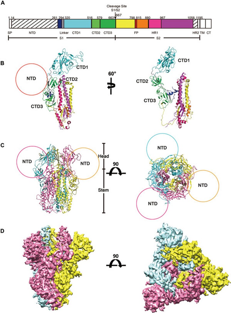
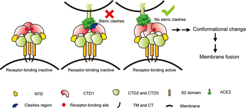
The global outbreak of SARS in 2002-2003 was caused by the infection of a new human coronavirus SARS-CoV. The infection of SARS-CoV is mediated mainly through the viral surface glycoproteins, which consist of S1 and S2 subunits and form trimer spikes on the envelope of the virions. Here we report the ectodomain structures of the SARS-CoV surface spike trimer in different conformational states determined by single-particle cryo-electron microscopy. The conformation 1 determined at 4.3 Å resolution is three-fold symmetric and has all the three receptor-binding C-terminal domain 1 (CTD1s) of the S1 subunits in "down" positions. The binding of the "down" CTD1s to the SARS-CoV receptor ACE2 is not possible due to steric clashes, suggesting that the conformation 1 represents a receptor-binding inactive state. Conformations 2-4 determined at 7.3, 5.7 and 6.8 Å resolutions are all asymmetric, in which one RBD rotates away from the "down" position by different angles to an "up" position. The "up" CTD1 exposes the receptor-binding site for ACE2 engagement, suggesting that the conformations 2-4 represent a receptor-binding active state. This conformational change is also required for the binding of SARS-CoV neutralizing antibodies targeting the CTD1. This phenomenon could be extended to other betacoronaviruses utilizing CTD1 of the S1 subunit for receptor binding, which provides new insights into the intermediate states of coronavirus pre-fusion spike trimer during infection.


Figures

Figure 1
Figure 1
Overall structure of the SARS-CoV S glycoprotein. (A) A schematic diagram showing the domain organization of the SARS-CoV S glycoprotein. SP: signal peptide; NTD: N-terminal domain; CTD1: C-terminal domain 1, cyan; Linker: the linker connecting NTD and CTD1, grey; CTD2: C-terminal domain 2, light green; CTD3: C-terminal domain 3, dark green; FP: fusion peptide, red; HR1: heptad repeat 1, pink; HR2: heptad repeat 2; TM: transmembrane domain; CT: cytoplasmic tail. The TM and CT regions are not included in the expression construct. The NTD and HR2 regions that are not resolved in the reconstruction are represented with diagonal stripes. (B) Ribbon diagrams showing the structure of one SARS-CoV S glycoprotein monomer with domains colored as the same as in A. (C) Ribbon diagrams showing the structure of the SARS-CoV S glycoprotein trimer. The three protomers are colored pink, yellow and cyan, respectively. The circles indicate the locations of the unmodeled NTD regions. (D) Surface shadowed diagrams showing the 4.3 Å resolution 3D density map of the SARS-CoV S trimer. The protomers are colored the same as in C.
Figure 2
Figure 2
Four different conformations of the SARS-CoV S glycoprotein trimer. Top: surface shadowed diagrams showing the four different conformations (conformations 1-4) of the S trimer. The CTD1s are colored pink. Bottom: ribbon diagrams showing S monomers with the semi-transparent CTD1 densities colored pink. The tilt angles of the CTD1s are defined by the angle between the long axis of the CTD1 (red cylinder) and its projection on the horizontal plane (grey ellipse). (A) Three-fold symmetric conformation 1 with all the three CTD1s in the “down” conformations. (B-D) Asymmetric conformations 2-4 with one CTD1 in the “up” conformation.
Figure 3
Figure 3
Models of the SARS-CoV S monomer and trimer bound with the receptor ACE2. (A) “Binding” of the receptor ACE2 (green) to one S monomer (pink) of the conformation 1 S trimer. The CTD1 is in the “down” conformation. (B) “Binding” of the receptor ACE2 (green) to the conformation 1 S trimer. Three CTD1s are all in the “down” conformations. The steric clashes between a neighboring CTD1 (grey) and ACE2 (green) are colored blue. (C) The same model as in A, with the S trimer density map presented. Only the boundary profile of the “bound” ACE2 is shown (green lines) for a better view of the clashes (volume: 10 696 Å3). (D) “Binding” of the receptor ACE2 (green) with the conformation 3 S monomer (pink) of which the CTD1 is in the “up” conformation. (E) “Binding” of the receptor ACE2 (green) to the “up” CTD1 (pink) of the conformation 3 S trimer showing no steric clashes with any neighboring “down” CTD1 (grey). (F) The same model as in D with the S trimer 3D density map presented. All models are generated by superimposing the CTD1-ACE2 complex crystal structure onto the CTD1 of the corresponding SARS-CoV S monomer or trimer. The NTD models are not shown.
Figure 4
Figure 4
Structural superimpositions showing the “binding” of neutralization antibodies to the SARS-CoV S trimers. (A) Structural superimposition of the CTD1-Fab m396 complex (PDB accession code: 2DD8) onto one CTD1 of the SARS-CoV S trimer in conformation 1, showing the “binding” of Fab m396 to the SARS-CoV S trimer. The EM densities of the S trimer are represented using shadowed surfaces in semi-transparent grey. (B-C) Similar structural superimpositions showing the “binding” of neutralization antibodies 80R (PDB accession code: 2GHW; B) and F26G19 (PDB accession code: 3BGF; C). The steric clashes with the “bound” Fab are colored blue and the corresponding volumes are shown in bracket. (D-F) Structural superimpositions of three CTD1-antibody complex structures with the SARS-CoV S trimer in conformation 3. No steric clashes between the “bound” Fab and the S trimer.
Figure 5
Figure 5
A cartoon model showing the transition of the S trimer spikes from receptor-binding inactive to active and subsequent fusogenic states. For betacoronavirus S glycoprotein utilizing the CTD1 as the receptor-binding domain, the state transition of the S trimer with the “down” to “up” conformational change of CTD1 would allow receptor binding and may initiate subsequent conformational changes in the S2 subunits to mediate membrane fusion.

References

    1. Su S, Wong G, Shi W, et al. Epidemiology, genetic recombination, and pathogenesis of coronaviruses. Trends Microbiol 2016; 24:490–502. - PMC - PubMed
    1. Ksiazek TG, Erdman D, Goldsmith CS, et al. A novel coronavirus associated with severe acute respiratory syndrome. N Engl J Med 2003; 348:1953–1966. - PubMed
    1. Drosten C, Gunther S, Preiser W, et al. Identification of a novel coronavirus in patients with severe acute respiratory syndrome. N Engl J Med 2003; 348:1967–1976. - PubMed
    1. Peiris JS, Lai ST, Poon LL, et al. Coronavirus as a possible cause of severe acute respiratory syndrome. Lancet 2003; 361:1319–1325. - PMC - PubMed
    1. Zaki AM, van Boheemen S, Bestebroer TM, Osterhaus AD, Fouchier RA. Isolation of a novel coronavirus from a man with pneumonia in Saudi Arabia. N Engl J Med 2012; 367:1814–1820. - PubMed

MeSH terms

Substances