. 2016 Dec 23:6:39775.
doi: 10.1038/srep39775.

Zika virus infection induces mitosis abnormalities and apoptotic cell death of human neural progenitor cells

Affiliations

Zika virus infection induces mitosis abnormalities and apoptotic cell death of human neural progenitor cells

Bruno S F Souza et al. Sci Rep. .

Abstract

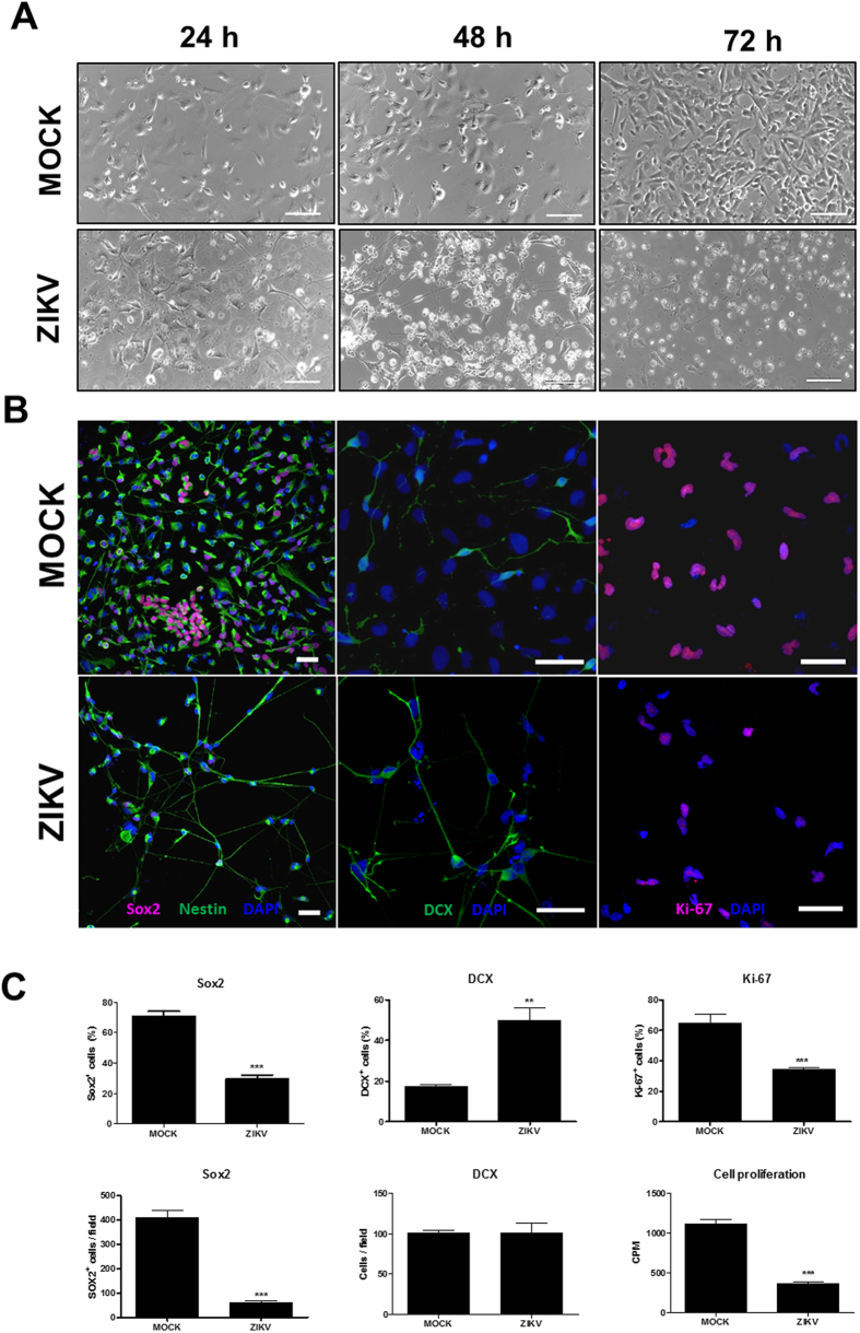
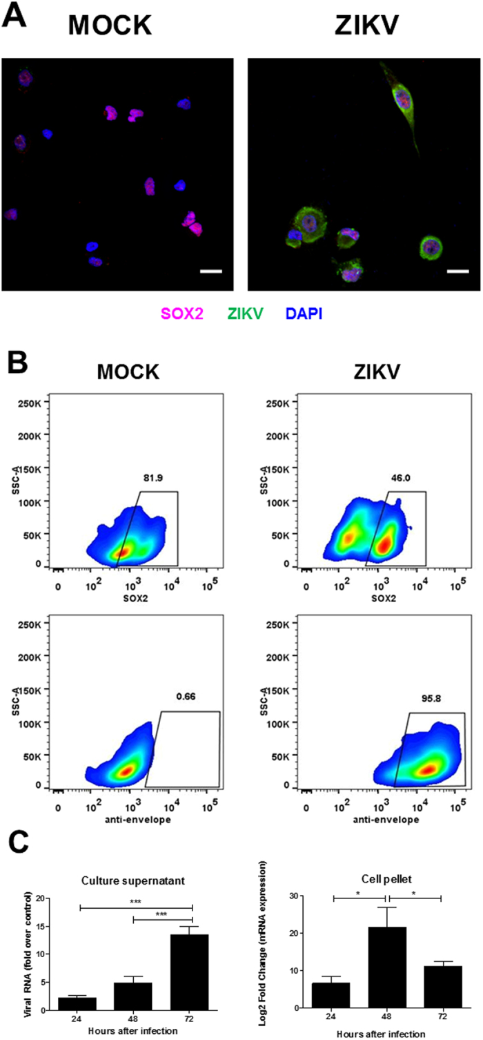
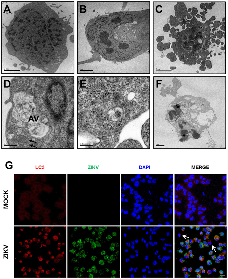
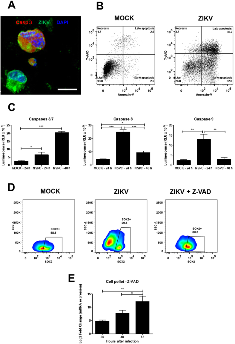
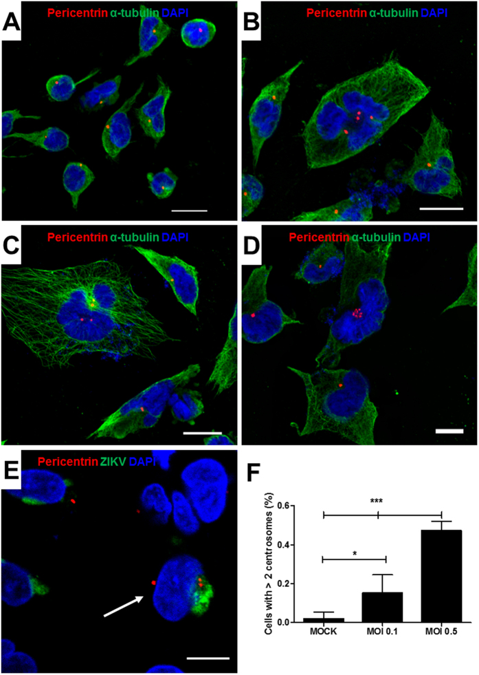
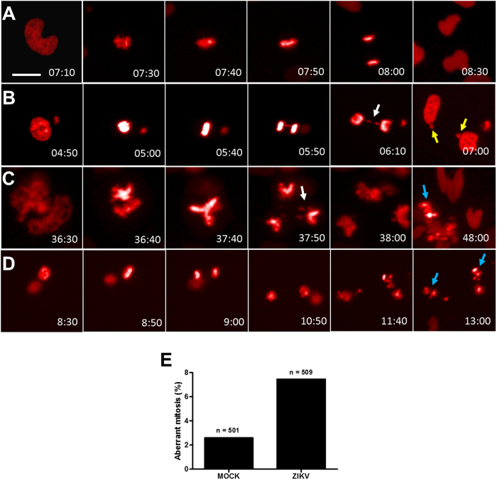
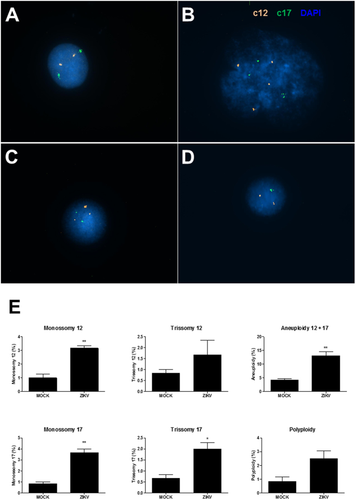
Zika virus (ZIKV) infection has been associated with severe complications both in the developing and adult nervous system. To investigate the deleterious effects of ZIKV infection, we used human neural progenitor cells (NPC), derived from induced pluripotent stem cells (iPSC). We found that NPC are highly susceptible to ZIKV and the infection results in cell death. ZIKV infection led to a marked reduction in cell proliferation, ultrastructural alterations and induction of autophagy. Induction of apoptosis of Sox2+ cells was demonstrated by activation of caspases 3/7, 8 and 9, and by ultrastructural and flow cytometry analyses. ZIKV-induced death of Sox2+ cells was prevented by incubation with the pan-caspase inhibitor, Z-VAD-FMK. By confocal microscopy analysis we found an increased number of cells with supernumerary centrosomes. Live imaging showed a significant increase in mitosis abnormalities, including multipolar spindle, chromosome laggards, micronuclei and death of progeny after cell division. FISH analysis for chromosomes 12 and 17 showed increased frequency of aneuploidy, such as monosomy, trisomy and polyploidy. Our study reinforces the link between ZIKV and abnormalities in the developing human brain, including microcephaly.


Figures

Figure 1
Figure 1. Human iPSC-derived NPC are highly sensitive to ZIKV infection.
(A) NPC cultures observed by phase contrast 24, 48 and 72 h after ZIKV infection, showing high loss of cells in culture when compared to mock infection. Scale bars = 100 μm. (B) Confocal microscopy analyses of mixed NPC cultures immunostained for Sox2, Nestin, DCX, and proliferation marker Ki-67, 72 h after ZIKV or mock infection. Scale bars = 50 μm. (C) Quantification of the percentage and density of Sox2+ cells, percentage and density of DCX+ cells, Ki-67+ cells 72 h after mock and ZIKV infection. Cell proliferation was evaluated by 3H-thymidine incorporation 48 h after infection. Data represented as mean ± SEM. **p < 0.01; ***p < 0.001.
Figure 2
Figure 2. Analysis of ZIKV infection in NPC cultures.
(A) Immunostaining for ZIKV and Sox2 in NPC cultures. Nuclei are stained with DAPI. Scale bars = 20 μm. (B) Flow cytometry analysis for quantification of Sox2+ and ZIKV+ cells 48 h after infection. (C) Quantification of viral RNA in culture supernatants and cell pellets by RT-qPCR 24, 48 and 72 h after ZIKV infection. Data normalized using the values obtained at 2 h time point (supernatant) or endogenous control gene (GAPDH; pellet), and are represented as mean ± SEM. *p < 0.05; ***p < 0.001.
Figure 3
Figure 3. Ultrastructural analysis of NPC cultures after ZIKV infection.
(A,B) Transmission electron micrographs of mock-infected cells, showing nuclei and organelles with normal aspect after 24 and 72 h of culture. (C–F) ZIKV-infected cells 24 (C,D), 48 (E) and 72 (F) h after infection. Cells in early (C) and late (F) apoptotic processes. (D) The presence of large perinuclear autophagic vacuoles (AV) can be observed. Black arrows indicate mitochondria with altered morphology. (E) Presence of viral capsid in intracellular vacuole. (G) Immunostaining for ZIKV (green) and the autophagic vacuole marker LC3 (red). Nuclei are stained with DAPI (blue). White arrows indicate cells with negative or low ZIKV staining, and absence of perinuclear LC3 staining. Scale bars = 20 μm.
Figure 4
Figure 4. ZIKV infection induces apoptotic cell death in NPC cultures.
(A) Confocal microscopy of mock and ZIKV-infected NPC stained for cleaved caspase-3 (Casp3; red), DAPI (nuclei; blue), and ZIKV (in green). Scale bars = 20 μm. (B) Flow cytometry analysis for detection of apoptosis by Annexin V and 7AAD staining 48 h after mock or ZIKV infection. (C) Activity of caspases 3/7, 8 and 9, in mock- and ZIKV-infected cultures was determined using luminescence assays after 24 or 48 h of culture. Data represented as mean ± SEM. *p < 0.05; **p < 0.01; ***p < 0.001. (D) Flow cytometry analysis showing the percentage of Sox2+ cells in mock-infected and in ZIKV-infected cultures, in the absence and in the presence of Z-VAD (50 μM), 48 h after infection. (E) Quantification of viral RNA in cell pellets by RT-qPCR 24, 48 and 72 h after ZIKV infection in the presence of Z-VAD (50 μM). Data were normalized using an endogenous control gene (GAPDH), and are represented as mean ± SEM. *p < 0.05; **p < 0.01.
Figure 5
Figure 5. Presence of supernumerary centrosomes in ZIKV-infected cultures.
Confocal microscopy of mock (A) or ZIKV (BD)-infected cells stained for alpha-tubulin (green), DAPI (nuclei; blue), and pericentrin (red), showing cells with more than 2 centrosomes in ZIKV-infected cultures. (E) ZIKV-infected cells co-stained with pericentrin (red), ZIKV (green) and DAPI (nuclei; blue), showing the presence of the virus in a cell with three centrosomes (arrow). Scale bars = 20 μm. (F) Quantification of cells with supernumerary centrosomes (>2) in mock-infected cultures or in cultures infected with ZIKV at M.O.I. 0.1 and 0.5, 48 h after infection. Data represented as mean ± SEM. *p < 0.05; ***p < 0.001.
Figure 6
Figure 6. ZIKV infection causes mitotic abnormalities in NPC cultures.
(AD) Still frames from live imaging experiment showing representative NPC undergoing a bipolar cell division (A); bipolar cell division with chromosome lagging and formation of micronuclei (B); multipolar cell division with chromosome lagging and loss of one progeny (C); and bipolar cell division followed by death in interphase (D). Coloured arrows indicate chromosome lagging (white), micronuclei (yellow) and progeny death (blue). (E) Percentage of aberrant mitosis in MOCK- and ZIKV-infected NPC cultures. Time, hours:minutes. Scale bars = 20 μm.
Figure 7
Figure 7. Chromosomal aberrations caused by ZIKV in NPC cultures.
Interphase FISH with centromeric probes on mock- (A) and ZIKV-infected (BD) NPC. (A) Euploid cell for chromosome 12 (two orange signals) and chromosome 17 (two green signals). (B) Polyploid cell carrying four copies of chromosome 12 and three copies of chromosome 17. (C) Trisomy of chromosome 12 (three orange signals). (D) Cell carrying a monosomy of chromosome 17. (E) Frequencies of numerical chromosomal abnormalities for the chromosomes 12 and 17 analyzed by FISH. The total aneuploidy included the numerical abnormalities (monosomies and trisomies) of chromosomes 12 and 17. Data represented as mean ± SEM. *p < 0.05; **p < 0.01.

References

    1. Dick G. W., Kitchen S. F. & Haddow A. J. Zika virus. I. Isolations and serological specificity. Trans. R. Soc. Trop. Med. Hyg 46, 509–520 (1952). - PubMed
    1. Duffy M. R. et al.. Zika virus outbreak on Yap Island, Federated States of Micronesia. N. Engl. J. Med 360, 2536–2543 (2009). - PubMed
    1. Cao-Lormeau V. M. et al.. Zika virus, French Polynesia, South Pacific, 2013. Emerg. Infect. Dis. 20, 1085–1086 (2014). - PMC - PubMed
    1. Hennessey M., Fischer M. & Staples J. E. Zika Virus Spreads to New Areas - Region of the Americas, May 2015-January 2016. MMWR Morb. Mortal. Wkly. Rep. 65, 55–58 (2016). - PubMed
    1. Kleber de Oliveira W. et al.. Increase in reported prevalence of microcephaly in infants born to women living in areas with confirmed Zika virus transmission during the first trimester of pregnancy - Brazil, 2015. MMWR Morb. Mortal. Wkly. Rep. 65, 242–247 (2016). - PubMed

Publication types