. 2016 Dec 23:6:39141.
doi: 10.1038/srep39141.

Drosophila microRNA-34 Impairs Axon Pruning of Mushroom Body γ Neurons by Downregulating the Expression of Ecdysone Receptor

Affiliations

Drosophila microRNA-34 Impairs Axon Pruning of Mushroom Body γ Neurons by Downregulating the Expression of Ecdysone Receptor

Yen-Wei Lai et al. Sci Rep. .

Abstract

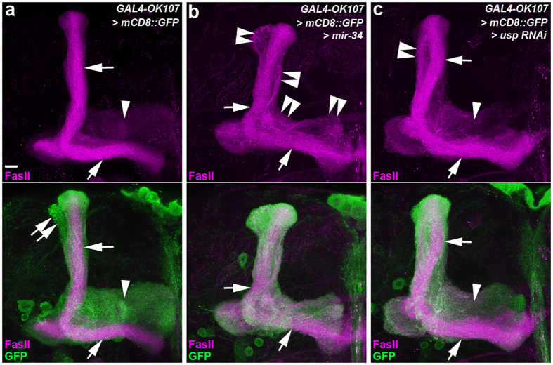
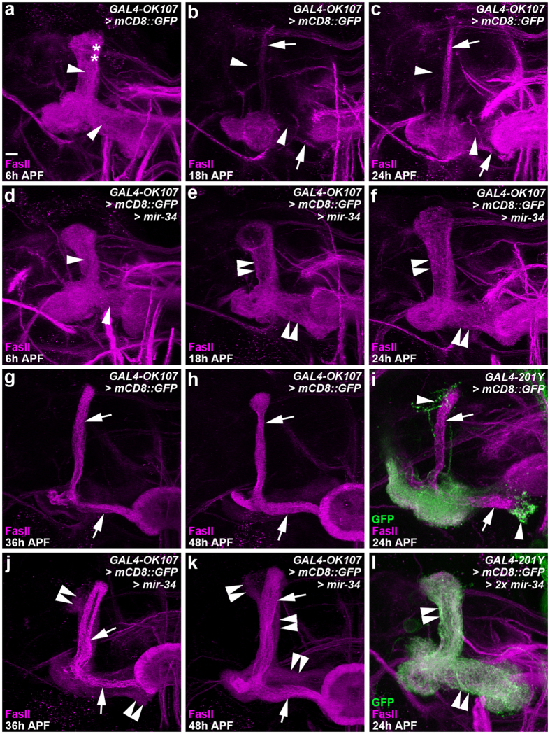
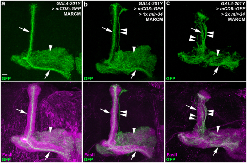
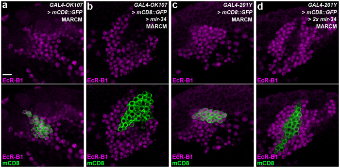
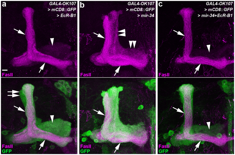
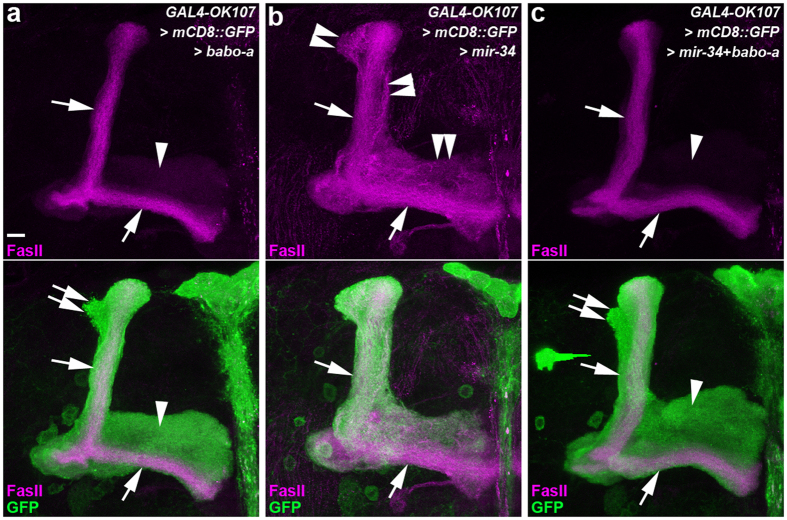
MicroRNA-34 (miR-34) is crucial for preventing chronic large-scale neurite degeneration in the aged brain of Drosophila melanogaster. Here we investigated the role of miR-34 in two other types of large-scale axon degeneration in Drosophila: axotomy-induced axon degeneration in olfactory sensory neurons (OSNs) and developmentally related axon pruning in mushroom body (MB) neurons. Ectopically overexpressed miR-34 did not inhibit axon degeneration in OSNs following axotomy, whereas ectopically overexpressed miR-34 in differentiated MB neurons impaired γ axon pruning. Intriguingly, the miR-34-induced γ axon pruning defect resulted from downregulating the expression of ecdysone receptor B1 (EcR-B1) in differentiated MB γ neurons. Notably, the separate overexpression of EcR-B1 or a transforming growth factor- β receptor Baboon, whose activation can upregulate the EcR-B1 expression, in MB neurons rescued the miR-34-induced γ axon pruning phenotype. Future investigations of miR-34 targets that regulate the expression of EcR-B1 in MB γ neurons are warranted to elucidate pathways that regulate axon pruning, and to provide insight into mechanisms that control large-scale axon degeneration in the nervous system.


Figures

Figure 1
Figure 1. Ectopic miR-34 overexpression caused aberrant lobe formation on mushroom body (MB) neurons.
Confocal images show the lobe phenotype of mushroom body (MB) neurons in wild-type flies (a) and flies with ectopic miR-34 overexpression (b) and RNAi knockdown of USP (c). Fasciculin II (FasII) staining (magenta) reveals the dorsal α and medial β lobes (arrows, strong magenta staining) and the medial γ lobes (arrowheads, faint magenta staining) on MB neurons. In the lower panels, GAL4-OK107-driven mCD8::GFP (GFP) expression (green) shows the morphology of α lobes (arrows), α′ lobes (double arrows), and γ lobes (arrowheads). (a–c) Compared to the α and β lobes of wild-type MB neurons (arrows, a), ectopic miR-34 expression resulted in aberrant axonal branches projecting adjacent to the α and β lobes (double arrowheads, b). This miR-34 induced lobe defect is similar to that of the γ lobe phenotype observed in MB neurons in which USP expression was knocked down by RNAi (double arrowhead, c). Fly genotypes are listed in Supplemental Table 2. Scale bar: 10 μm for all panels.
Figure 2
Figure 2. Ectopic miR-34 overexpression inhibited γ lobe pruning in MB neurons.
Confocal images show γ lobes of MB neurons in wild-type flies (a–c,g–i) and flies that were for ectopic miR-34 overexpression (one copy of miR-34 in (d–f,j,k); two copies of miR-34 in l) driven by GAL4-OK107 (a–h,j,k) or GAL4-201Y (i and l). FasII staining (magenta) and mCD8::GFP (GFP) expression (green) of MB neurons reveal the morphology of the larval-specific γ lobes (arrowheads) and α and β lobes (arrows) at various time points after puparium formation (APF). In MB neurons of wild-type flies (a–c and g–i), the process of pruning larval-specific γ lobes (arrowheads) appeared to be initiated in the γ lobes themselves, where the lack of FasII staining was observed from 6 h APF (asterisks, a) to the near completion of axon pruning at 18 h APF (arrowheads). The developing α and β lobes were observed from 24 h APF onward (arrows, c,g–i). By contrast, in the MB neurons with ectopic miR-34 overexpression, a significant fraction of larval-specific γ lobes was remained at 18–24 h APF (double-arrowheads, e,f,i), and aberrant axonal braches (most likely γ lobe-derived) were located adjacent to the developing α and β lobes at 36-48 h APF (double-arrowheads, j,k). Fly genotypes are listed in Supplemental Table 2. Scale bar: 10 μm for all panels.
Figure 3
Figure 3. Ectopic miR-34 overexpression in differentiated MB neurons disrupted γ axon pruning.
Confocal images show axon pruning of MB neurons in wild-type flies (a) and flies with ectopic miR-34 overexpression using the mosaic analysis with a repressible cell marker system (b,c). GAL4-201Y-driven mCD8::GFP (GFP) expression (green) reveals the morphology of γ lobes (arrowheads) and a subset of α and β lobes (arrows). In the lower panels, FasII staining (magenta) reveals the α and β lobes (strong magenta staining; arrows) and the medial γ lobes (faint magenta staining; arrowheads). A subset of the dorsal α lobe was observed on MB neurons in wild-type flies (upper arrow, a), whereas an aberrant axonal bundle (likely unpruned γ lobe) projected outside the MB α lobe (double arrowhead, b) and most of the γ lobes were intact in the sample with ectopic expression of one copy of mir-34 transgene (arrowhead, b). Defective γ lobe pruning was more severe in flies for ectopic overexpression of two copies of miR-34 transgenes in MB γ neurons (double arrowheads, c). Fly genotypes are listed in Supplemental Table 2. Scale bar: 10 μm for all panels.
Figure 4
Figure 4. Ectopic miR-34 overexpression downregulated EcR-B1 expression in MB neurons.
Confocal images show EcR-B1 expression in MB neurons in wild-type flies (a,c) and flies for ectopic miR-34 overexpression (one copy of miR-34 in b; two copies of miR-34 in d) using the mosaic analysis with a repressible cell marker (MARCM) system. The MARCM clones were induced as newly hatched larvae, and the brains were dissected at the wandering larval stage (WL) or the white pupal stage (WP). In the lower panels, the expression of mCD8::GFP (green) driven by GAL4-OK107 (a,b) or GAL4-201Y (c,d) outlines the cell bodies of MARCM clones, whereas EcR-B1 staining (magenta) reveals ecdysone-responsive neurons. The expression of EcR-B1 was significantly reduced by ectopic miR-34 overexpression in the MARCM clones (b,d), compared to that of the wild-type MARCM clones (a,c). Fly genotypes are listed in Supplemental Table 2. Scale bar: 10 μm for all panels.
Figure 5
Figure 5. Overexpression of EcR-B1 rescued the miR-34-induced defective γ lobe phenotype in MB neurons.
Confocal images show the lobe pruning phenotypes of MB neurons in flies with ectopic overexpression of EcR-B1 (a), ectopic overexpression of miR-34 (b), or the overexpression of both (c). FasII staining reveals the medial γ lobes (arrowheads, faint magenta staining) and dorsal α and medial β lobes (arrows, strong magenta staining). In the lower panels, GAL4-OK107-driven mCD8::GFP (GFP) expression (green) shows the morphology of α lobes (arrows), α′ lobes (double arrows), and γ lobes (arrowheads). Overexpression of EcR-B1 in MB neurons had no observable effect on the MB lobe pruning phenotype (a). Ectopic miR-34 overexpression in MB neurons caused defective γ lobe pruning (double-arrowheads, b). The defective γ lobe pruning phenotype was rescued by overexpression of EcR-B1 in MB neurons with ectopic miR-34 overexpression (c). Fly genotypes are listed in Supplemental Table 2. Scale bar: 10 μm for all panels.
Figure 6
Figure 6. Overexpression of Babo-a rescued the miR-34-induced defective γ lobe phenotype in MB neurons.
Confocal images show lobe pruning phenotypes of MB neurons in flies with ectopic overexpression of Babo-a (a), ectopic overexpression of miR-34 (b), or the overexpression of both (c). FasII staining reveals the medial γ lobes (arrowheads, faint magenta staining) and dorsal α and medial β lobes (arrows, strong magenta staining). In the lower panels, GAL4-OK107-driven mCD8::GFP (green) shows the morphology of α lobes (arrows), α′ lobes (double arrows), and γ lobes (arrowheads). Overexpression of Babo-a in MB neurons had no observable effect on the MB lobe pruning phenotype (a). Ectopic miR-34 overexpression in MB neurons caused defective γ lobe pruning (double-arrowheads, b). The defective γ lobe pruning phenotype was rescued by overexpression of Babo-a in MB neurons with ectopic miR-34 overexpression (c). Fly genotypes are listed in Supplemental Table 2. Scale bar: 10 μm for all panels.

Similar articles

Cited by

References

    1. Kandel E. R. Principles of neural science. 5th edn, (McGraw-Hill, 2013).
    1. Im H. I. & Kenny P. J. MicroRNAs in neuronal function and dysfunction. Trends Neurosci 35, 325–334, doi: 10.1016/j.tins.2012.01.004 (2012). - DOI - PMC - PubMed
    1. Chen W. & Qin C. General hallmarks of microRNAs in brain evolution and development. RNA Biol 12, 701–708, doi: 10.1080/15476286.2015.1048954 (2015). - DOI - PMC - PubMed
    1. Cao D. D., Li L. & Chan W. Y. MicroRNAs: Key Regulators in the Central Nervous System and Their Implication in Neurological Diseases. Int J Mol Sci 17, doi: 10.3390/ijms17060842 (2016). - DOI - PMC - PubMed
    1. Ballatore C., Lee V. M. & Trojanowski J. Q. Tau-mediated neurodegeneration in Alzheimer’s disease and related disorders. Nat Rev Neurosci 8, 663–672, doi: 10.1038/nrn2194 (2007). - DOI - PubMed

Publication types

MeSH terms

LinkOut - more resources