. 2016 Dec 23:6:39738.
doi: 10.1038/srep39738.

Antiretroviral Treatment with Efavirenz Disrupts the Blood-Brain Barrier Integrity and Increases Stroke Severity

Affiliations

Antiretroviral Treatment with Efavirenz Disrupts the Blood-Brain Barrier Integrity and Increases Stroke Severity

Luc Bertrand et al. Sci Rep. .

Abstract

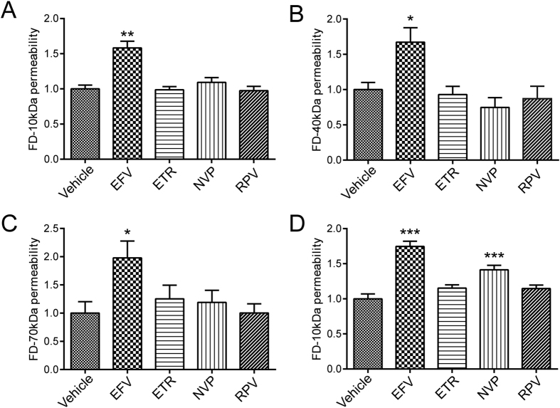
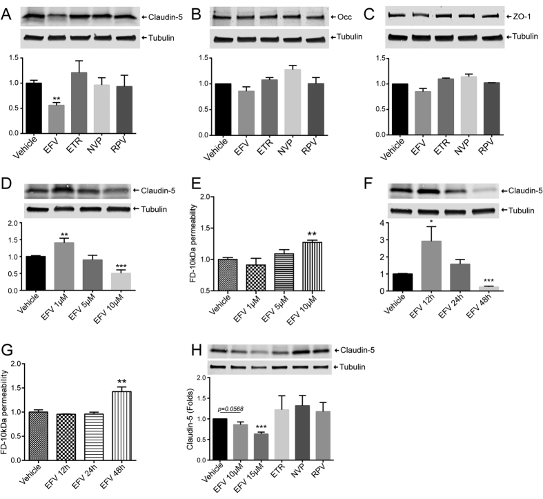
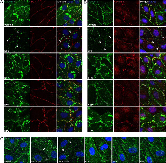
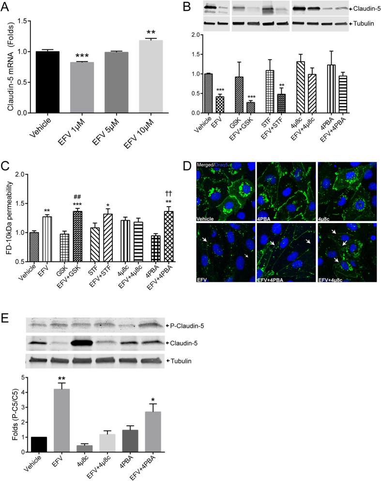
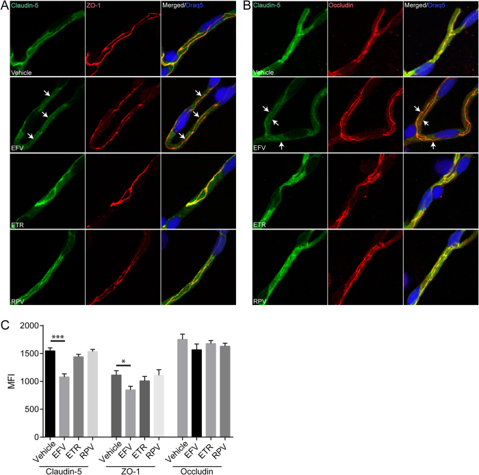
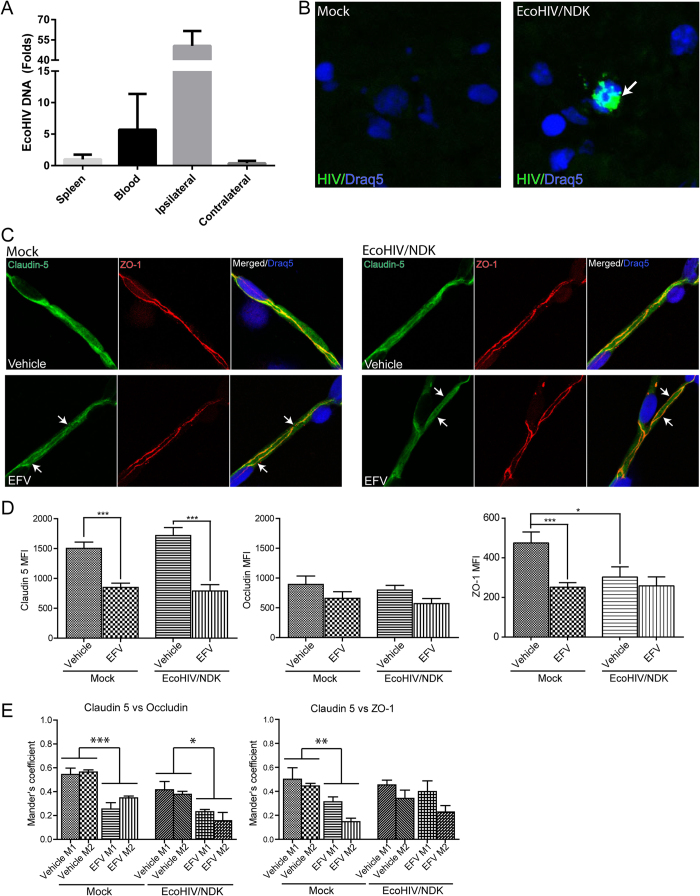
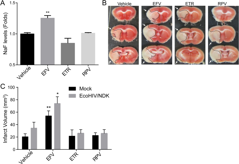
The introduction of antiretroviral drugs (ARVd) changed the prognosis of HIV infection from a deadly disease to a chronic disease. However, even with undetectable viral loads, patients still develop a wide range of pathologies, including cerebrovascular complications and stroke. It is hypothesized that toxic side effects of ARVd may contribute to these effects. To address this notion, we evaluated the impact of several non-nucleoside reverse transcriptase inhibitors (NNRTI; Efavirenz, Etravirine, Rilpivirine and Nevirapine) on the integrity of the blood-brain barrier, and their impact on severity of stroke. Among studied drugs, Efavirenz, but not other NNRTIs, altered claudin-5 expression, increased endothelial permeability, and disrupted the blood-brain barrier integrity. Importantly, Efavirenz exposure increased the severity of stroke in a model of middle cerebral artery occlusion in mice. Taken together, these results indicate that selected ARVd can exacerbate HIV-associated cerebrovascular pathology. Therefore, careful consideration should be taken when choosing an anti-retroviral therapy regimen.


Figures

Figure 1
Figure 1. Impact of NNRTIs on endothelial permeability.
hCMEC (A–C) and human primary brain endothelial cells (D), grown to confluence on Transwells, were incubated with vehicle (DMSO), Efavirenz (Efa), Etravirine (ETR), Nevirapine (NVP) or Rilpivirine (RPV) for 48 h. Culture media in the apical compartment of the Transwell system was replaced with medium containing fluorescently tagged dextran (FD) of 10 kDa (A and D), 40 kDa (B) or 70 kDa (C). Basolateral levels of FD were assayed 90 min post exposure. Data are mean ± SEM, expressed as fold increase over vehicle control; three independent experiments, each with n = 4; *p < 0.05 vs. Vehicle; **p < 0.01 vs. Vehicle; ***p < 0.001 vs. Vehicle.
Figure 2
Figure 2. Impact of NNRTIs on tight junction protein expression.
hCMEC (A–G) and human primary brain endothelial cells (H) were grown to confluence, followed by exposure to vehicle (DMSO), Efavirenz (Efa), Etravirine (ETR), Nevirapine (NVP) or Rilpivirine (RPV) for 48 h. Expression of claudin-5 (A,D,F,H), occludin (B), and ZO-1 (C) was assessed by western blotting. Dose-dependent effects of Efavirenz on claudin-5 levels (D,H) and endothelial permeability (E). Time-dependent impact of Efavirenz on claudin-5 levels (F) and endothelial permeability (G). Presented blots are cropped from the originals, full size blots available in Supplementary Fig. S1. Data are mean ± SEM, expressed as fold increase over vehicle control; three or four independent experiments, each with n = 4. *p < 0.05 vs. Vehicle; **p < 0.01 vs. Vehicle; ***p < 0.001 vs. Vehicle.
Figure 3
Figure 3. Treatment with Efavirenz, but not with other NNRTIs, decreases claudin-5 immunoreactivity and localization.
hCMEC were grown on coverslips to confluence, exposed to NNRTIs as in Fig. 1, and immunostained for claudin-5 (green), ZO-1 (red), or occludin (red). Draq5 (blue) visualizes nuclei. Left and middle columns in A and B are staining for specific tight junction proteins. The right column in (A) is colocalization of claudin-5 with ZO-1, and the right column in (B) is colocalization of claudin-5 with occludin. (C) Localization of claudin-5 in primary human brain endothelial cells. Arrows indicate discontinuous, absence of claudin-5 immunoreactivity and lack of ZO-1 or Occludin colocalization. EFV: Efavirenz; ETR: Etravirine; NVP: Nevirapine; RPV: Rilpivirine.
Figure 4
Figure 4. Inhibition of ER stress restores Efavirenz-induced alterations of claudin-5 protein levels but not claudin-5 phosphorylation and disrupted endothelial permeability.
(A) Dose-depended effects of Efavirenz on claudin-5 mRNA levels as analyzed by real-time PCR. (B) Cells were pretreated with various ER stress inhibitors for 6 h before a 48 h exposure to 10 μM Efavirenz, followed by immunoblotting for claudin-5 protein expression. Tubulin was assessed as house-keeping reference. (C) Cells were treated as in (B) and endothelial permeability was evaluated using FD-10 kDa dye transfer as in Fig. 1. (D) Cells were pretreated with selected inhibitors of ER, followed by exposure to Efavirenz as in (C), and followed by staining for claudin-5 immunoreactivity (green). Draq5 (blue) visualizes nuclei. Arrows indicate discontinuous or absence of claudin-5 immunoreactivity. (E) Claudin-5 phosphorylation levels in cells exposed as in (D). Graphs represent ratio of phosphorylated claudin-5 (P-Claudin-5) compared to total claudin-5 levels. Presented blots are cropped from the originals, full size blots available in Supplementary Fig. S1. Data are mean ± SEM, expressed as fold increase over vehicle control; three or four independent experiments, each with n = 4. GSK: GSK2606414; STF: STF-083010; 4PBA: 4-Phenylbutyrate; EFV: Efavirenz. *p < 0.05 vs. Vehicle; **p < 0.01 vs. Vehicle; ***p < 0.001 vs. Vehicle; ##p < 0.01 vs. GSK; ††p < 0.01 vs. 4PBA.
Figure 5
Figure 5. Treatment with Efavirenz, but not other NNRTIs, affects claudin-5 immunoreactivity and localization in brain microvessels.
Mice were treated with various NNRTIs or vehicle for 30 days as described in the Materials and Methods. Claudin-5 (green), ZO-1 (red), and occludin (red) were analyzed by immunostaining in isolated brain microvessels. Nuclei were stained with Draq5 (blue). Left and middle columns in A and B are staining for specific tight junction proteins. The right column in (A) is colocalization of claudin-5 with ZO-1 and the right column in (B) is colocalization of claudin-5 with occludin. Arrows indicate discontinuous, absence of claudin-5 immunoreactivity and lack of ZO-1 or Occludin colocalization. (C) Quantification of mean fluorescence index (MFI) signal for tight junction proteins analyzed in isolated microvessels. Data obtained from 3 mice per group, 5–7 microvessels per mice. *p < 0.05; ***p < 0.001. EFV: Efavirenz; ETR: Etravirine; RPV: Rilpivirine.
Figure 6
Figure 6. Tight junction protein expression and colocalization in EcoHIV/NDK-infected brains.
Mice were infected with the mouse adapted strain EcoHIV/NDK infused into the common carotid artery. (A) Real-time PCR analysis of HIV DNA content one week post infection. Bars represent fold changed compared to spleen HIV DNA content (n = 5). (B) In situ PCR analysis of HIV presence in brain sections of mock or EcoHIV/NDK infected mice. Positive HIV amplification is indicated in green (arrow). Nuclei are stained with Draq5 (blue). (C) Immunofluorescence of tight junction proteins in brain microvessels isolated from mock (left panel) or EcoHIV/NDK-infected (right panel) mice and treated with vehicle or Efavirenz (10 mg/kg for 30 days). Slides were immunostained for claudin-5 (green) and ZO-1 (red); Draq5 (blue) was used to stain nuclei. Arrows indicate discontinuous, absence of claudin-5 immunoreactivity and lack of ZO-1 colocalization. (D) Mean fluorescence index analysis of tight junction protein staining in isolated brain microvessels as in (C). Claudin-5, ZO-1, and occludin immunoreactivity was analyzed. (E) Analysis of colocalization of claudin-5 with ZO-1 or claudin-5 with occludin using the Mander’s coefficient. (C–E) Data obtained from 5 mice per groups, 5–9 microvessels per mice. Data are mean ± SEM. Analysis was performed using Image J and JACoP plugin. EFV: Efavirenz. *p < 0.05; **p < 0.01; ***p < 0.001.
Figure 7
Figure 7. Efavirenz increases BBB permeability and stroke tissue injury.
(A) Quantification of BBB permeability. Mice were treated for 30 days with either vehicle or NNRTIs as described in the Material and Methods. Translocation of sodium fluorescein (NaF) from plasma into the brain parenchyma was used as the indicator of BBB integrity. Data are mean ± SEM, expressed as fold change compared to vehicle, n = 5 per group. To analyze infarct volume, mice were either mock or EcoHIV/NDK infected and treated as in (A). Brains were stained with TTC 24 h after a 60 min occlusion of the middle cerebral artery occlusion (MCAO). (B) Representative images of infarct areas in brain slices from NNRTI-treated mock-infected mice. Infarct areas are highlighted and indicated by arrows. (C) Quantification of total infarct volume from all animal groups. Data are mean ± SEM, n = 4–7 animals per group. *p < 0.05 vs. Vehicle; **p < 0.01 vs. Vehicle.

Similar articles

Cited by

References

    1. Vance D. E. Aging with HIV: clinical considerations for an emerging population. Am J Nurs 110, 42–47; quiz 48–49, doi: 10.1097/01.NAJ.0000368952.80634.42 (2010). - DOI - PMC - PubMed
    1. Costagliola D. Demographics of HIV and aging. Curr Opin HIV AIDS 9, 294–301, doi: 10.1097/COH.0000000000000076 (2014). - DOI - PubMed
    1. Bhatia N. S. & Chow F. C. Neurologic Complications in Treated HIV-1 Infection. Curr Neurol Neurosci Rep 16, 62, doi: 10.1007/s11910-016-0666-1 (2016). - DOI - PubMed
    1. Martin-Iguacel R., Negredo E., Peck R. & Friis-Moller N. Hypertension Is a Key Feature of the Metabolic Syndrome in Subjects Aging with HIV. Curr Hypertens Rep 18, 46, doi: 10.1007/s11906-016-0656-3 (2016). - DOI - PMC - PubMed
    1. Nasi M. et al.. Aging and inflammation in patients with HIV infection. Clin Exp Immunol, doi: 10.1111/cei.12814 (2016). - DOI - PMC - PubMed

Publication types