. 2017 Mar;103(6):1034-1045.
doi: 10.1111/mmi.13606. Epub 2017 Jan 10.

Structural basis for rifamycin resistance of bacterial RNA polymerase by the three most clinically important RpoB mutations found in Mycobacterium tuberculosis

Affiliations

Structural basis for rifamycin resistance of bacterial RNA polymerase by the three most clinically important RpoB mutations found in Mycobacterium tuberculosis

Vadim Molodtsov et al. Mol Microbiol. 2017 Mar.

Abstract

Since 1967, Rifampin (RMP, a Rifamycin) has been used as a first line antibiotic treatment for tuberculosis (TB), and it remains the cornerstone of current short-term TB treatment. Increased occurrence of Rifamycin-resistant (RIFR ) TB, ∼41% of which results from the RpoB S531L mutation in RNA polymerase (RNAP), has become a growing problem worldwide. In this study, we determined the X-ray crystal structures of the Escherichia coli RNAPs containing the most clinically important S531L mutation and two other frequently observed RIFR mutants, RpoB D516V and RpoB H526Y. The structures reveal that the S531L mutation imparts subtle if any structural or functional impact on RNAP in the absence of RIF. However, upon RMP binding, the S531L mutant exhibits a disordering of the RIF binding interface, which effectively reduces the RMP affinity. In contrast, the H526Y mutation reshapes the RIF binding pocket, generating significant steric conflicts that essentially prevent any RIF binding. While the D516V mutant does not exhibit any such gross structural changes, certainly the electrostatic surface of the RIF binding pocket is dramatically changed, likely resulting in the decreased affinity for RIFs. Analysis of interactions of RMP with three common RIFR mutant RNAPs suggests that modifications to RMP may recover its efficacy against RIFR TB.


Figures

Figure 1
Figure 1. The RIF binding pocket of bacterial RNAP
(A) Chemical structure of Rifampin (RMP). The five oxygen atoms forming hydrogen bonds with the RIF binding pocket of RNAP are shown by red circles and labeled. (B) Schematic drawing of RNAP β subunit interactions with RMP (left, side vide of the ansa-bridge; right, top view of the naphthalene ring). Residues participating in hydrogen bonds are shown in stick models, with hydrogen bonds depicted as dashed lines. D516 and H526 are also shown as stick models. Three amino acid residues investigated in this study are highlighted by red.
Figure 2
Figure 2. Sequence alignment spanning RIF resistance-determining regions (RRDRs) of theE. coli, T. thermophilus and MTB RpoB (β subunit) of RNAP
(A) RRDRs are indicated above the amino acid sequences. Amino acids that are identical among the three species are shown as gray background. Mutations that confer RIFR in E. coli (above) and MTB (below) are indicated (Zhou et al., 2013, Sandgren et al., 2009). Three major RIFR mutation sites are labeled. Mutations unique for E. coli RNAP are shown in blue, mutations unique for MTB RNAP are shown in red, and mutations found in both RNAPs are shown in black. (B) The structure of the RIF binding pocket. For clarity, only the β subunit (depicted as a ribbon model) is shown with the RMP stick model. The RRDRs and fork loop 2 are labeled. Three RIFR mutation sites investigated in this work are shown as black spheres at their Cα atoms and labeled.
Figure 3
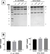
Figure 3. Preparations of RIFR E. coli and MTB RNAPs
(A) SDS-PAGE of purified WT and RIFR E. coli (left) and MTB RNAPs (right). (B) Determination of contaminating endogenous E. coli RNAP found in the E. coli RIFR RNAPs (left) and the MTB RIFR RNAPs (right) estimated by the in vitro transcription assay in the presence of 200 nM RMP.
Figure 4
Figure 4. Structural basis of the RIF resistance by the S531L mutation
(A) Fork loop 2 disordering upon RMP binding to S531L RNAP. The RIF binding sites of the WT RNAP•RMP (left) and the S531L RNAP•RMP (right) complexes are shown with the β subunit (cyan, transparent molecular surfaces plus cartoon models) and the RMP (sphere models). In the left panel, fork loop 2 is colored blue and residue R540 is shown as a stick model. In the right panel, amino acid residues 532 and 541 that connect the disordered fork loop 2 are indicated as blue circles. Area of the RMP naphthalene ring exposure to solvent due to disordering of fork loop 2 is indicated by a red arrow. (B) Proposed mechanism for RIF resistance by the S531L mutation in which RMP binding induces disordering of fork loop 2.
Figure 5
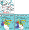
Figure 5. Structural basis of the RIF resistance by the H526Y mutation
(A) Comparison of the WT and H526Y mutant RNAPs. RNAP structures are depicted as ribbon models (WT, the same color as in Fig. 2B; H526Y, gray) and were superposed at their RRDRs. The H526Y side chain is shown as a stick model and labeled. Two regions of the β subunit that change their conformations between the WT and mutant RNAPs are shown as dashed ovals and labeled. (B) Steric hindrance of RMP binding to the H526Y mutant (left: the WT RNAP•RMP complex; right: the H526Y RNAP•RMP complex model). The β subunit and RMP are shown by molecular surface (cyan) and stick models (yellow), respectively. The four amino acid residues (Q513, F514, D516 and H526/H526Y) forming the binding surface of the ansa bridge (C16 to C25 side) of RMP are indicated. The WT RNAP•RMP complex and the H526Y structures were superposed with their RRDRs and RMP is overlaid on the H526Y structure to make the H526Y RNAP•RMP complex model (right). The locations of the clash between the ansa bridge of RMP with H526Y are indicated by arrows.
Figure 6
Figure 6. Structural basis of RIF resistance by the D516V mutation
(A) Electrostatic surfaces of the RIF binding pocket of the WT (left) and D516V mutant (right) RNAPs complexed with RMP (stick models). RNAP surfaces are colored with positive (blue), negative (red) and neutral (white) electrostatic potentials. Positions of the D516 residue in WT RNAP (left) and V516 residue in the mutant (right) are indicated by red circles.

Similar articles

Cited by

References

    1. Afonine PV, Mustyakimov M, Grosse-Kunstleve RW, Moriarty NW, Langan P, Adams PD. Joint X-ray and neutron refinement with phenix.refine. Acta crystallographica. Section D, Biological crystallography. 2010;66:1153–1163. - PMC - PubMed
    1. Alekshun M, Kashlev M, Schwartz I. Molecular cloning and characterization of Borrelia burgdorferi rpoB. Gene. 1997;186:227–235. - PubMed
    1. Aristoff PA, Garcia GA, Kirchhoff PD, Showalter HD. Rifamycins - Obstacles and Opportunities. Tuberculosis. 2010;90:94–118. - PubMed
    1. Artsimovitch I, Vassylyeva MN, Svetlov D, Svetlov V, Perederina A, Igarashi N, Matsugaki N, Wakatsuki S, Tahirov TH, Vassylyev DG. Allosteric modulation of the RNA polymerase catalytic reaction is an essential component of transcription control by rifamycins. Cell. 2005;122:351–363. - PubMed
    1. Bae B, Davis E, Brown D, Campbell EA, Wigneshweraraj S, Darst SA. Phage T7 Gp2 inhibition of Escherichia coli RNA polymerase involves misappropriation of sigma70 domain 1.1. Proc Natl Acad Sci U S A. 2013;110:19772–19777. - PMC - PubMed

MeSH terms