. 2017 Mar;35(3):626-640.
doi: 10.1002/stem.2558. Epub 2017 Jan 19.

New Monoclonal Antibodies to Defined Cell Surface Proteins on Human Pluripotent Stem Cells

Affiliations

New Monoclonal Antibodies to Defined Cell Surface Proteins on Human Pluripotent Stem Cells

Carmel M O'Brien et al. Stem Cells. 2017 Mar.

Abstract

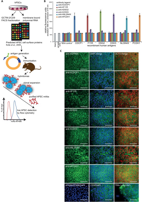
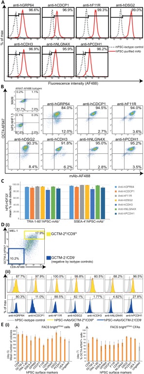
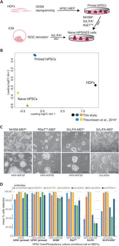
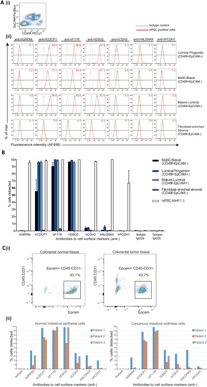
The study and application of human pluripotent stem cells (hPSCs) will be enhanced by the availability of well-characterized monoclonal antibodies (mAbs) detecting cell-surface epitopes. Here, we report generation of seven new mAbs that detect cell surface proteins present on live and fixed human ES cells (hESCs) and human iPS cells (hiPSCs), confirming our previous prediction that these proteins were present on the cell surface of hPSCs. The mAbs all show a high correlation with POU5F1 (OCT4) expression and other hPSC surface markers (TRA-160 and SSEA-4) in hPSC cultures and detect rare OCT4 positive cells in differentiated cell cultures. These mAbs are immunoreactive to cell surface protein epitopes on both primed and naive state hPSCs, providing useful research tools to investigate the cellular mechanisms underlying human pluripotency and states of cellular reprogramming. In addition, we report that subsets of the seven new mAbs are also immunoreactive to human bone marrow-derived mesenchymal stem cells (MSCs), normal human breast subsets and both normal and tumorigenic colorectal cell populations. The mAbs reported here should accelerate the investigation of the nature of pluripotency, and enable development of robust cell separation and tracing technologies to enrich or deplete for hPSCs and other human stem and somatic cell types. Stem Cells 2017;35:626-640.

Keywords: Breast; Cancer; Cell surface markers; Colorectal; Human embryonic stem cells; Human iPS cells; Monoclonal antibodies; Naive; Pluripotency.


Figures

Figure 1
Figure 1
Monoclonal antibodies (mAbs) raised to candidate cell‐surface antigens detect epitopes on human pluripotent stem cells (hPSCs). (A): A schematic overview for the approach used to generate new mAbs capable of recognising hPSC extracellular proteins. Target proteins were identified from fluorescence activated cell sorting (FACS)‐based CD9/GCTM‐2 immunotranscriptional profiling of hPSC cultures bioinformatically combined with membrane polysome translation state array analyses 25, and antigens generated by peptide synthesis or cloning into mammalian expression vectors. Hybridomas raised and initially validated by solid‐state antigen array were screened by high throughput flow cytometry for detection of live hPSCs, prior to cloning, expansion, mAb purification and final validation by detection of live hPSCs by flow cytometry. (B): ELISA fluorescence measurement at 488 nm excitation and emission 519 nm validated specific detection of recombinant antigen immunogens corresponding to purified anti‐hCDCP1 (blue bar), anti‐hF11R (red bar), anti‐hDSG2 (green bar), anti‐hCDH3 (purple bar), anti‐hNLGN4X (light blue bar), anti‐hPCDH1 (orange bar) antibodies compared with control wells without antigen (blank), secondary antibody only (AF488 only) and BSA protein. Error bars depict SEM, n = 3. (C): Representative images of undifferentiated MEL1 human ES cell cultured on a feeder layer of mouse embryonic fibroblasts in hPSC medium for 4‐7 days, showing surface immunostaining using purified anti‐hGPR64, anti‐hCDCP1, anti‐hF11R, anti‐hDSG2, anti‐hCDH3, anti‐hNLGN4X, anti‐hPCDH1 (green, AF488), merged with 4′,6‐diamidino‐2phenylindole (DAPI) counterstained nuclei (blue) and colocalizing with OCT4‐positive cells (red), compared with CD9, TRA‐1‐60 (AF488/DAPI), and isotype controls for the fluorochromes AF488 and AF647. All images shown are for fixed cell mAb staining, except for anti‐hGPR64‐AF488 that was live cell stained prior to fixation for OCT4 detection. Scale bars = 100 µm. Abbreviations: DAPI, 4′,6‐diamidino‐2phenylindole; FACS, fluorescence activated cell sorting; hPSCs, human pluripotent stem cells; mAbs, monoclonal antibodies.
Figure 2
Figure 2
New monoclonal antibodies (mAbs) enable live cell detection and culture of self‐renewing human pluripotent stem cells (hPSCs). For undifferentiated hPSC cultures immunolabeled with new purified mAbs anti‐hGPR64, anti‐hCDCP1, anti‐hF11R, anti‐hDSG2, anti‐hCDH3, anti‐hNLGN4X, anti‐hPCDH1 (A) Representative flow cytometric histogram plots showing live cell fluorescence detection (AF488) of protein epitopes corresponding to all mAbs for a high percentage (%) of total MEL1 cells analyzed (hiPS‐NHF1.3 cells for anti‐hGPR64), (red histogram), against isotype controls (gray histogram). (B): Representative flow cytometric dot plots showing high coexpression of mAb detected hPSC‐surface proteins (AF488) and OCT4 (AF647) against isotype controls following sequential live and fixed cell immunolabeling of hiPS‐NHF1.3 cells (anti‐hGPR64) and WA09 cells (all other mAbs). (C): Multicolor immunostaining and flow cytometric replicate analyses shown graphically for the live cell codetection (mean %) of human iPS cells‐HDF32f cells by each mAb with the TRA‐1‐60 or SSEA‐4 pluripotency‐associated antibodies, compared with isotype controls (not shown). Bars represent the mean percentage of cells staining positively for each mAb within the TRA‐1‐60 or SSEA‐4 positive cell population (n = 3, mean +/− SEM). (D): Triple color flow cytometric analyses demonstrates varying coexpression profiles of each mAb with the GCTM‐2/CD9 gradient in hPSC cultures; (i) Representative flow cytometric analysis for GCTM‐2 (AF647) and CD9 (AF488) coexpression gradient in live MEL1 cell cultures. Population gates are set against isotype controls for negative (GCTM‐2‐AF647/CD9‐AF488 blue gate) and high (GCTM‐2‐AF647hi/CD9‐AF488hi yellow gate) cells; (ii) Representative flow cytometric histogram plots showing the percentage of mAb‐detected cells overlapping with the GCTM‐2/CD9 high (yellow) and negative (blue) populations against isotype controls (gray). (E): Colony‐forming assays (CFAs) for the postfluorescence activated cell sorting (FACS) culture (5,000 cells/3.8cm2 well) of hiPS‐NHF1.3 cells gated for replating the top 25% (FACS bright25%hi, orange bars) and ungated (gray bar) cells detected by mAbs compared with CD9, GCTM‐2, TRA‐1‐60, SSEA‐4, and UEA‐1 lectin; (i) Mean colony counts per well after 5 days culture in hPSC conditions (n = 3). (ii) Percentage OCT4‐AF647 positive cells detected by flow cytometry for the preceding CFAs harvested at 7 days of post‐FACS culture for FACS bright25%hi (orange bars) and ungated (gray bar) cells (n = 3). All Error bars depict SEM. Abbreviation: hPSCs, human pluripotent stem cells.
Figure 3
Figure 3
Cell surface antigens are detected on naive state human pluripotent cells. Transcriptional and protein analyses showing the expression of cell surface epitopes on human iPS cells (hiPSCs) and human ES cells (hESCs) cultured in conditions supporting a naive state of pluripotency. (A): Schematic depicting the generation of naive human cell cultures both from lineage primed hiPSCs and blastocyst epiblast cells. Human dermal fibroblasts (HDFs) were reprogrammed to primed hiPSCs in standard human pluripotent stem cell (hPSC)‐MEF culture then coaxed to a naive pluripotent state in NHSMa, RSET, and 5i/L/FAb defined media‐MEF supported culture conditions. Naive state hESCs were derivedc and maintained following the direct culture of preimplantation blastocyst in 5i/L/FA‐MEF conditions. (B): PCA of RNA sequencing data for the primed and naive (5i/L/FA) state hiPSCs from this study and the microarray data extracted from the published report of Theunissen et al. (2014), 38 confirms differential clustering of two parental HDFs (black dot), lineage primed hiPSCs from this study (dark blue dot) and Theunissen et al. (2014), (light blue dot), and naive hiPSCs from this study (dark yellow dot) clustering with the naive hiPSCs from Theunissen et al. (2014) (gold dot). (C): Representative images for naive hiPSC colonies originating from two parental HDF cell lines (HDF32f, HDF55f) following culture in NHSMa‐MEF, RSeT‐MEF, and 5i/L/FAb‐MEF conditions and preimplantation blastocyst‐derived hES naive cell colonies (UCLA20nc) generated directly in 5i/L/FA‐MEF culture conditions. HiPS and hESC naive cell cultures show a domed colony morphology by bright field phase contrast microscopy (BF). (D): Flow cytometric replicate analyses shown graphically for the live cell detection of naive state hiPS‐HDF32f cells maintained on MEFs in NHSMa, 5i/L/FAb, and RSeT culture media, and naive UCLA20n hESCs in 5i/L/FA‐MEF culture, by the monoclonal antibodies anti‐hGPR64 (light blue bars), anti‐hCDCP1 (orange bars), anti‐hF11R (gray bars), anti‐hDSG2 (yellow bars), anti‐hCDH3 (mid blue bars), anti‐hNLGN4X (green bars), anti‐hPCDH1 (dark blue bars), compared with lineage primed WA09 cells (hESC primed) and parental hiPS‐HDF32f cells (hiPSC primed) cultured on MEFs in hPSC medium, against isotype controls (not shown), (n = 3, except UCLA20n in 5i/L/FA n = 2). Error bars depict SEM. Scale bars = 500 µm (white) and 200 µm (black) for BF images. aGafni O et al. Nature 2013;504:282‐286. bTheunissen TW et al. Cell Stem Cell 2014;15:471‐487. cPastor WA et al. Cell Stem Cell 2016;18:323‐329. Abbreviations: hESCs, human ES cells; hiPSCs, human iPS cells.
Figure 4
Figure 4
New monoclonal antibodies (mAbs) determine downregulation of cell surface epitopes during human pluripotent stem cells (hPSC) differentiation. (A): Flow cytometric replicate analyses shown graphically for the detection of live cells by immunostaining with purified mAbs in differentiating embryoid body (EB) cultures for hPSC lines MEL1 (blue plotted lines), WA09 (red plotted lines), hiPS‐NHF1.3 (green plotted lines), hiPS‐PDL‐D1C6 (purple plotted lines) on the day of harvesting routine undifferentiated cultures (day 0), and following induction of differentiation in EB suspension cultures (days 7, 14, 28). Comparative analyses show detection of live cells for the same cultures and time points using CD9, TRA‐1‐60, GCTM‐2, and biotinylated UEA‐1 lectin, and fixed cell detection for OCT4 (n = 3). (B): For the analyses shown in (A), data displayed graphically for % of total residual OCT4‐positive cells detected by each mAb and pluripotency associated marker in differentiating cultures for each cell line at each time point (n = 3). All error bars depict SEM.
Figure 5
Figure 5
Expression of pluripotency‐associated antigens in human breast tissue and on normal and cancerous intestinal epithelial cells. (A): Human CD31CD45CD235a cell populations isolated from mammary tissue were (i) fluorescence activated cell sorting (FACS)‐delineated using EpCAM and CD49f to resolve each of the four breast epithelial cell subsets:‐ luminal progenitor (CD49f+ EpCAM+), MaSC and basal (CD49f+ EpCAM), mature luminal (CD49fEpCAM+), and fibroblast‐enriched stromal (CD49fEpCAM); (ii) Representative flow histogram plots show the presence or absence of antigens detected by the purified monoclonal antibodies (mAbs) anti‐hGRP64, anti‐hCDCP1, anti‐hF11R, anti‐hDSG2, anti‐hCDH2, anti‐hNLGN4X, and anti‐hPCDH1 in these cell subsets, compared with isotype controls. (B): Bar graphs show the detection of pluripotency‐associated antigens for mammary cell subsets from four donor specimens as mean +/− SEM across two technical replicates compared with isotype controls and NHF1‐3 hiPS positive control cells. (C): (i) Human intestinal epithelial cells were isolated from normal and cancerous colorectal tissues by FACS selection for EpCAM+ CD31CD45 cells. (ii) Bar graphs show the variation in detection of pluripotency‐associated antigens by the panel of mAbs (A‐B) on normal and cancerous intestinal epithelial cells from three patient donor samples compared with isotype controls. Abbreviation: hiPSCs, human iPS cells.

Similar articles

Cited by

References

    1. Thomson JA, Itskovitz‐Eldor J, Shapiro SS et al. Embryonic stem cell lines derived from human blastocysts. Science 1998;282:1145–1147. - PubMed
    1. Yu J, Vodyanik MA, Smuga‐Otto K et al. Induced pluripotent stem cell lines derived from human somatic cells. Science 2007;318:1917–1920. - PubMed
    1. Takahashi K, Tanabe K, Ohnuki M et al. Induction of pluripotent stem cells from adult human fibroblasts by defined factors. Cell 2007;131:861–872. - PubMed
    1. Fox IJ, Daley GQ, Goldman SA et al. Use of differentiated pluripotent stem cells as replacement therapy for treating disease. Science 2014;345:889–900. - PMC - PubMed
    1. Kleitman N, Rao MS, Owens DF. Pluripotent stem cells in translation: A Food and Drug Administration‐National Institutes of Health collaboration. Stem Cells Transl Med 2013;2:483–487. - PMC - PubMed

Publication types