

Disruptions in the left frontoparietal network underlie resting state endophenotypic markers in schizophrenia
- PMID: 28009080
- PMCID: PMC6866857
- DOI: 10.1002/hbm.23477
Disruptions in the left frontoparietal network underlie resting state endophenotypic markers in schizophrenia
Abstract
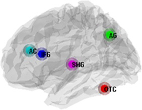
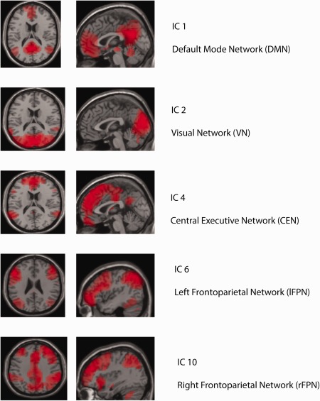
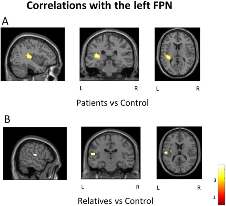
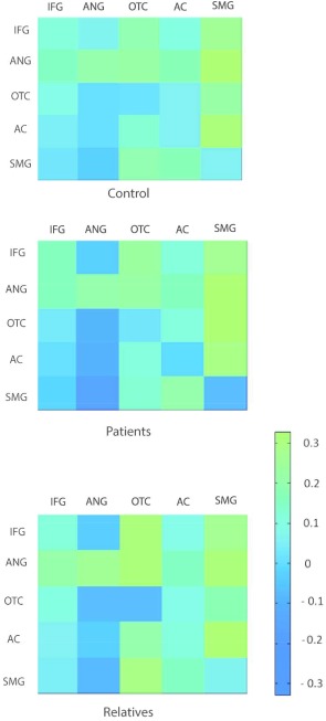
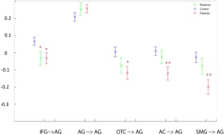
Advances in functional brain imaging have improved the search for potential endophenotypic markers in schizophrenia. Here, we employed independent component analysis (ICA) and dynamic causal modeling (DCM) in resting state fMRI on a sample of 35 schizophrenia patients, 20 first-degree relatives and 35 control subjects. Analysis on ICA-derived networks revealed increased functional connectivity between the left frontoparietal network (FPN) and left temporal and parietal regions in schizophrenia patients (P < 0.001). First-degree relatives shared this hyperconnectivity, in particular in the supramarginal gyrus (SMG; P = 0.008). DCM analysis was employed to further explore underlying effective connectivity. Results showed increased inhibitory connections to the left angular gyrus (AG) in schizophrenia patients from all other nodes of the left FPN (P < 0.001), and in particular from the left SMG (P = 0.001). Relatives also showed a pattern of increased inhibitory connections to the left AG (P = 0.008). Furthermore, the patient group showed increased excitatory connectivity between the left fusiform gyrus and the left SMG (P = 0.002). This connection was negatively correlated to inhibitory afferents to the left AG (P = 0.005) and to the negative symptom score on the PANSS scale (P = 0.001, r = -0.51). Left frontoparietotemporal dysfunction in schizophrenia has been previously associated with a range of abnormalities, including formal thought disorder, working memory dysfunction and sensory hallucinations. Our analysis uncovered new potential endophenotypic markers of schizophrenia and shed light on the organization of the left FPN in patients and their first-degree relatives. Hum Brain Mapp 38:1741-1750, 2017. © 2017 Wiley Periodicals, Inc.
Keywords: biomarker; endophenotypes; neuroimaging; schizophrenia.
© 2016 Wiley Periodicals, Inc.
Figures
References
-
- American Psychiatric Association (1994): Diagnostic and Statistical Manual of Mental Disorders, 4th ed.
-
- Beckmann CF, Mackay CE, Filippini N, Smith SM (2009): Group comparison of resting‐state FMRI data using multi‐subject ICA and dual regression. Neuroimage 47(Suppl. 1), S148.
-
- Bhojraj TS, Francis AN, Rajarethinam R, Eack S, Kulkarni S, Prasad KM, Montrose DM, Dworakowski D, Diwadkar V, Keshavan MS (2009): Verbal fluency deficits and altered lateralization of language brain areas in individuals genetically predisposed to schizophrenia. Schizophr Res 115:202–208. - PMC - PubMed
Publication types
MeSH terms
Substances
LinkOut - more resources
Full Text Sources
Other Literature Sources
Medical
Miscellaneous
