. 2017 Apr;35(4):1053-1064.
doi: 10.1002/stem.2559. Epub 2017 Jan 19.

Protein Tyrosine Phosphatase PRL2 Mediates Notch and Kit Signals in Early T Cell Progenitors

Affiliations

Protein Tyrosine Phosphatase PRL2 Mediates Notch and Kit Signals in Early T Cell Progenitors

Michihiro Kobayashi et al. Stem Cells. 2017 Apr.

Abstract

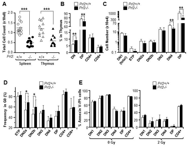
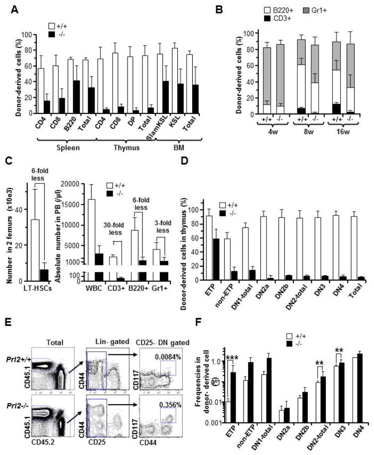
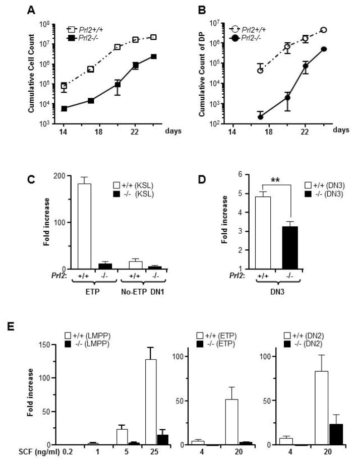
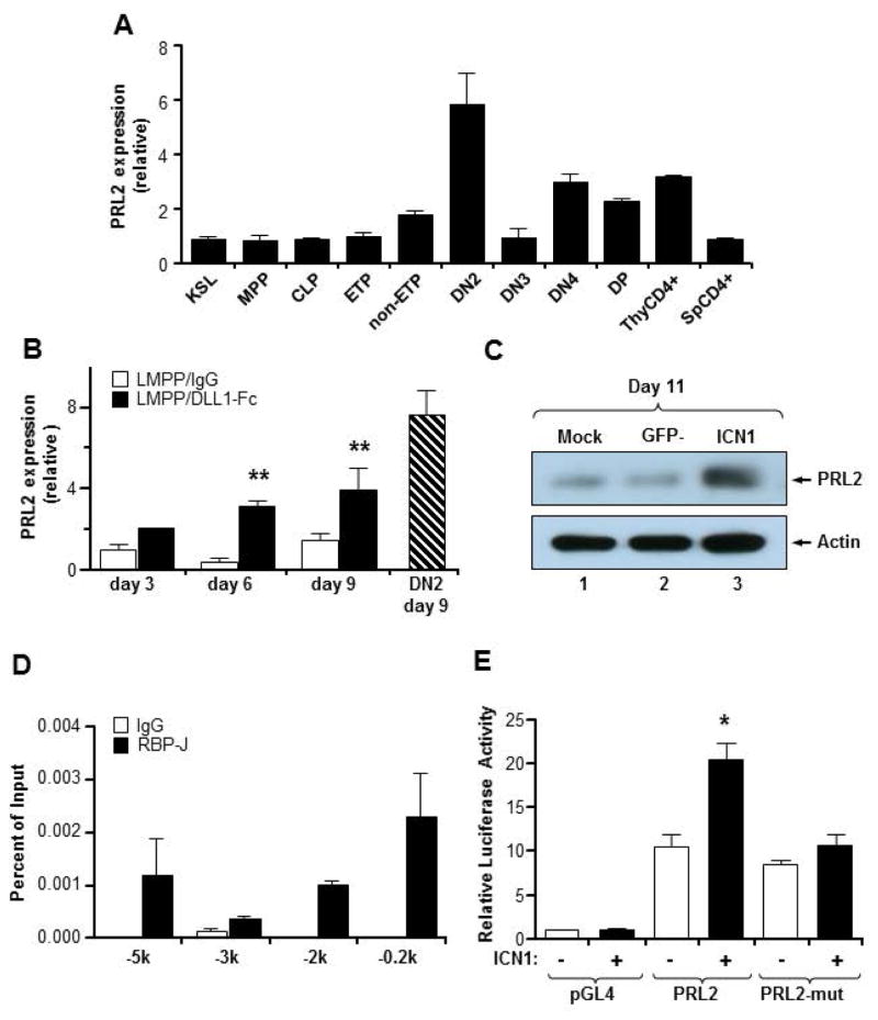
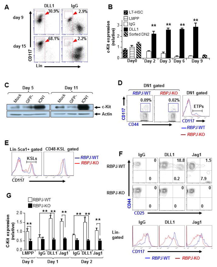
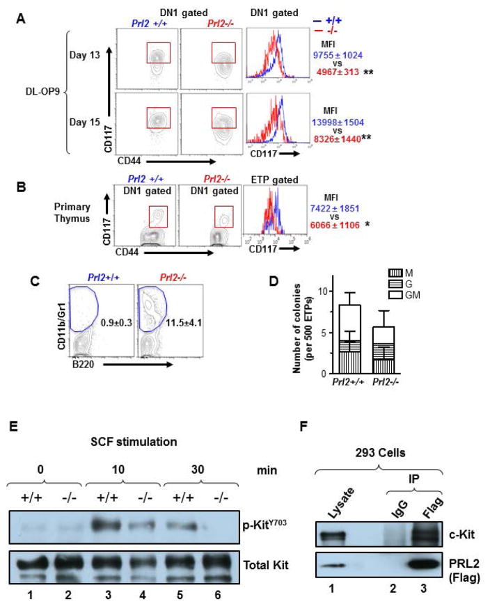
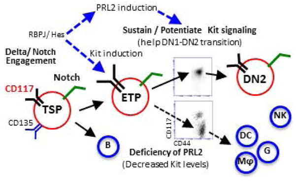
The molecular pathways regulating lymphoid priming, fate, and development of multipotent bone marrow hematopoietic stem and progenitor cells (HSPCs) that continuously feed thymic progenitors remain largely unknown. While Notch signal is indispensable for T cell specification and differentiation, the downstream effectors are not well understood. PRL2, a protein tyrosine phosphatase that regulates hematopoietic stem cell proliferation and self-renewal, is highly expressed in murine thymocyte progenitors. Here we demonstrate that protein tyrosine phosphatase PRL2 and receptor tyrosine kinase c-Kit are critical downstream targets and effectors of the canonical Notch/RBPJ pathway in early T cell progenitors. While PRL2 deficiency resulted in moderate defects of thymopoiesis in the steady state, de novo generation of T cells from Prl2 null hematopoietic stem cells was significantly reduced following transplantation. Prl2 null HSPCs also showed impaired T cell differentiation in vitro. We found that Notch/RBPJ signaling upregulated PRL2 as well as c-Kit expression in T cell progenitors. Further, PRL2 sustains Notch-mediated c-Kit expression and enhances stem cell factor/c-Kit signaling in T cell progenitors, promoting effective DN1-DN2 transition. Thus, we have identified a critical role for PRL2 phosphatase in mediating Notch and c-Kit signals in early T cell progenitors. Stem Cells 2017;35:1053-1064.

Keywords: Early T lineage progenitors; Notch; PRL2; T cell progenitors; Thymopoiesis; c-Kit.


Conflict of interest statement

DISCLOSURE OF POTENTIAL CONFLICTS OF INTEREST

The authors declared that no conflict interest exists.

Figures

Figure 1
Figure 1. Prl2−/− mice have moderate T cell defects in steady state
(A) Splenocytes and thymocytes from 8-week-old Prl2+/+ and Prl2−/− mice were isolated and counted. Absolute splenocyte and thymocyte numbers from Prl2−/− mice are reduced to 50% of Prl2+/+ mice. ***P<0.001, n=10. (B) Analysis of CD4+, CD8+, DP, and DN thymocytes in Prl2+/+ and Prl2−/− mice by flow cytometry. The frequency of DN cells is increased in Prl2−/− thymus compared to Prl2+/+ thymus. **P<0.01, n=7. (C) Analysis of DN1, ETP, DN2, DN3, DN4 and DP thymocytes in Prl2+/+ and Prl2−/− mice by flow cytometry. Absolute number of DN4 and DP cells is decreased in Prl2−/− thymus compared to Prl2+/+ thymus. **P<0.01, n=7. (D) Steady state cell cycle status of thymocytes was determined by Ki67 and DAPI staining. The percentage of cells at the G0 phase of the cell cycle was shown. Percentage of quiescent (G0) Prl2−/− DN2a and DN2b thymocytes is slightly increased compared to Prl2+/+ cells. *P<0.05, n=6. (E) Staining for Annexin V and propidium iodide (PI) in thymocytes isolated from Prl2+/+ and Prl2−/− mice after 48 h culture in serum-free medium, in the presence or the absence of irradiation (2Gy).
Figure 2
Figure 2. PRL2 deficiency impairs T cell development following hematopoietic stem cell transplantation
(A) The frequency of donor-derived cells in spleen, thymus, and bone marrow of recipient mice was determined 18 weeks following bone marrow transplantation. Prl2−/− cells show decreased engraftment in thymus compared to Prl2+/+ cells. (B) The frequency of donor-derived cells in the peripheral blood (PB) of recipient mice was determined every 4 weeks following transplant Prl2+/+ or Prl2−/− HSCs into lethally-irradiated recipient mice. CD3+ T cell recovery was significantly lower in recipients repopulated with Prl2−/− HSCs than that of the Prl2+/+ HSCs. (C) Absolute number of donor derived LT-HSCs in 2 femurs (left panel). Absolute number of PB T cells generated from Prl2−/− HSCs was remarkably reduced compared with that of the Prl2+/+ HSCs. (D) Achievement of donor-derived chimerism in each differentiation stage at 16 weeks after transplantation is shown. (E) Representative flow cytometry plot of donor-derived ETPs. Number indicated the percentage of ETPs in total CD45.2 positive cells. (F) The frequency of different T cell population in donor-derived cells is shown (***P<0.001, **P<0.01, n=8).
Figure 3
Figure 3. PRL2 deficiency impairs T cell development in vitro
(A and B) Purified Prl2+/+ and Prl2−/− KSL cells were cultured on DL-OP9 cells in the presence of IL-7. Cells were then harvested at different time point and stained with various markers to measure T cell differentiation. Prl2−/− KSLs show marked reduction of cumulative total cell count (A) and double positive T cell count (B) compared with Prl2+/+ cells. (C and D) To determine stage specificity, sorted KSLs and DN3 were cultured on OP-DL1 cells for 8 days or 15 days, respectively. Production of ETPs from KSLs and production of total nucleated cells (TNCs) from DN3 cells were calculated. Prl2−/− cells show profound expansion defects at early stage (C) than that of the later stage (D). (E) Sorted LMPPs, ETPs, or DN2 cells were cultured on DLL1-Fc coated plate in the presence of graded dose of SCF. Total cell count was determined at day 12. Prl2−/− cells show defective expansion compared with Prl2+/+ cells, even in the presence of high dose of SCF.
Figure 4
Figure 4. Notch signaling regulates PRL2 expression during T cell differentiation
(A) Quantitative real-time PCR analysis of PRL2 expression in sorted MPPs, CLPs, ETPs, DN2, DN3, DN4, DP, and CD4+ thymocytes, presented relative to expression in KSLs, set as 1. PRL2 is highly expressed in DN2 cells. n = three biological replicates. (B) Sorted Prl2+/+ LMPPs were cultured in the presence or the absence of DLL1-Fc. Cells were collected at each time point and PRL2 expression was determined by quantitative real-time PCR analysis. **P<0.01, n = three biological replicates. (C) Notch-ICN1 was introduced into Prl2+/+ LinSca1+ cells and the levels of PRL2 protein were determined by western blot analysis. (D) Chromatin-bound DNAs from Tail-7 cells were immunoprecipitated with a RBPJ-antibody or normal mouse IgG. Quantitative real-time PCR amplification was performed on corresponding templates using primers for PRL2 gene. (E) Notch-ICN1 transactivates PRL2 promoter. 293 cells were transfected with PRL2 promoter driven luciferase reporter plasmids containing either RBPJ binding sites or mutant RBPJ binding sites. Luciferase activity was assayed 24 hours after transfection. *P<0.05, n=3.
Figure 5
Figure 5. PRL2 sustains Notch mediated c-Kit expression in T cell progenitors
(A) Expression of c-Kit was induced by DLL1-Fc during T cell differentiation, assessed by flow cytometry. (B) Sorted Prl2+/+ LMPPs and DN2 cells were cultured in the presence or the absence of DLL1-Fc. Cells were collected at each time point and c-Kit expression was determined by quantitative real-time PCR analysis, presented relative to expression in sorted LMPPs. **P<0.01, n = three biological replicates. (C) Notch-ICN1 was introduced into wild type LinSca1+ cells and the levels of c-Kit protein were determined by western blot analysis. (D, E) Flow cytometry analysis of c-Kit expression in primary RBPJ-KO thymus (D) or BM (E) at 3 weeks post pI:pC injection. The mean fluorescent intensity (MFI) of c-Kit was greatly reduced in RBPJ-deficient cells compared with that of the WT cells. (F) T cell induction from RBPJ-KO or WT LMPPs. Sorted LMPPs were cultured with control IgG, DLL1, or Jag1. The MFI of c-Kit was measured at day 7. (G) Kinetic study of c-Kit expression in T cell cultures of LMPPs isolated from WT and RBPJ-KO mice. Relative c-Kit mRNA levels were measured by quantitative real-time PCR analysis. **P<0.01, n = three biological replicates.
Figure 6
Figure 6. PRL2 sustains Notch-mediated c-Kit expression in T cell progenitors
(A) Expression of c-Kit in DN1-gated Prl2+/+ and Prl2−/− cells in OP9-DL1 cultures, assessed by flow cytometry. Right panels show the mean fluorescent intensity (MFI) of c-Kit in DN1 cells. **P<0.01, n=3. (B) Expression of c-Kit in DN1 thymocytes isolated from Prl2+/+ and Prl2−/− mice, assessed by flow cytometry. Right panels show the mean fluorescent intensity (MFI) of c-Kit in ETP gated thymocytes. **P<0.01, n=10. (C) Myeloid differentiation was evaluated in DLL1 cultures of LMPPs. Prl2−/− LMPPs differentiated into CD11b+/Gr1+ myeloid cells at day 16 (0.5±0.3 vs 11.5±4.1 %, **P<0.01, n=3). (D) Myeloid progenitors were quantified by methylcellulose culture using ETPs from Prl2+/+ and Prl2−/− mice. (E) PRL2 is important for KIT activation following SCF stimulation. Immunoblot analysis of KIT phosphorylation in WT and Prl2 null hematopoietic progenitor cells following SCF stimulation. (F) PRL2 interacts with KIT in 293 cells.
Figure 7
Figure 7. Protein tyrosine phosphatase PRL2 mediates Notch and Kit signals in early T cell progenitors
(A) Working model. Cooperation of PRL2 phosphatase with Notch and c-Kit signals is critical for early T cell differentiation. Delta/Notch engagement induces the expression of both c-Kit and PRL2 in ETPs. PRL2 then sustains and/or potentiates Kit signaling to facilitate DN1-DN2 transition. In the absence of PRL2, c-Kit expression is decreased and decreased c-Kit expression promotes ETP differentiation toward myeloid lineages. Thus, sustaining high level of c-Kit expression by PRL2 is crucial for ETP expansion, survival, and commitment during early stage T cell development.

Similar articles

Cited by

References

    1. Rothenberg EV. Transcriptional control of early T and B cell developmental choices. Annu Rev Immunol. 2014;32:283–321. - PMC - PubMed
    1. Rothenberg EV, Moore JE, Yui MA. Launching the T-cell-lineage developmental programme. Nat Rev Immunol. 2008;8:9–21. - PMC - PubMed
    1. Matsuzaki Y, Gyotoku J, Ogawa M, et al. Characterization of c-kit positive intrathymic stem cells that are restricted to lymphoid differentiation. J Exp Med. 1993;178:1283–1292. - PMC - PubMed
    1. Godfrey DI, Kennedy J, Suda T, Zlotnik A. A developmental pathway involving four phenotypically and functionally distinct subsets of CD3-CD4-CD8- triple-negative adult mouse thymocytes defined by CD44 and CD25 expression. J Immunol. 1993;150:4244–4252. - PubMed
    1. Waskow C, Paul S, Haller C, Gassmann M, Rodewald HR. Viable c-Kit(W/W) mutants reveal pivotal role for c-kit in the maintenance of lymphopoiesis. Immunity. 2002;17:277–288. - PubMed

Publication types

MeSH terms

Substances