

Distribution and characteristics of transactive response DNA binding protein 43 kDa pathology in progressive supranuclear palsy
- PMID: 28009087
- PMCID: PMC5933946
- DOI: 10.1002/mds.26809
Distribution and characteristics of transactive response DNA binding protein 43 kDa pathology in progressive supranuclear palsy
Abstract
Background: This study aimed to determine the frequency of transactive response DNA binding protein 43 kDa pathology in PSP, the clinical features of patients with this pathology, and genetic risk factors for it.
Methods: Hippocampal sections were screened with immunohistochemistry for transactive response DNA binding protein 43 kDa in 945 PSP cases. A subset of 261 cases that were negative in hippocampus was screened in the amygdala. The density and disruption of this pathology, as well as regional tau burden, and clinical and genetic characteristics were analyzed.
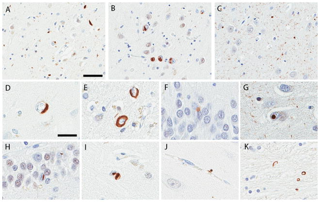
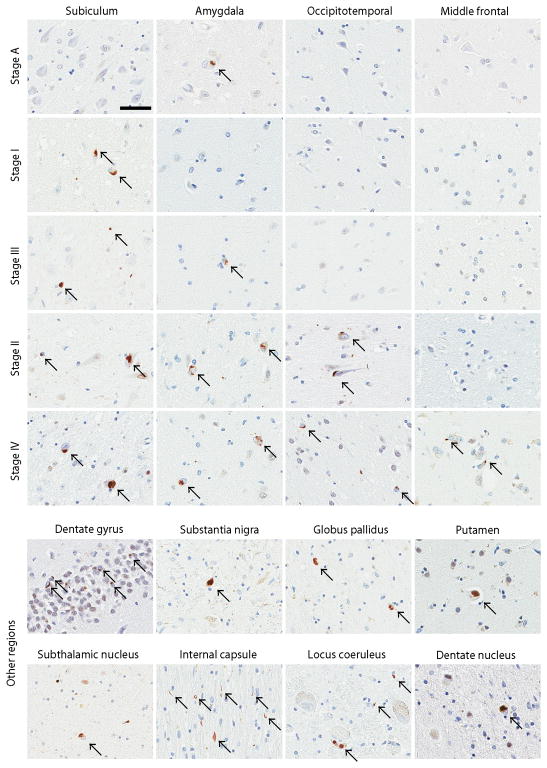
Results: We observed transactive response DNA binding protein 43 kDa pathology in 47 cases in the hippocampus and an additional 9 cases that only affected the amygdala. Hippocampal sclerosis was the strongest risk factor, followed by Alzheimer's disease pathology, argyrophilic grain disease, and older age at death. Five stages of transactive response DNA binding protein 43 kDa pathology were identified in PSP: Stage A had pathology only in the amygdala (16%); stage I had pathology confined to the hippocampus and entorhinal cortex (9%); stage II included both regions of stage A and I (38%); stage III spread further to medial occipitotemporal gyrus (20%); and stage IV had pathology in the dorsolateral frontal lobe (18%). Anatomical areas vulnerable to PSP pathology had varying degrees of this pathology in stage II and later. PSP with transactive response DNA binding protein 43 kDa pathology were older at disease onset and had lower median MMSE scores; however, the latter was driven by concurrent pathologies.
Conclusions: Distribution and clinical characteristics of transactive response DNA binding protein 43 kDa pathology in PSP were influenced by concurrent pathologies. This is the first study to observe that PSP-vulnerable regions are also susceptible to this non-tau pathology. © 2016 International Parkinson and Movement Disorder Society.
Keywords: TDP-43; hippocampal sclerosis; progressive supranuclear palsy.
© 2016 International Parkinson and Movement Disorder Society.
Figures
References
-
- Arai T, Hasegawa M, Akiyama H, et al. TDP-43 is a component of ubiquitin-positive tau-negative inclusions in frontotemporal lobar degeneration and amyotrophic lateral sclerosis. Biochem Biophys Res Commun. 2006;351(3):602–611. - PubMed
-
- Neumann M, Sampathu DM, Kwong LK, et al. Ubiquitinated TDP-43 in frontotemporal lobar degeneration and amyotrophic lateral sclerosis. Science. 2006;314(5796):130–133. - PubMed
MeSH terms
Substances
Grants and funding
LinkOut - more resources
Full Text Sources
Other Literature Sources
Medical
Miscellaneous
