Review
. 2017 Feb;69(1):38-55.
doi: 10.23736/S0393-2249.16.02823-X. Epub 2016 Nov 8.

Robot assisted lymphadenectomy in urology: pelvic, retroperitoneal and inguinal

Affiliations
Review

Robot assisted lymphadenectomy in urology: pelvic, retroperitoneal and inguinal

Giovannalberto Pini et al. Minerva Urol Nefrol. 2017 Feb.

Abstract

Lymph node dissection represents an essential surgical step in the treatment of the most commonly treated urological cancers. The introduction of robotic surgery has lead to the possibility of treating these diseases with a minimally invasive surgical approach, but the surgical principles of open surgery need to be carefully respected in order to achieve comparable oncological results. Therefore, the robotic approach to urological cancers must include a carefully performed lymph node dissection when indicated. In the current manuscript we reviewed the current indications and extensions of lymph node dissection in prostate, bladder, testicular, upper urinary tract, renal and penile cancers respectively, with a special focus on the state of the art surgical technique for each procedure.


Conflict of interest statement

Conflicts of interest.—The authors certify that there is no conflict of interest with any financial organization regarding the material discussed in the manuscript.

Figures

Figure 1.—
Figure 1.—
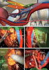
Template in PLND dissection in prostate cancer. A) Lymph node dissection templates in prostate cancer (courtesy of F. Gaboardi); B-E) boundaries of extended PLND. Caudally Cloquet’s lymph node, laterally genitofemoral nerve, medially umbilical obliterated artery, peritoneal sheet and bladder; cranially ureter crossing at level of common iliac artery. 1: Limited - External iliac above the obturator nerve; 2: standard - external iliac, obturator, hypogastric (pelvic sidewall laterally, the bladder wall medially, the floor of the pelvis posteriorly, Cooper’s ligament distally and the internal iliac artery proximally); 3: extended - previous template and common iliac vessels at level of ureter crossing; 4: super-extended – previous template and presacral, aortic bifurcation, eventually preaortic and precaval.
Figure 2.—
Figure 2.—
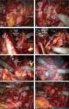
Steps in robot-assisted PLND dissection in prostate cancer. A) lateral lymph node package dissection (left sided) at level of external iliac artery (Courtesy of G. Pini); B) external iliac vein dissection (left sided) and approaching the obturator fossa; C) superficial obturator nerve lymph nodes package (left sided); D) deep obturator nerve lymph nodes package (left sided); E) Marcille’s triangle/fossa exposition (left sided): after detaching left external iliac vessels from psoas muscle the procedure allows for a safer dissection of obturator nerve emergency from lateral wall of pelvic floor. Vessel loop secures ureter; F) sacral lymphnode dissection follows a proper dissection of bilateral common iliac artery; G) final result of an extended-PLND (right sided); H) salvage lymph node dissection (right sided) following biochemical relapse and positive single node at c-11 choline PET-CT scan.
Figure 3.—
Figure 3.—
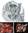
Robot-assisted PLND dissection in bladder cancer. A) High extended pelvic lymph node dissection (courtesy of M. Desai); B) final view following a high extended pelvic lymph node dissection (courtesy of M. Desai). The distal aorta, common iliac arteries (CIAs), and presacral (PS) regions are completely dissected off lymphatic tissue; C) Pre- and paracaval lymph node dissection dissection (courtesy of G. Pini), lumbar vein are easily visible. 1: right external iliac; 2) right internal iliac/obturator; 3) right common iliac; 4) preparacaval and preparaaortic; 5) presacral (PS); 6) left common iliac; 7) left external iliac; and 8) left internal iliac/obturator.
Figure 4.—
Figure 4.—
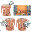
Robotic-assisted nephroureterectomy, bladder cuff excision and RPLND in upper tract urothelial carcinoma (courtesy of SF Matin). A-D) Port placement and robot docking for a single-docking approach with the da Vinci Si. The robotic cart is placed over the patient’s hip at a 45° angle, facing the contralateral shoulder. The patient is positioned in a modified flank position (45–60°) with the disease side up, in a slight Trendelenburg position (15°) and table flexed; E, F) modified template suggested in right- and left-sided UTUC; G) bladder cuff excision. The single-docking approach allows for an easy ureteral dissection. Before excision of the bladder cuff, the ureter is secured with clips to prevent tumor migration. Cystostomy is then closed with running suture. α. Cystostomy β. Pelvic ureter; H) peri, pre- and interaorto-caval RPLND in right sided tumor. Following circumferential cava dissection, a vessel loop helps for retrocaval RPLND. Ω. Cava; γ. Peri-, pre- and interaorto-caval LN package; I) aorta and cava completely skeletonized. Ω. Cava; μ: Aorta; J) interaortocaval RPLND in left sided tumor. Ω. Cava; μ: Aorta; €: Interaortocaval LN package; $: clipped left renal artery; K) para- and peri-aortic RPLND from left side. μ: aorta; ¥: LN package, $: clipped left renal artery.
Figure 5.—
Figure 5.—
Robot-assisted RPLND in testicular cancer. A, B) Port placement for a supine approach with the da Vinci Si for right, left, and bilateral templates (courtesy of J. Porter). Four arms approach and inverted U-shaped configuration. Two 8-mm robotic ports are placed in the left lower quadrant, while an 8-mm robotic port and a 12-mm assistant port are placed in the right lower quadrant; C, D) port placement for a supine approach with the da Vinci Xi (courtesy of Porter J). Four arms approach with a linear port configuration. The assistant port placed in the lower quadrant opposite the side of the template.
Figure 6.—
Figure 6.—
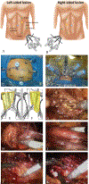
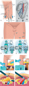
RAVEIL in penile cancer. A) Left surgical procedures template (courtesy of R. Sotelo). The femoral (inverted) triangle delineate port positioning and dissection template; B) schematic view of a deep dissection results on left side (courtesy of S. F. Matin); C) comparison of standard open incision and RAVEIL (courtesy of R. Sotelo); D, E) patient position, robot docking and assistant position (courtesy of R. Sotelo). Split-leg and low lithotomy position (Allen-Stirrups) allows bilateral dissection without repositioning the robot. For the right RAVEIL, the robot is located at 45° contralateral to procedure and bed-side assistant and the right side. For the left RAVEIL cart is parallel to the table and assistant between the patient’s leg; F, G) schematic and intraoperative blunt subcutaneous dissection (courtesy of R. Sotelo). A 2-cm incision is made 3 cm below the apex of femoral triangle, (25 cm below the inguinal ligament). Scarpa’s fascia is identified and finger dissection or balloon dissection allow to to develop the potential space to insert two additional robotic (8-mm) ports. Further subcutaneous workspace is obtained under vision by sweeping the endoscope cranially; H) dissection of the superficial LND package (courtesy of R. Sotelo). With blunt dissection, the node packet can be rolled inwards on both sides. This manoeuvre is continued inferiorly as much as possible from both sides to define inferior apex of the nodal packet. The saphenous vein will be identified, section when necessary, even if its preservation reduce the risk of postoperative lymphedema; I) dissection of the deep LND package (courtesy of R. Sotelo). Fascia lata is opened medial to the saphenous arch and the saphenofemoral junction isexposed. Deep inguinal nodes dissection should be continued to the level of the femoral canal and until the pectineus muscle is seen to insure complete nodal retrieval.

References

    1. Guillonneau B, Cappèle O, Martinez JB, Navarra S, Vallancien G. Robotic assisted, laparoscopic pelvic lymph node dissection in humans. J Urol 2001;165:1078–81. - PubMed
    1. Suardi N, Larcher A, Haese A, Ficarra V, Govorov A.Buffi NM, et al.; EAU Young Academic Urologists-Robotic Section. Indication for and extension of pelvic lymph node dissection during robot-assisted radical prostatectomy: an analysis of five European institutions. Eur Urol 2014;66:635–43. - PubMed
    1. Salminen AP, Jambor I, Syvanen KT, Bostrom PJ. Update on novel imaging techniques for the detection of lymph node metastases in bladder cancer. Minerva Urol Nefrol 2016;68:138–49. - PubMed
    1. Heidenreich A, Bastian PJ, Bellmunt J, Bolla M, Joniau S, van der Kwast T, et al. EAU guidelines on prostate cancer. part 1: screening, diagnosis, and local treatment with curative intent-update 2013.; European Association of Urology. Eur Urol 2014;65:124–37. - PubMed
    1. National Comprehensive Cancer Network, Prostate cancer guideline version 1.2015; [Internet]. Available from: http://www.nccn.org/professionals/physician_gls/pdf/prostate.pdf. Latest NCCN guideline for PCa [cited 2016, Nov 30].

LinkOut - more resources