. 2017 Mar;24(3):492-499.
doi: 10.1038/cdd.2016.148. Epub 2016 Dec 23.

A negative feedback loop of ICER and NF-κB regulates TLR signaling in innate immune responses

Affiliations

A negative feedback loop of ICER and NF-κB regulates TLR signaling in innate immune responses

Sihan Lv et al. Cell Death Differ. 2017 Mar.

Abstract

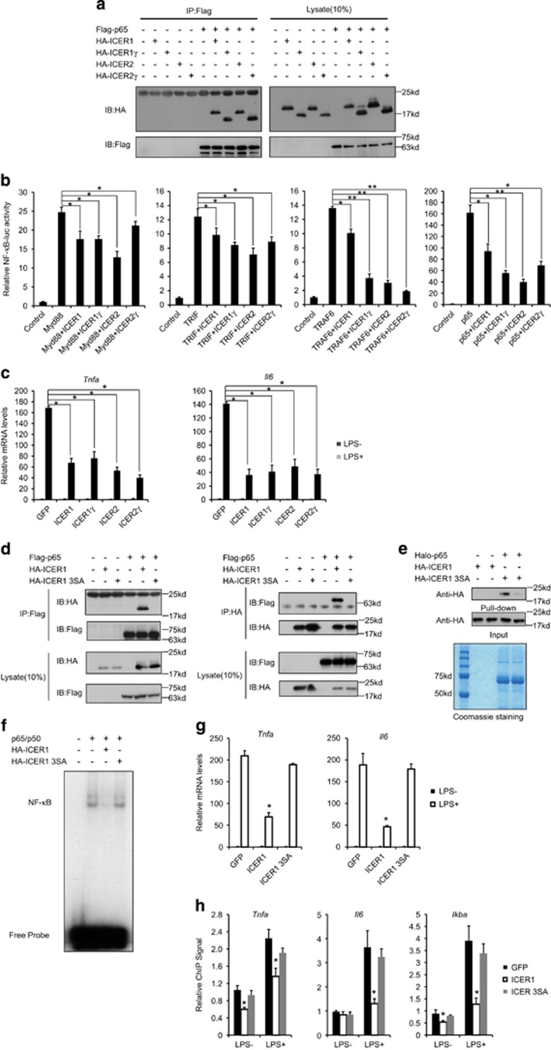
The NF-κB pathway has important roles in innate immune responses and its regulation is critical to maintain immune homeostasis. Here, we report a newly discovered feedback mechanism for the regulation of this pathway by TLR ligands in macrophages. Lipopolysaccharide (LPS) induced the expression of ICER via p38-mediated activation of CREB in macrophages. ICER, in turn, inhibited the transcriptional activity of NF-κB by direct interaction with the p65 subunit of NF-κB. Deficiency in ICER elevated binding of NF-κB to promoters of pro-inflammatory genes and their subsequent gene expression. Mice deficient in ICER were hypersensitive to LPS-induced endotoxic shock and showed propagated inflammation. Whereas ICER expression in ICER KO bone marrow transplanted mice rescued the ultra-inflammation phenotype, expression of a p65 binding-deficient ICER mutant failed to do so. Our results thus establish p38-CREB-ICER as key components of a negative feedback mechanism necessary to regulate TLR-driven inflammation.


Conflict of interest statement

The authors declare no conflict of interest.

Figures

Figure 1
Figure 1
LPS stimulates ICER expression via p38-CREB pathway. (a) Effect of intraperitoneal (i.p.) LPS on Icer mRNA levels in peripheral whole blood cells, spleen, and liver. Data are represented as mean±S.E.M. *P<0.05 and **P<0.01. (b) Effect of LPS on Icer mRNA levels in peritoneal macrophages at indicated times. Data are represented as mean±S.E.M. *P<0.05. (c) Effect of MAPK and IKK inhibitors on Icer mRNA levels in peritoneal macrophages. Data are represented as mean±S.E.M. *P<0.05. (d) Effect of LPS on JNK, P38, IKK, and CREB phosphorylation in peritoneal macrophages. (e) Effect of MAPK and IKK inhibitors on LPS-induced CREB phosphorylation in peritoneal macrophages. (f) Effect of LPS on Icer mRNA levels in peritoneal macrophages from mice with a deletion of the CREB gene in macrophages (CREB MKO) or control littermates. Data are represented as mean±S.E.M. *P<0.05. (g) Effect of LPS on Icer mRNA levels in peritoneal macrophages infected with either GFP or ACREB lentivirus. Data are represented as mean±S.E.M. *P<0.05. (h) Chromatin immunoprecipitation (ChIP) assay of phosph-CREB recruitment over Icer promoter in peritoneal macrophages exposed to LPS. Data are represented as mean±S.E.M. *P<0.05. (i) Effect of p38 inhibitors on phosph-CREB recruitment over Icer promoter in peritoneal macrophages exposed to LPS. Data are represented as mean±S.E.M. *P<0.05
Figure 2
Figure 2
ICER deficiency promotes p65 DNA-binding and inflammatory responses in macrophage. (a and b) Effect of LPS on pro-inflammatory genes mRNA (Tnfa and Il6) levels (a) and cytokine (TNFa and IL-6) secretion in peritoneal macrophages from Icer KO mice or control littermates. Data are represented as mean±S.E.M. *P<0.05. (c and d) Effect of LPS on JNK, P38, IKK, and CREB phosphorylation, IκBα degradation (c) and p65 nuclear translocation (d) in peritoneal macrophages from Icer KO mice or control littermates. (e) ChIP assay of p65 recruitment over Tnfa, Il6, and IκBα promoters in peritoneal macrophages from Icer KO mice or control littermates with two different p65 antibodies. Data are represented as mean±S.E.M. *P<0.05
Figure 3
Figure 3
ICER inhibits NF-κB activation via binding to p65. (a) Interaction between Flag-tagged p65 and HA-tagged ICER isoforms in HEK293T cells. (b) Transient assay of HEK293T cells showing NF-κB reporter activity in cells co-transfected with ICER isoforms together with MyD88, TRIF, TRAF6, or p65. Data are represented as mean±S.E.M. *P<0.05 and **P<0.01. (c) Effect of LPS on pro-inflammatory cytokine genes (Tnfa and Il6) mRNA levels in peritoneal macrophages infected with either GFP or ICER isoforms lentivirus. Data are represented as mean±S.E.M. *P<0.05. (d) Interaction between Flag-tagged p65 and HA-tagged ICER1 or ICER1 3SA in HEK293T cells. (e) Pull-down assay showing interaction between purified p65 and IPed HA-tagged ICER1 or ICER1 3SA. (f) Effect of ICER1 or ICER1 3SA on p65 DNA-binding activity examined by EMSA. (g) Effect of LPS on pro-inflammatory cytokine genes (Tnfa and Il6) mRNA levels in peritoneal macrophages infected with GFP, ICER1, or ICER1 3SA lentivirus. Data are represented as mean±S.E.M. *P<0.05. (h) ChIP assay of p65 recruitment over Tnfa, Il6, and IκBα promoters in peritoneal macrophages infected with GFP, ICER1, or ICER1 3SA lentivirus. Data are represented as mean±S.E.M. *P<0.05. See also Supplementary Figures S1
Figure 4
Figure 4
Anti-inflammatory effects of ICER in mice. (a) Effect of LPS i.p. (30 mg/kg) injection on survival in Icer KO mice or control littermates. Data are represented as mean±S.E.M. *P<0.05. (b) Effect of LPS i.p. (10 mg/kg and 20mglkg) injection on circulating cytokine concentrations in Icer KO mice or control littermates. Data are represented as mean±S.E.M. *P<0.05. (c) Pro-inflammatory genes (Tnfa and Il6) mRNA levels in spleen and liver from Icer KO mice or control littermates injected with indicated amount of LPS. Data are represented as mean±S.E.M. *P<0.05. (d and e) Effect of LPS i.p. injection on survival (30 mg/kg) (d) and circulating cytokine concentrations (20 mg/kg) (e) in lethally irradiated mice transplanted with indicated bone marrow. Data are represented as mean±S.E.M. *P<0.05

Similar articles

Cited by

References

    1. Akira S, Hemmi H. Recognition of pathogen-associated molecular patterns by TLR family. Immunol Lett 2003; 85: 85–95. - PubMed
    1. Miggin SM, O'Neill LA. New insights into the regulation of TLR signaling. J Leukoc Biol 2006; 80: 220–226. - PubMed
    1. Moresco EM, LaVine D, Beutler B. Toll-like receptors. Curr Biol 2011; 21: R488–R493. - PubMed
    1. Kawai T, Akira S. TLR signaling. Semin Immunol 2007; 19: 24–32. - PubMed
    1. Han J, Lee JD, Bibbs L, Ulevitch RJ A. MAP kinase targeted by endotoxin and hyperosmolarity in mammalian cells. Science 1994; 265: 808–811. - PubMed

MeSH terms

LinkOut - more resources