

Dynamic X-ray diffraction sampling for protein crystal positioning
- PMID: 28009558
- PMCID: PMC5182024
- DOI: 10.1107/S160057751601612X
Dynamic X-ray diffraction sampling for protein crystal positioning
Abstract
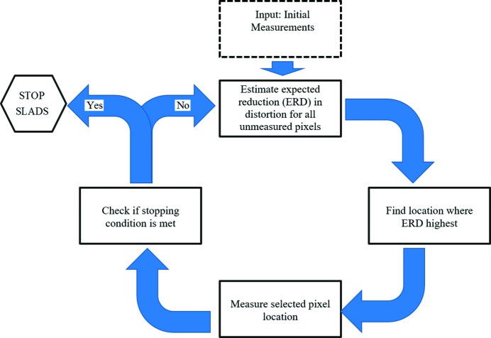
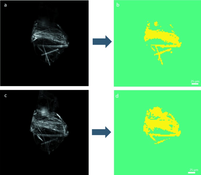
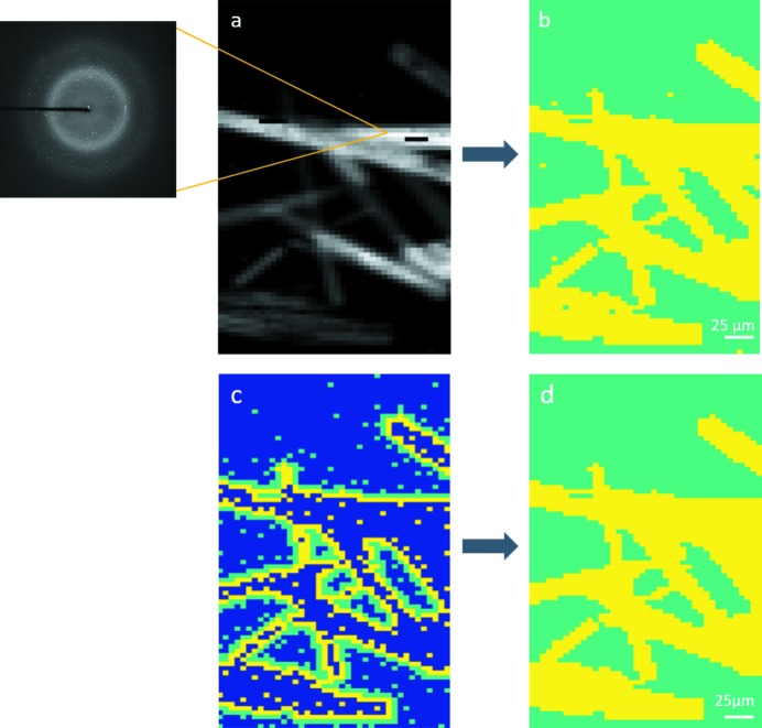
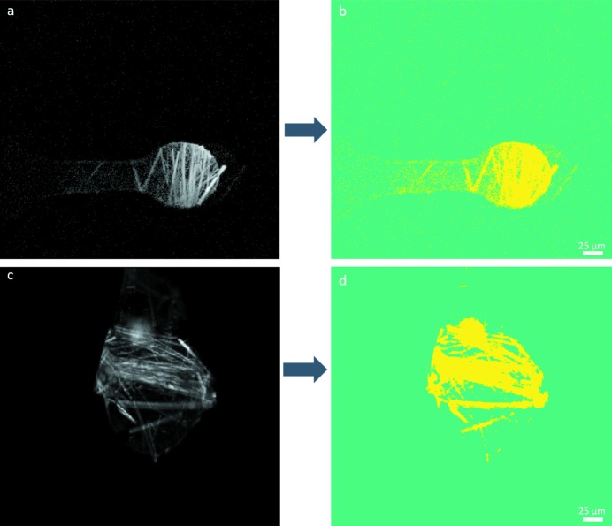
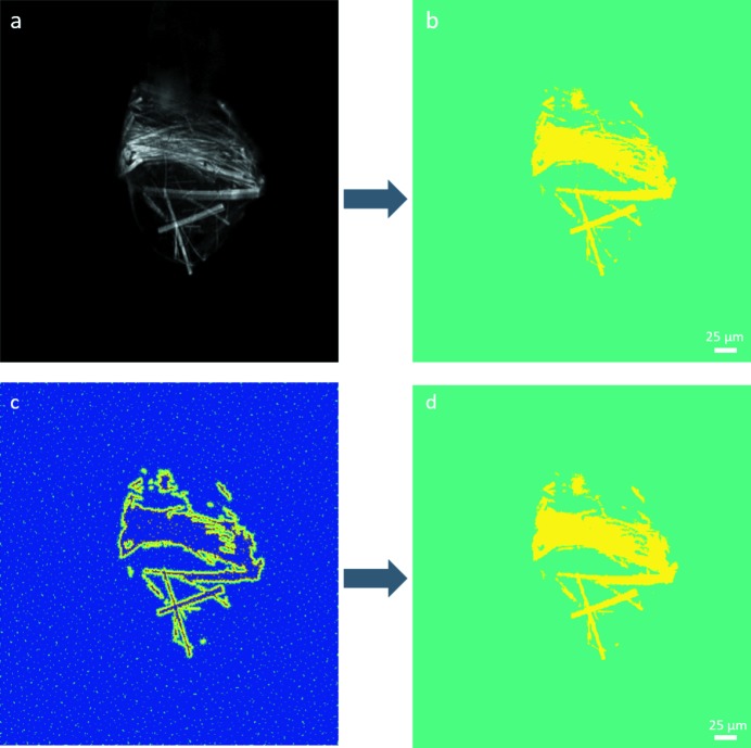
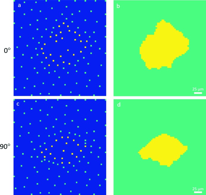
A sparse supervised learning approach for dynamic sampling (SLADS) is described for dose reduction in diffraction-based protein crystal positioning. Crystal centering is typically a prerequisite for macromolecular diffraction at synchrotron facilities, with X-ray diffraction mapping growing in popularity as a mechanism for localization. In X-ray raster scanning, diffraction is used to identify the crystal positions based on the detection of Bragg-like peaks in the scattering patterns; however, this additional X-ray exposure may result in detectable damage to the crystal prior to data collection. Dynamic sampling, in which preceding measurements inform the next most information-rich location to probe for image reconstruction, significantly reduced the X-ray dose experienced by protein crystals during positioning by diffraction raster scanning. The SLADS algorithm implemented herein is designed for single-pixel measurements and can select a new location to measure. In each step of SLADS, the algorithm selects the pixel, which, when measured, maximizes the expected reduction in distortion given previous measurements. Ground-truth diffraction data were obtained for a 5 µm-diameter beam and SLADS reconstructed the image sampling 31% of the total volume and only 9% of the interior of the crystal greatly reducing the X-ray dosage on the crystal. Using in situ two-photon-excited fluorescence microscopy measurements as a surrogate for diffraction imaging with a 1 µm-diameter beam, the SLADS algorithm enabled image reconstruction from a 7% sampling of the total volume and 12% sampling of the interior of the crystal. When implemented into the beamline at Argonne National Laboratory, without ground-truth images, an acceptable reconstruction was obtained with 3% of the image sampled and approximately 5% of the crystal. The incorporation of SLADS into X-ray diffraction acquisitions has the potential to significantly minimize the impact of X-ray exposure on the crystal by limiting the dose and area exposed for image reconstruction and crystal positioning using data collection hardware present in most macromolecular crystallography end-stations.
Keywords: X-ray diffraction; dynamic sampling; nonlinear optical microscopy; second-harmonic generation; supervised learning approach; two-photon-excited fluorescence.
Figures
References
-
- Andrey, P., Lavault, B., Cipriani, F. & Maurin, Y. (2004). J. Appl. Cryst. 37, 265–269.
-
- Broennimann, Ch., Eikenberry, E. F., Henrich, B., Horisberger, R., Huelsen, G., Pohl, E., Schmitt, B., Schulze-Briese, C., Suzuki, M., Tomizaki, T., Toyokawa, H. & Wagner, A. (2006). J. Synchrotron Rad. 13, 120–130. - PubMed
-
- Burmeister, W. P. (2000). Acta Cryst. D56, 328–341. - PubMed
Publication types
MeSH terms
Substances
Grants and funding
LinkOut - more resources
Full Text Sources
Other Literature Sources
