

EWSR1 Fusions With CREB Family Transcription Factors Define a Novel Myxoid Mesenchymal Tumor With Predilection for Intracranial Location
- PMID: 28009602
- PMCID: PMC5350023
- DOI: 10.1097/PAS.0000000000000788
EWSR1 Fusions With CREB Family Transcription Factors Define a Novel Myxoid Mesenchymal Tumor With Predilection for Intracranial Location
Abstract
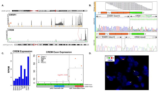
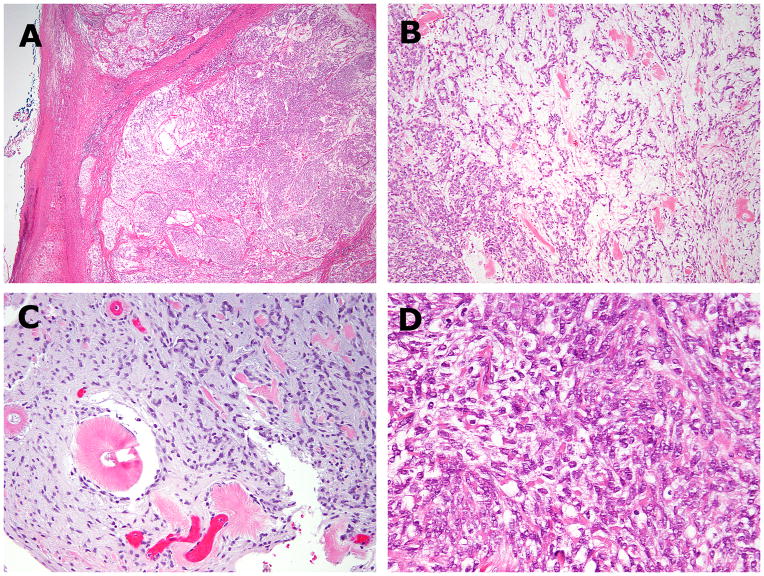
Recurrent gene fusions involving EWSR1 with members of the cAMP response element binding protein (CREB) family (ATF1 and CREB1) have been reported in a diverse group of tumors including angiomatoid fibrous histiocytoma (AFH), soft tissue and gastrointestinal clear cell sarcoma, primary pulmonary myxoid sarcoma, and hyalinizing clear cell carcinoma of salivary gland. We have recently encountered a group of 5 myxoid mesenchymal tumors positive for EWSR1 fusions with one of the CREB family member (ATF1, CREB1, and CREM), with histologic features distinct from any of the previously described pathologic entities. Tumors occurred in children or young adults (12 to 23 y; mean, 18 y), with equal sex distribution. All except 1 were intracranial (intra-axial, 2; meningeal, 2), whereas 1 was perirectal. Histologically, the tumors were well circumscribed, often lobulated, composed of uniform ovoid to round cells, and arranged in cord-like or reticular structures in a myxoid background. All except 1 displayed unique sunburst amianthoid fibers. Immunohistochemically, tumors were positive for epithelial membrane antigen (5/5; 4 focal, 1 diffuse) and desmin (3/5). A novel EWSR1-CREM fusion was identified by RNA sequencing in the perirectal tumor, which was further confirmed by fluorescence in situ hybridization (FISH) and reverse transcription-polymerase chain reaction (RT-PCR). A second case with similar EWSR1-CREM fusion was identified by RT-PCR and FISH in a meningeal tumor. The remaining cases studied by FISH showed the presence of EWSR1-CREB1 fusion in 2 cases and EWSR1-ATF1 in 1. In conclusion, we report a distinct group of myxoid mesenchymal neoplasms occurring in children or young adults with a predilection for intracranial locations. Although the immunoprofile [epithelial membrane antigen (EMA), desmin] and the fusion type raise the possibility of a myxoid AFH, none of the typical histologic findings of AFH were present, suggesting a novel entity.
Conflict of interest statement
Figures

References
-
- Thway K, Fisher C. Tumors with EWSR1-CREB1 and EWSR1-ATF1 fusions: the current status. Am J Surg Pathol. 2012;36:e1–e11. - PubMed
-
- Antonescu CR, Dal Cin P, Nafa K, et al. EWSR1-CREB1 is the predominant gene fusion in angiomatoid fibrous histiocytoma. Genes Chromosomes Cancer. 2007;46:1051–1060. - PubMed
-
- Antonescu CR, Nafa K, Segal NH, et al. EWS-CREB1: a recurrent variant fusion in clear cell sarcoma--association with gastrointestinal location and absence of melanocytic differentiation. Clin Cancer Res. 2006;12:5356–5362. - PubMed
-
- Hisaoka M, Ishida T, Kuo TT, et al. Clear cell sarcoma of soft tissue: a clinicopathologic, immunohistochemical, and molecular analysis of 33 cases. Am J Surg Pathol. 2008;32:452–460. - PubMed
MeSH terms
Substances
Grants and funding
LinkOut - more resources
Full Text Sources
Other Literature Sources
Medical
Research Materials
