Case Reports
. 2016 Dec 21;3(4):44.
doi: 10.3390/children3040044.

Natural History of Asymptomatic and Unrepaired Vascular Rings: Is Watchful Waiting a Viable Option? A New Case and Review of Previously Reported Cases

Affiliations
Case Reports

Natural History of Asymptomatic and Unrepaired Vascular Rings: Is Watchful Waiting a Viable Option? A New Case and Review of Previously Reported Cases

Rohit S Loomba. Children (Basel). .

Abstract

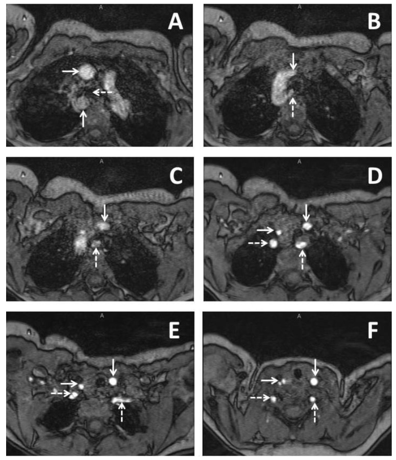
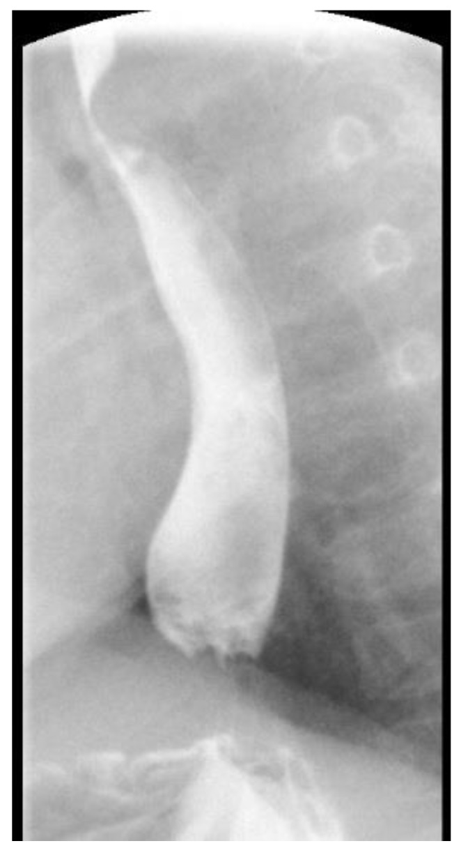
Vascular rings are a rare form of congenital heart disease in which abnormal aortic arch anatomy leads to encircling of the esophagus and/or trachea by the aortic vasculature. Symptoms can develop from this and prompt the need for surgery. A natural history study has been done on mildly symptomatic patients but no such study has been done on asymptomatic patients. We present a case report of three children with asymptomatic vascular rings who continue to receive follow-up without intervention and review all published cases of asymptomatic or unrepaired vascular rings. Clinical observation of asymptomatic and mildly symptomatic vascular rings, regardless of aortic arch anatomy, seems to be a safe approach. Children with mild symptoms almost invariably seem to have resolution of their symptoms by four years of age.

Keywords: asymptomatic; double aortic arch; right aortic arch; unrepaired; vascular anomaly; vascular ring.


Conflict of interest statement

The authors declare no conflict of interest. This research received no specific grant from any funding agency, commercial or not-for-profit sectors.

Figures

Figure 1
Figure 1
Imaging from a three months old baby girl with DiGeorge syndrome who had some issues with feeding. Series of axial slices from magnetic resonance imaging presented in an inferior to superior progression. Panel A demonstrates a right sided aortic arch with the ascending aorta (solid horizontal arrow) coursing anterior to the right bronchus (dashed horizontal arrow) and the descending aorta (solid vertical arrow) coursing immediately posterior to the right bronchus; Panel B demonstrates the left common carotid (solid vertical arrow) as the first branching head and neck vessel and arises from the ascending aorta. An aberrant left subclavian artery (dashed vertical arrow) is seen as the last branching head and neck vessel and arises from the descending aorta; Panel C demonstrates the course of the left common carotid (solid vertical arrow) and aberrant left subclavian artery (dashed vertical arrow); Panel D demonstrates bifurcation of the brachiocephalic trunk into the right common carotid (solid horizontal arrow) and the right subclavian artery (dashed horizontal arrow). The left common carotid (solid vertical arrow) and the left subclavian artery (dashed vertical arrow) are seen demonstrated as well; Panel E demonstrates the right vertebral artery arising from the right subclavian artery (dashed horizontal arrow). The right common carotid (solid horizontal line), left common carotid (solid vertical arrow, and aberrant left subclavian (dashed vertical arrow) are also demonstrated; Panel F demonstrates early bifurcation of the right common carotid into the right internal and external carotid arteries (solid horizontal arrow). The right vertebral artery (dashed horizontal arrow), the left common carotid (solid vertical arrow), and the left vertebral artery are also demonstrated (dashed vertical arrow).
Figure 2
Figure 2
A lateral projection of an upper gastrointestinal imaging study in a 21 months old girl with posterior fossa malformations–hemangiomas–arterial anomalies–cardiac defects–eye abnormalities–sternal cleft and supraumbilical raphe (PHACES) syndrome who had a right aortic arch which demonstrates posterior impingement of the esophagus.

References

    1. Keane J.F., Lock J.E., Fyler D.C., Nadas A.S. Nadas’ Pediatric Cardiology. 2nd ed. Saunders; Philadelphia, PA, USA: 2006.
    1. Marmon L.M., Bye M.R., Haas J.M., Balsara R.K., Dunn J.M. Vascular rings and slings: Long-term follow-up of pulmonary function. J. Pediatr. Surg. 1984;19:683–692. doi: 10.1016/S0022-3468(84)80353-X. - DOI - PubMed
    1. Van Son J.A., Julsrud P.R., Hagler D.J., Sim E.K., Pairolero P.C., Puga F.J., Schaff H.V., Danielson G.K. Surgical treatment of vascular rings: The Mayo Clinic experience. Mayo Clin. Proc. 1993;68:1056–1063. doi: 10.1016/S0025-6196(12)60898-2. - DOI - PubMed
    1. Bonnard A., Auber F., Fourcade L., Marchac V., Emond S., Revillon Y. Vascular ring abnormalities: A retrospective study of 62 cases. J. Pediatr. Surg. 2003;38:539–543. doi: 10.1053/jpsu.2003.50117. - DOI - PubMed
    1. Woods R.K., Sharp R.J., Holcomb G.W., 3rd, Snyder C.L., Lofland G.K., Ashcraft K.W., Holder T.M. Vascular anomalies and tracheoesophageal compression: A single institution’s 25-year experience. Ann. Thorac. Surg. 2001;72:434–438; discussion 438–439. doi: 10.1016/S0003-4975(01)02806-5. - DOI - PubMed

Publication types

LinkOut - more resources