. 2017 Mar;103(6):931-957.
doi: 10.1111/mmi.13613. Epub 2017 Feb 7.

Suppression and synthetic-lethal genetic relationships of ΔgpsB mutations indicate that GpsB mediates protein phosphorylation and penicillin-binding protein interactions in Streptococcus pneumoniae D39

Affiliations

Suppression and synthetic-lethal genetic relationships of ΔgpsB mutations indicate that GpsB mediates protein phosphorylation and penicillin-binding protein interactions in Streptococcus pneumoniae D39

Britta E Rued et al. Mol Microbiol. 2017 Mar.

Abstract

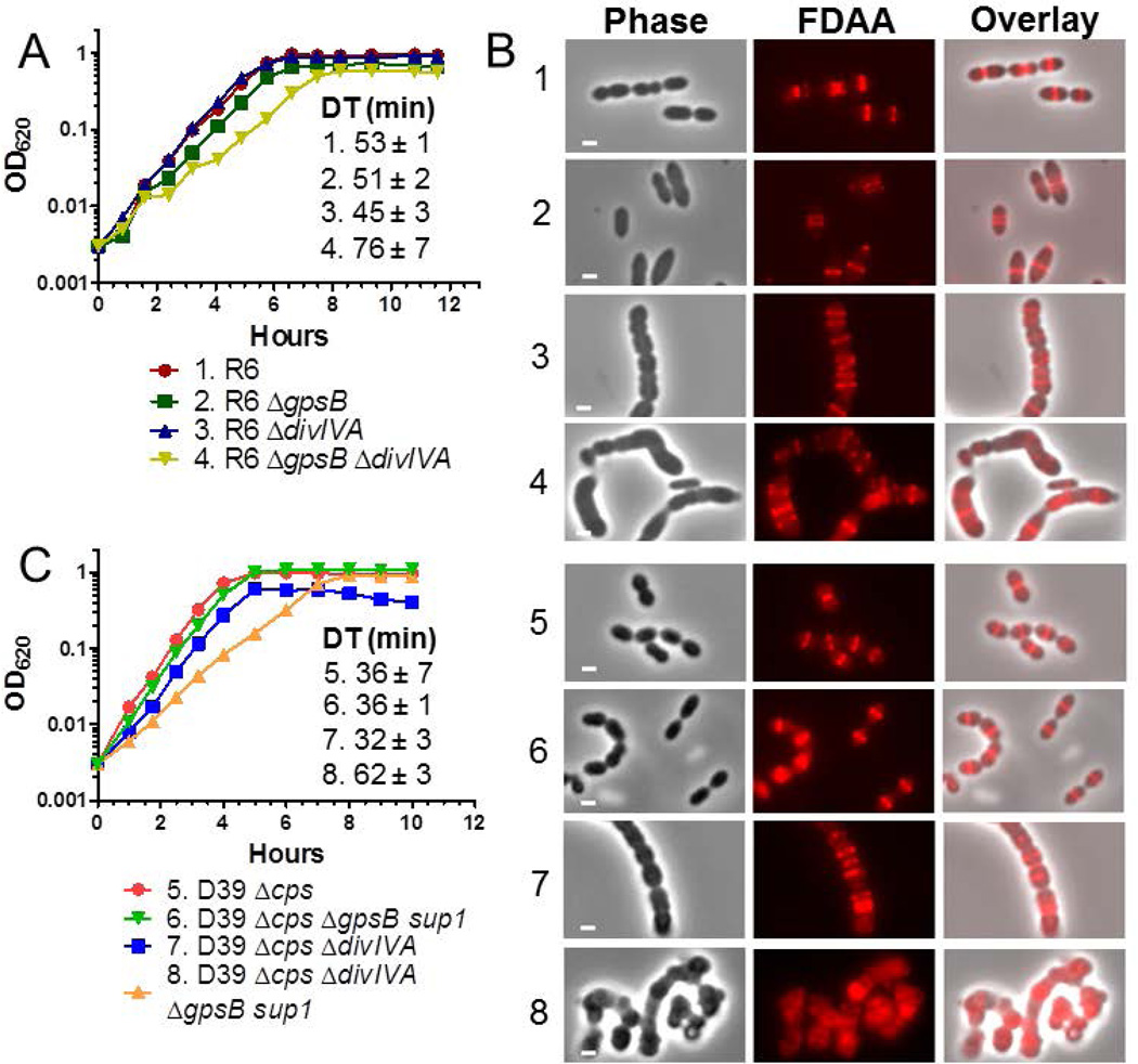
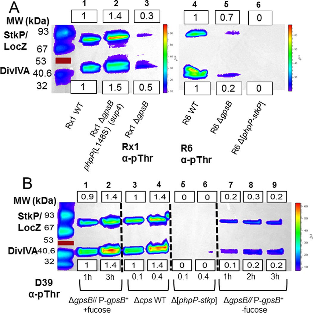
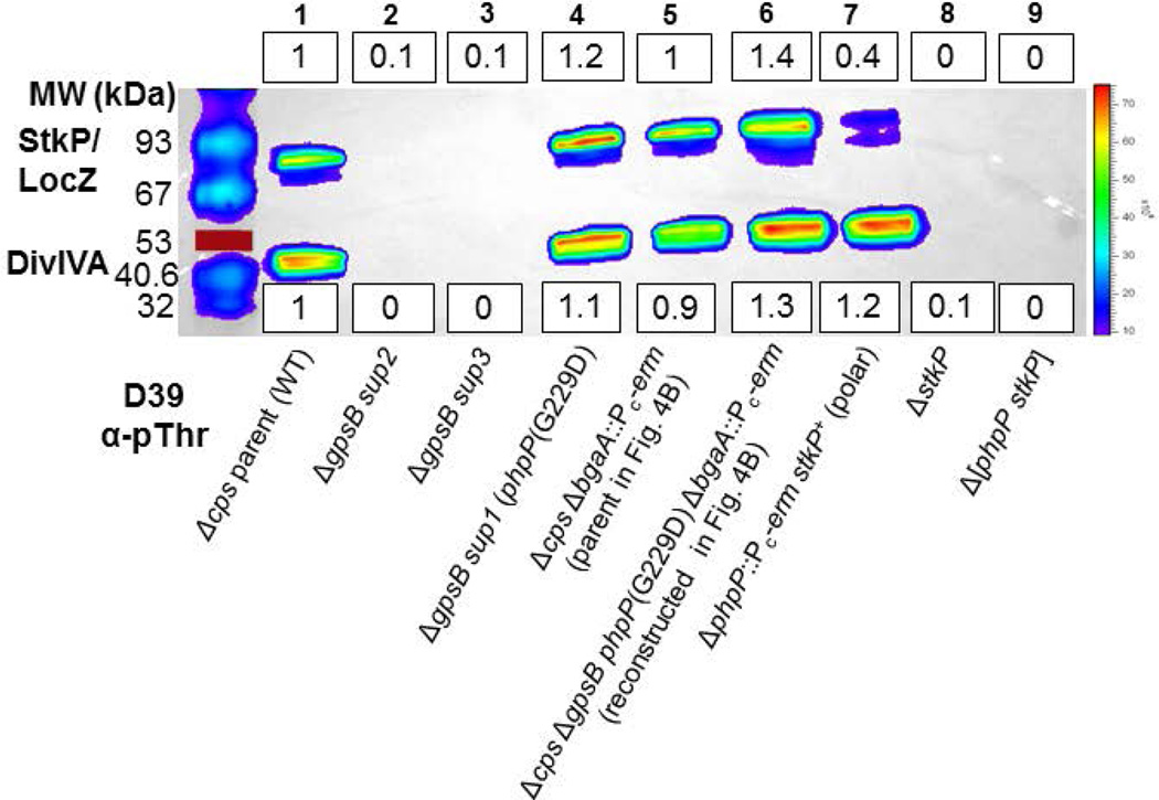
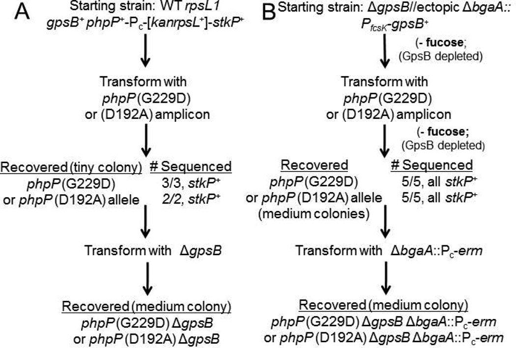
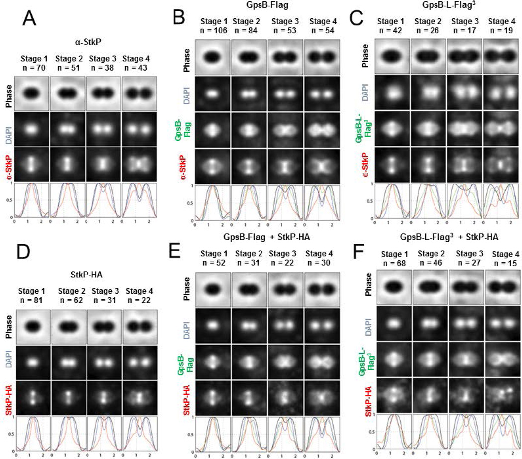
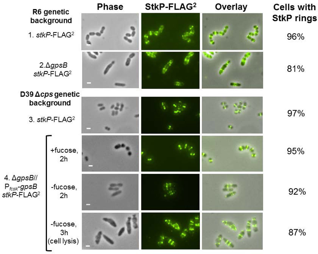
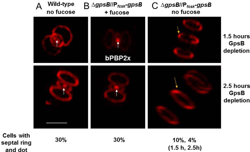
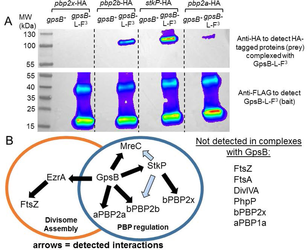
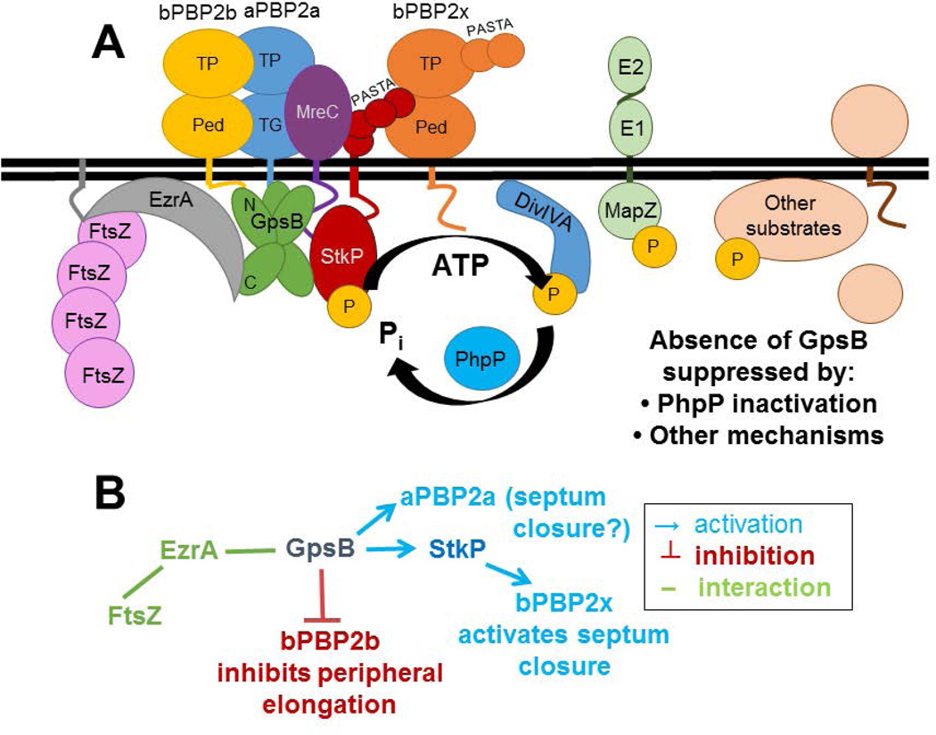
GpsB regulatory protein and StkP protein kinase have been proposed as molecular switches that balance septal and peripheral (side-wall like) peptidoglycan (PG) synthesis in Streptococcus pneumoniae (pneumococcus); yet, mechanisms of this switching remain unknown. We report that ΔdivIVA mutations are not epistatic to ΔgpsB division-protein mutations in progenitor D39 and related genetic backgrounds; nor is GpsB required for StkP localization or FDAA labeling at septal division rings. However, we confirm that reduction of GpsB amount leads to decreased protein phosphorylation by StkP and report that the essentiality of ΔgpsB mutations is suppressed by inactivation of PhpP protein phosphatase, which concomitantly restores protein phosphorylation levels. ΔgpsB mutations are also suppressed by other classes of mutations, including one that eliminates protein phosphorylation and may alter division. Moreover, ΔgpsB mutations are synthetically lethal with Δpbp1a, but not Δpbp2a or Δpbp1b mutations, suggesting GpsB activation of PBP2a activity. Consistent with this result, co-IP experiments showed that GpsB complexes with EzrA, StkP, PBP2a, PBP2b and MreC in pneumococcal cells. Furthermore, depletion of GpsB prevents PBP2x migration to septal centers. These results support a model in which GpsB negatively regulates peripheral PG synthesis by PBP2b and positively regulates septal ring closure through its interactions with StkP-PBP2x.


Figures

Fig. 1
Fig. 1
ΔdivIVA mutations are not epistatic to ΔgpsB in pneumococcal strains R6 and D39. A) Representative growth curves of R6 strains. Isogenic strains listed as 1–4 are: 1, R6 (EL59); 2, R6 ΔgpsB (IU8224); 3, R6 ΔdivIVA (IU8371); 4, R6 ΔgpsB ΔdivIVA (IU8369). Average doubling times (±SEM) from 2 independent experiments were calculated for OD620 ≈ 0.015 to 0.2 using a nonlinear regression exponential growth curve program (GraphPad Prism). B) Fluorescent D-amino acid (FDAA) staining and microscopy of live cells labeled with FDAA for 5 min were performed as described in Experimental procedures. The panels shown from left to right are: phase, FDAA, and a phase/FDAA overlay. Genotypes are indicated according to the numbers in panels A and C. Representative images are shown of ≥95% of the cells (n>50 for R6 strains; n>70 for D39 strains) examined manually of each strain. C) Representative growth curves of D39 Δcps strains and D39 Δcps ΔgpsB sup1 strains, which contain a suppressor mutation (phpP(G229D)) of ΔgpsB as described in the text. Isogenic strains listed as 5–8 are: 5, D39 Δcps (IU1945); 6, D39 Δcps ΔgpsB phpP(G229D) (IU6442); 7, D39 Δcps ΔdivIVA (IU8496); 8, D39 Δcps ΔgpsB phpP(G229D) ΔdivIVA (IU11205). Doubling times were calculated as described above. Independent experiments were performed two to three times with similar results. Scale bar = 1 micron.
Fig. 2
Fig. 2
GpsB deletion or depletion results in decreased threonine (Thr) phosphorylation of proteins. Proteins phosphorylated on Thr residues were detected by Western blotting with α-pThr antibody, and blots were imaged and quantitated as described in Experimental procedures. Equal protein loading was confirmed by amido-black staining of membranes after blotting. Representative blots are shown, with background-subtracted luminescence values relative to the wild-type (wt) strain indicated for the StkP/LocZ and DivIVA bands in boxes above or below the blots, respectively. Relative values of band intensities were calculated as described in Experimental procedures, and mean relative intensities (±SEM) are compiled for all experiments in Table S5 and S6. A) ΔgpsB mutants of laboratory strains Rx1 and R6 harvested at OD620 ≈ 0.4. Lane 1, wild-type Rx1 parent (IU9256); lane 2, suppressed Rx1 ΔgpsB phpP(L148S) (IU9262); lane 3, Rx1 ΔgpsB phpP+ stkP+ (IU11574); lane 4 wild-type R6 parent (EL59); lane 5, R6 ΔgpsB (IU8224); and lane 6, R6 Δ[phpP-stkP] control (IU8419). The experiment was performed twice independently with similar results. B) GpsB depletion of D39 Δcps strains as described in Experimental procedures. Lane 1–2, merodiploid strain Δcps ΔgpsB//PfcsK-gpsB+ (IU4888) grown with fucose for the times indicated; lane 3–4, wild-type parent strain D39 Δcps (IU1945) grown to OD620 ≈ 0.1 or 0.4; lanes 5–6, D39 Δcps Δ[phpP-stkP] (E739), and lanes, 7–9, merodiploid strain Δcps ΔgpsB//PfcsK-gpsB+ (IU4888) incubated without fucose for the times indicated. The experiment was performed three times independently with similar results. See text for additional details. Red lines marks a colored 53 kDa standard that did not transfer.
Fig. 3
Fig. 3
phpP(G229D) restores wild-type levels of protein phosphorylation to D39 Δcps ΔgpsB mutants in originally isolated and reconstructed suppressor strains. A representative Western blot of phosphorylated proteins was performed and quantitated as described in Figure 2 and Experimental procedures. Mean relative values (±SEM) of band intensities are compiled for all experiments in Table S7. Strains were harvested at OD620 ≈ 0.4. Lane 1, wild-type parent D39 Δcps (IU1945); lane 2, D39 Δcps ΔgpsB Δ[spd_1026–spd_1037] Ω[spd_0889–spd_1026] (IU5845, sup2) (Table 2, line 2); lane 3, D39 Δcps ΔgpsB Δ[spd_1026–spd_1037] Ω[spd_0889–spd_1026] (IU6441, sup3); (Table 2, line 3); lane 4, D39 Δcps ΔgpsB phpP(G229D) (IU6442, sup1) (Table 2, line 1); Lane 5, D39 Δcps ΔbgaA::Pc-erm (E46) (strain used in reconstruction; Fig. 4B); lane 6, D39 Δcps ΔbgaA::Pc-erm ΔgpsB phpP(G229D) (IU11221) (reconstructed suppressor; Fig. 4B); lane 7, D39 Δcps ΔphpP-Pc-erm (IU11442) (polar mutant with reduced StkP expression); lane 8, D39 Δcps ΔstkP (IU11460) control; and lane 9, D39 Δcps Δ[phpP-stkP] (IU11462) control. The experiment was performed twice independently with similar results. The red line marks a colored 53 kDa standard that did not transfer.
Fig. 4
Fig. 4
Scheme for reconstruction of phpP(G229D) or phpP(D192A) mutants in D39 Δcps rpsL1 gpsB+ and D39 Δcps gpsB+ genetic backgrounds. See Experimental procedures for details. These strains were constructed twice with similar results, and resulting strains are listed in Table S1.
Fig. 5
Fig. 5
D39 Δcps ΔgpsB phpP(G229D) suppressor strains grow similarly to wild-type parent strains, but form slightly rounder, smaller-sized cells. A) Representative growth curves of the originally isolated ΔgpsB sup1 (phpP(G229D)) (Table 2, line 1) and reconstructed strain (Fig. 4B). Strain 1, D39 Δcps wild-type parent (IU1945); strain 2, original ΔgpsB sup1 strain (D39 Δcps ΔgpsB phpP(G229D); IU6442, Table 2, line 1); strain 3, E46 used in reconstruction (D39 Δcps ΔbgaA::Pc-erm; Fig. 4B); strain 4, reconstructed suppressor (D39 Δcps ΔgpsB phpP(G229D) ΔbgaA::Pc-erm; IU11221; Fig. 4B). Doubling times were calculated as described for Figure 1. B) Representative images of live cells at OD620 ≈ 0.1–0.2 from growth curves in panel A. C) Length, width, aspect ratio, and cell volume relative to the median value of IU1945 for each strain listed in panel A. Over 50 cells were measured per strain in two experimental replicates. Scale bar = 1 micron.
Fig. 6
Fig. 6
GpsB and StkP have different, but overlapping localization patterns at each division stage. Comparison of GpsB and StkP localization by immunofluorescence (IFM) of double- and single-tagged strains and image averaging and quantitation were performed as detailed in Experimental procedures. Averaged IFM images of the indicated number of cells at each division stage (n) and fluorescence intensity traces of protein localization are shown for the following strains: A) IU1945 (D39 Δcps) probed with DAPI and anti-StkP antibody; B) IU5838 (D39 Δcps gpsB-FLAG) probed with anti-FLAG and anti-StkP antibodies; C) IU5458 (D39 Δcps gpsB-L-FLAG3) probed with anti-FLAG and anti-StkP antibodies; D) IU7438 (D39 Δcps stkP-HA) probed with DAPI and anti-HA antibody; E) IU11716 (D39 Δcps gpsB-FLAG stkP-HA) probed with anti-FLAG and anti-HA antibody; and F) IU11412 (D39 Δcps gpsB-L-FLAG3 stkP-HA) probed with anti-FLAG and anti-HA antibodies. See text for additional information.
Fig.7
Fig.7
2D IFM microscopy demonstrates that the absence or depletion of GpsB does not abolish StkP ring formation. 2D IFM was performed as outlined in Experimental procedures. Panels shown from left to right are: phase, FITC antibody labeled FLAG-tagged StkP, and phase/FITC overlay. 1) R6 stkP-FLAG2 (IU8819, sampled at OD620 ≈ 0.2); 2) R6 ΔgpsB stkP-FLAG2 (IU8311, sampled at OD620 ≈ 0.2); 3) D39 Δcps stkP-FLAG2 (IU7434, sampled at OD620 ≈ 0.2); 4) merodiploid strain D39 Δcps stkP-FLAG2 Δcps ΔgpsB//PfcsK-gpsB+ (IU8230) grown for 2 h with fucose addition or without fucose for 2 h or 3 h to deplete GpsB, eventually causing cell lysis. Representative images of each strain are shown for each experiment, which were performed three times independently with similar results. Percentages of cells with StkP rings are based on 100 manually examined cells of each strain. Scale bar = 1 micron.
Fig. 8
Fig. 8
GpsB depletion prevents FDAA labeling of septal centers, indicative of bPBP2x migration. Wild-type parent strain IU1945 (D39 Δcps) and gpsB merodiploid strain IU4888 (D39 Δcps ΔgpsB//PfcsK-gpsB+) were grown and labeled with FDAA (TADA) as described in Experimental procedures. The parent strain and gpsB merodiploid strain grown in fucose to induce GpsB expression were growing exponentially at the time of FDAA labeling (A and B), whereas the gpsB merodiploid switched to medium lacking fucose was depleted for GpsB for 1.5 h or 2.5 h at the time of FDAA labeling (C). White arrows point to the presence of the central septal spot of FDAA labeling within the septal outer ring in growing cells (A and B). Previous work has shown that this central spot corresponds to bPBP2x TP activity (see text). Yellow arrows point to septal outer rings without central septal spots in elongated cells depleted for GpsB (C). A minimum of 100 cells was observed per condition and strain. Wild-type cells and gpsB merodiploid cells grown in fucose had a central septal spot within the septal outer ring ≈ 30% of the time, whereas cells depleted for GpsB for 1.5 h or 2.5 h had a central septal spot within the septal outer ring only 10% or 4% of the time, respectively. The experiment was performed independently twice with similar results. All images are at the same magnification, and scale bar = 1 micron.
Fig. 9
Fig. 9
A) Pairwise co-IP of GpsB-L-FLAG3 with bPBP2b-HA, StkP-HA, or aPBP2a-HA4, but not with bPBP2x-HA. Co-IP experiments were performed as described in Experimental procedures. Top blot was probed with anti-HA primary antibody for HA-tagged prey proteins, using GpsB-L-FLAG3 as bait protein. 57 µg of each lysate sample were loaded on the input gel, while 20 µL of each elution sample was loaded on to the elution gel, after mixing 1:1 with 2× Laemlli sample buffer. Predicted molecular weight (MW) of bPBP2x-HA, bPBP2b-HA, StkP-HA, and aPBP2a-HA4 are 83.5 kDa, 75.7 kDa, 73.5 kDa, and 85.2 kDa, respectively. Bottom blot was probed with anti-FLAG primary antibody for GpsB-L-FLAG3 (bait). Two major bands are detected by anti-FLAG primary antibody in strains expressing GpsB-L-FLAG3. The bottom band correlates to GpsB-L-FLAG3 monomer (≈ 16.4 kDa), whereas the top band is likely a GpsB-L-FLAG3 trimer based on MW. Lanes shown on blot are as follows (all strains were constructed in the D39 Δcps background, IU1945): lane 1, pbp2x-HA gpsB+ (IU6929); lane 2, gpsB-L-FLAG3 pbp2x-HA (IU11314); lane 3, pbp2b-HA gpsB+ (IU6933); lane 4, gpsB-L-FLAG3 pbp2b-HA (IU11316); lane 5, stkP-HA gpsB+ (IU7438); lane 6, gpsB-L-FLAG3 stkP-HA (IU11412); lane 7, pbp2a-HA4 gpsB+ (IU11560); and lane 8 pbp2a-HA4 gpsB-L-FLAG3 (IU11516). This experiment was performed twice with similar results. B) Map of interactions found by in vivo co-IP that are proposed to coordinate divisome assembly with PBP regulation. GpsB was detected in complexes with EzrA, StkP, aPBP2a, bPBP2b, and/or MreC at stages of the division cycle (above; Fig. S10, S11, and S14). StkP was detected in complexes with bPBP2x, bPBP2b, and MreC (Fig. S13 and S14), although complexes with bPBP2b and MreC could be indirect (blue arrows) via interactions of these proteins with GpsB. EzrA is in complexes with FtsZ and GpsB (Fig. S11 and S12) and other division proteins not shown (Amilcar Perez, in preparation for submission). GpsB did not pull down detectable levels of FtsZ, FtsA, DivIVA, PhpP, bPBP2x, or aPBP1a by this in vivo co-IP method (above; Fig. S10–S12 and S14).
Fig. 10
Fig. 10
Model of GpsB interactions and coordination of septal and peripheral PG synthesis in Spn strain D39. A) Complexes containing EzrA, which binds to FtsZ, and GpsB link FtsZ-divisome dynamics (which are not shown) with GpsB regulation of downstream functions. Wild-type levels of GpsB mediate the normal protein phosphorylation cycle by StkP kinase and PhpP phosphatase of numerous division proteins, including DivIVA, MapZ(LocZ), whose extracellular E1 and E2 domains are labeled, and other proteins. Septal and peripheral PG synthesis are coordinated by GpsB complexed with aPBP2a, bPBP2b, MreC, and StkP, which interacts with bPBP2x. bPBP2x and possibly aPBP2a catalyze septal ring closure, whereas bPBP2b and MreC catalyze peripheral PG synthesis. Deletion of gpsB is lethal and can be suppressed by non-polar mutations that inactivate the PhpP phosphatase, thereby implicating maintenance of protein phosphorylation levels as an important regulatory function of GpsB; however, the critical phosphorylated protein(s) remain to be determined. B) Genetic scheme of PBP activation by GpsB that can account for the enlarged, elongated cells with unconstricted septa caused by GpsB depletion. According to this scheme, which is based on phenotypes, genetic relationships, microscopy, and interaction maps, GpsB positively regulates septum closure by activating aPBP2a directly and bPBP2x indirectly, via an interaction between GpsB and StkP, whereas GpsB directly or indirectly inhibits bPBP2b/MreC and peripheral PG elongation. See text for additional details.

Comment in

Similar articles

Cited by

References

    1. Agarwal S, Agarwal S, Pancholi P, Pancholi V. Strain-specific regulatory role of eukaryote-like serine/threonine phosphatase in pneumococcal adherence. Infect Immun. 2012;80:1361–1372. - PMC - PubMed
    1. Altschul SF, Madden TL, Schaffer AA, Zhang J, Zhang Z, Miller W, Lipman DJ. Gapped BLAST and PSI-BLAST: a new generation of protein database search programs. Nuc Acids Res. 1997;25:3389–3402. - PMC - PubMed
    1. Altschul SF, Wootton JC, Gertz EM, Agarwala R, Morgulis A, Schaffer AA, Yu YK. Protein database searches using compositionally adjusted substitution matrices. FEBS J. 2005;272:5101–5109. - PMC - PubMed
    1. Archambaud C, Gouin E, Pizarro-Cerda J, Cossart P, Dussurget O. Translation elongation factor EF-Tu is a target for Stp, a serine-threonine phosphatase involved in virulence of Listeria monocytogenes. Molec Microbiol. 2005;56:383–396. - PubMed
    1. Ashkenazy H, Abadi S, Martz E, Chay O, Mayrose I, Pupko T, Ben-Tal N. ConSurf 2016: an improved methodology to estimate and visualize evolutionary conservation in macromolecules. Nuc Acids Res. 2016;44:W344–W350. - PMC - PubMed

Publication types

MeSH terms

LinkOut - more resources