

Covalent Modulators of the Vacuolar ATPase
- PMID: 28010062
- PMCID: PMC5274637
- DOI: 10.1021/jacs.6b12511
Covalent Modulators of the Vacuolar ATPase
Abstract
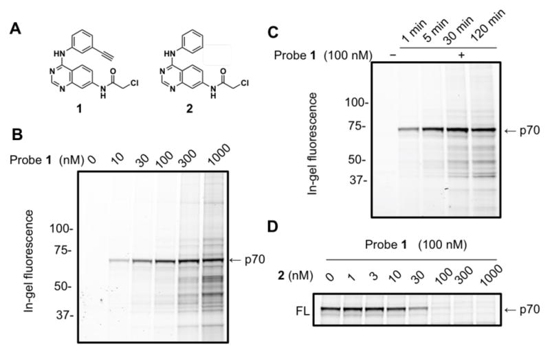
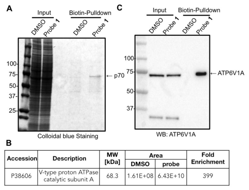
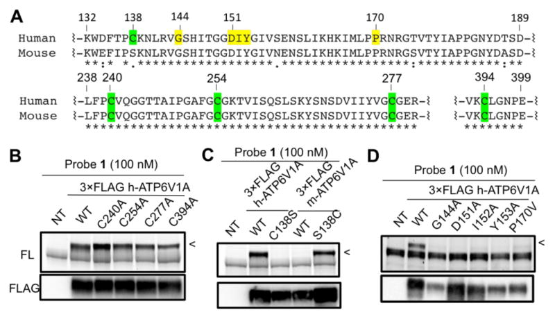
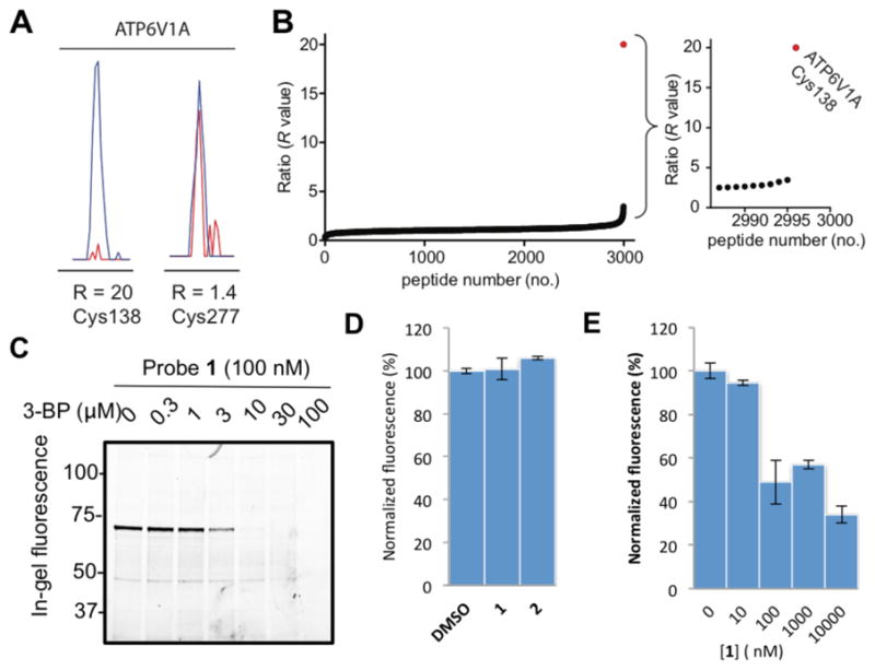
The vacuolar H+ ATPase (V-ATPase) is a complex multisubunit machine that regulates important cellular processes through controlling acidity of intracellular compartments in eukaryotes. Existing small-molecule modulators of V-ATPase either are restricted to targeting one membranous subunit of V-ATPase or have poorly understood mechanisms of action. Small molecules with novel and defined mechanisms of inhibition are thus needed to functionally characterize V-ATPase and to fully evaluate the therapeutic relevance of V-ATPase in human diseases. We have discovered electrophilic quinazolines that covalently modify a soluble catalytic subunit of V-ATPase with high potency and exquisite proteomic selectivity as revealed by fluorescence imaging and chemical proteomic activity-based profiling. The site of covalent modification was mapped to a cysteine residue located in a region of V-ATPase subunit A that is thought to regulate the dissociation of V-ATPase. We further demonstrate that a previously reported V-ATPase inhibitor, 3-bromopyruvate, also targets the same cysteine residue and that our electrophilic quinazolines modulate the function of V-ATPase in cells. With their well-defined mechanism of action and high proteomic specificity, the described quinazolines offer a powerful set of chemical probes to investigate the physiological and pathological roles of V-ATPase.
Conflict of interest statement
The authors declare no competing financial interest.
Figures
Similar articles
-
The vacuolar (H+)-ATPases--nature's most versatile proton pumps.Nat Rev Mol Cell Biol. 2002 Feb;3(2):94-103. doi: 10.1038/nrm729. Nat Rev Mol Cell Biol. 2002. PMID: 11836511 Review.
-
The cellular energization state affects peripheral stalk stability of plant vacuolar H+-ATPase and impairs vacuolar acidification.Plant Cell Physiol. 2011 May;52(5):946-56. doi: 10.1093/pcp/pcr044. Epub 2011 Apr 6. Plant Cell Physiol. 2011. PMID: 21474463
-
Vacuolar ATPases: rotary proton pumps in physiology and pathophysiology.Nat Rev Mol Cell Biol. 2007 Nov;8(11):917-29. doi: 10.1038/nrm2272. Nat Rev Mol Cell Biol. 2007. PMID: 17912264 Review.
-
Previously Uncharacterized Vacuolar-type ATPase Binding Site Discovered from Structurally Similar Compounds with Distinct Mechanisms of Action.ACS Chem Biol. 2019 Jan 18;14(1):20-26. doi: 10.1021/acschembio.8b00656. Epub 2018 Dec 18. ACS Chem Biol. 2019. PMID: 30461263
-
Structural bases of physiological functions and roles of the vacuolar H(+)-ATPase.Cell Signal. 2011 Aug;23(8):1244-56. doi: 10.1016/j.cellsig.2011.03.003. Epub 2011 Mar 17. Cell Signal. 2011. PMID: 21397012 Review.
Cited by
-
Activity-based protein profiling guided new target identification of quinazoline derivatives for expediting bactericide discovery: Activity-based protein profiling derived new target discovery of antibacterial quinazolines.J Adv Res. 2025 Aug;74:57-72. doi: 10.1016/j.jare.2024.10.002. Epub 2024 Oct 9. J Adv Res. 2025. PMID: 39389307 Free PMC article.
-
Reactive-cysteine profiling for drug discovery.Curr Opin Chem Biol. 2019 Jun;50:29-36. doi: 10.1016/j.cbpa.2019.02.010. Epub 2019 Mar 18. Curr Opin Chem Biol. 2019. PMID: 30897495 Free PMC article. Review.
-
Schisandrol A protects AGEs-induced neuronal cells death by allosterically targeting ATP6V0d1 subunit of V-ATPase.Acta Pharm Sin B. 2022 Oct;12(10):3843-3860. doi: 10.1016/j.apsb.2022.06.013. Epub 2022 Jun 30. Acta Pharm Sin B. 2022. PMID: 36213534 Free PMC article.
-
Electron-Deficient Alkynes as Powerful Tools against Root-Knot Nematode Melodogyne incognita: Nematicidal Activity and Investigation on the Mode of Action.J Agric Food Chem. 2020 Oct 7;68(40):11088-11095. doi: 10.1021/acs.jafc.0c00835. Epub 2020 Sep 24. J Agric Food Chem. 2020. PMID: 32924513 Free PMC article.
-
Multisite Labeling of Proteins Using the Ligand-Directed Reactivity of Triggerable Michael Acceptors.Bioconjug Chem. 2023 Jun 21;34(6):1130-1138. doi: 10.1021/acs.bioconjchem.3c00155. Epub 2023 May 23. Bioconjug Chem. 2023. PMID: 37220065 Free PMC article.
References
-
- Forgac M. Nat Rev Mol Cell Biol. 2007;8:917–929. - PubMed
-
- Karet FE, Finberg KE, Nelson RD, Nayir A, Mocan H, Sanjad SA, Rodriguez-Soriano J, Santos F, Cremers CWRJ, Di Pietro A, Hoffbrand BI, Winiarski J, Bakkaloglu A, Ozen S, Dusunsel R, Goodyer P, Hulton SA, Wu DK, Skvorak AB, Morton CC, Cunningham MJ, Jha V, Lifton RP. Nat Genet. 1999;21:84–90. - PubMed
-
- Toyomura T, Murata Y, Yamamoto A, Oka T, Sun-Wada GH, Wada Y, Futai M. J Biol Chem. 2003;278:22023–22030. - PubMed
-
- Sennoune SR, Bakunts K, Martinez GM, Chua-Tuan JL, Kebir Y, Attaya MN, Martinez-Zaguilan R. Am J Physiol Cell Physiol. 2004;286:C1443–C1452. - PubMed
-
- Maxson ME, Grinstein S. J Cell Sci. 2014;127:4987–4993. - PubMed
Publication types
MeSH terms
Substances
Grants and funding
LinkOut - more resources
Full Text Sources
Other Literature Sources
