. 2017 Sep 1;27(7):379-397.
doi: 10.1089/ars.2016.6642. Epub 2017 Jan 30.

Lysyl Oxidase Induces Vascular Oxidative Stress and Contributes to Arterial Stiffness and Abnormal Elastin Structure in Hypertension: Role of p38MAPK

Affiliations

Lysyl Oxidase Induces Vascular Oxidative Stress and Contributes to Arterial Stiffness and Abnormal Elastin Structure in Hypertension: Role of p38MAPK

Sonia Martínez-Revelles et al. Antioxid Redox Signal. .

Erratum in

Abstract

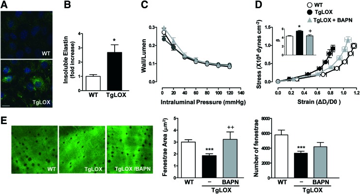
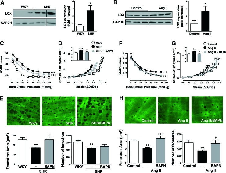
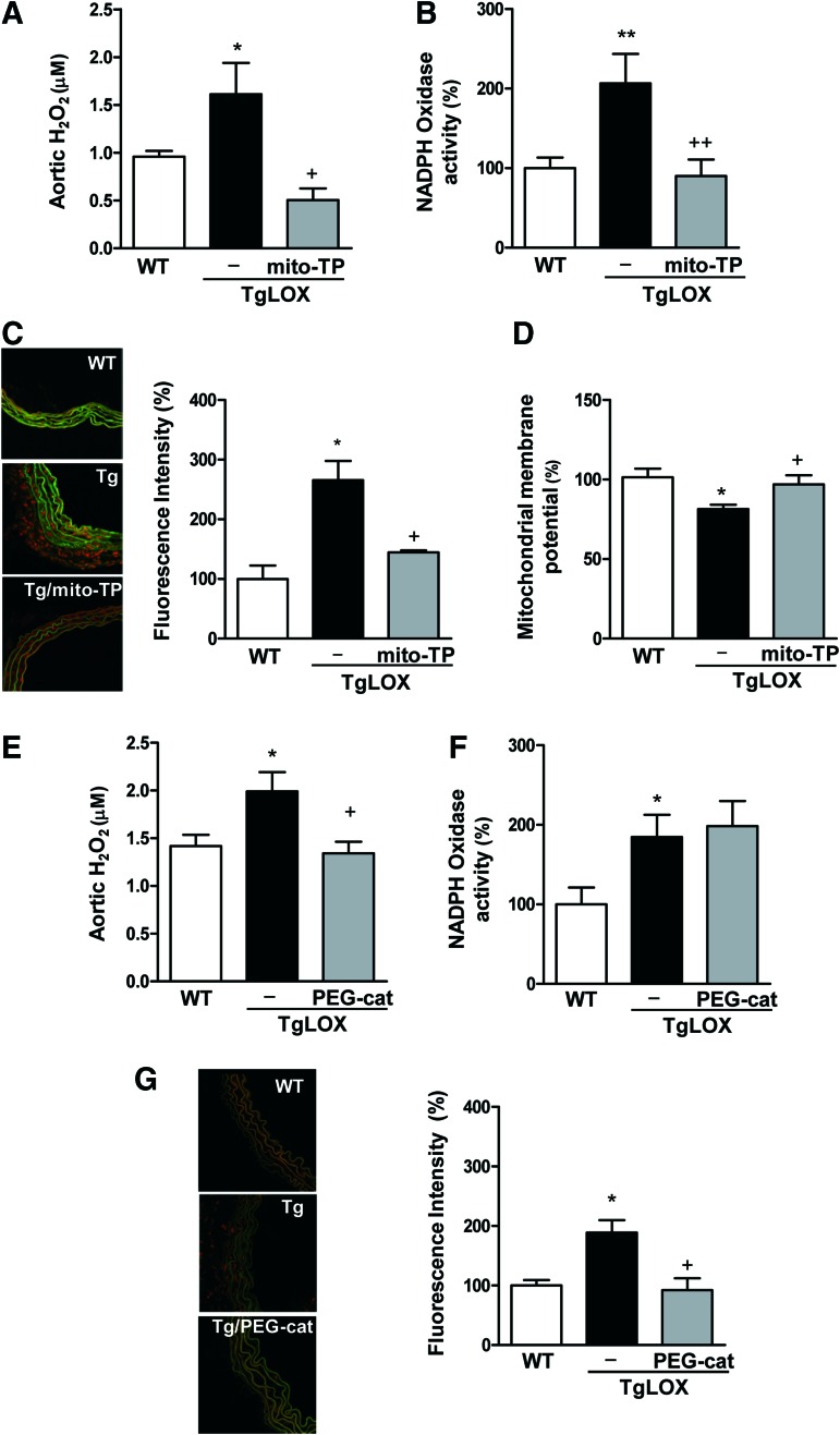
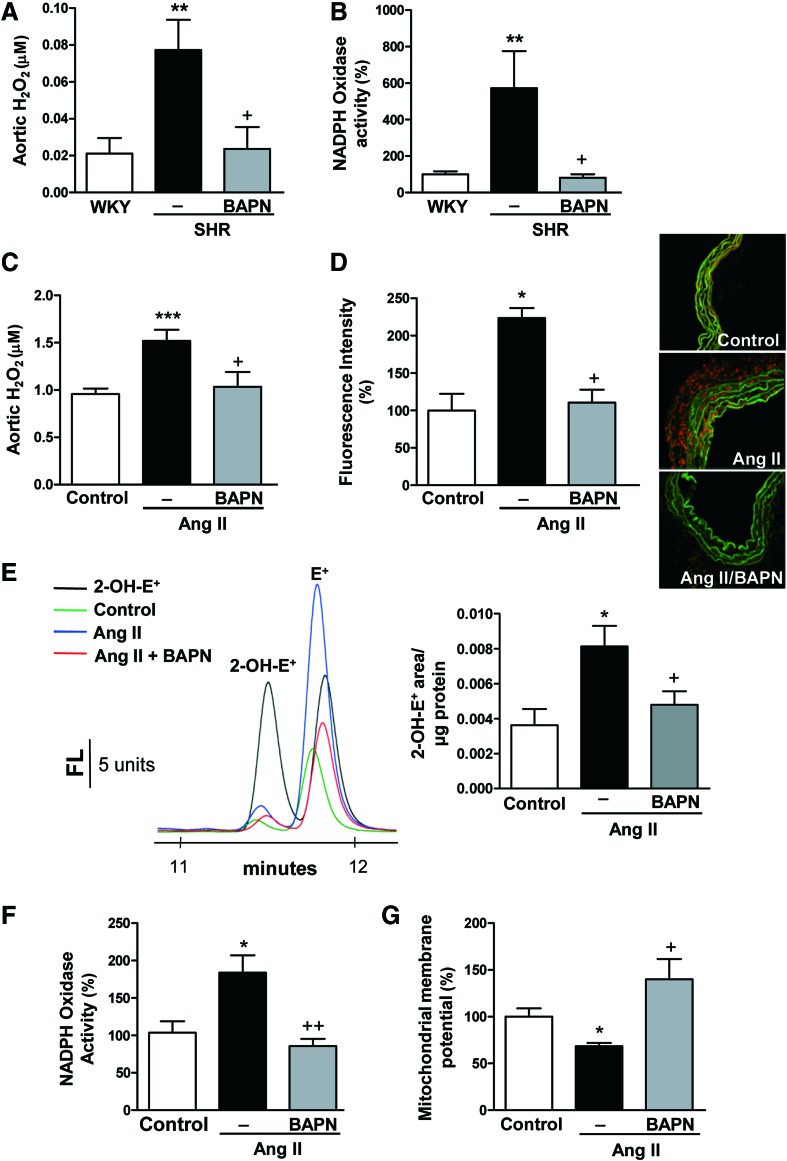
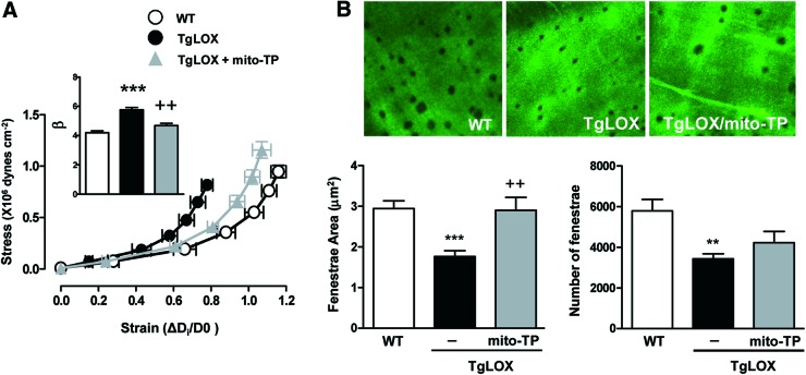
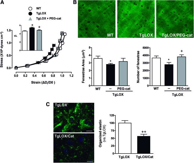
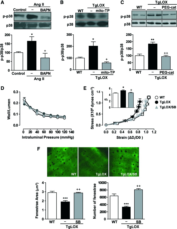
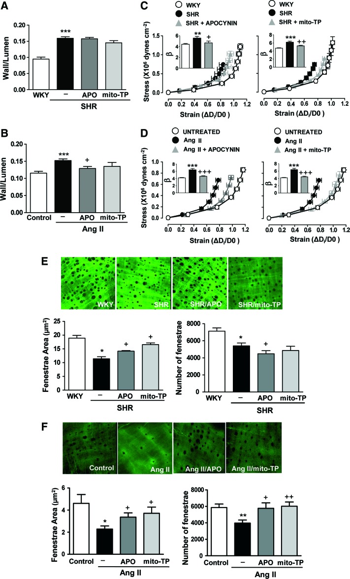
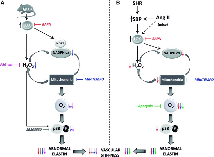
Aims: Vascular stiffness, structural elastin abnormalities, and increased oxidative stress are hallmarks of hypertension. Lysyl oxidase (LOX) is an elastin crosslinking enzyme that produces H2O2 as a by-product. We addressed the interplay between LOX, oxidative stress, vessel stiffness, and elastin.

Results: Angiotensin II (Ang II)-infused hypertensive mice and spontaneously hypertensive rats (SHR) showed increased vascular LOX expression and stiffness and an abnormal elastin structure. Mice over-expressing LOX in vascular smooth muscle cells (TgLOX) exhibited similar mechanical and elastin alterations to those of hypertensive models. LOX inhibition with β-aminopropionitrile (BAPN) attenuated mechanical and elastin alterations in TgLOX mice, Ang II-infused mice, and SHR. Arteries from TgLOX mice, Ang II-infused mice, and/or SHR exhibited increased vascular H2O2 and O2.- levels, NADPH oxidase activity, and/or mitochondrial dysfunction. BAPN prevented the higher oxidative stress in hypertensive models. Treatment of TgLOX and Ang II-infused mice and SHR with the mitochondrial-targeted superoxide dismutase mimetic mito-TEMPO, the antioxidant apocynin, or the H2O2 scavenger polyethylene glycol-conjugated catalase (PEG-catalase) reduced oxidative stress, vascular stiffness, and elastin alterations. Vascular p38 mitogen-activated protein kinase (p38MAPK) activation was increased in Ang II-infused and TgLOX mice and this effect was prevented by BAPN, mito-TEMPO, or PEG-catalase. SB203580, the p38MAPK inhibitor, normalized vessel stiffness and elastin structure in TgLOX mice.

Innovation: We identify LOX as a novel source of vascular reactive oxygen species and a new pathway involved in vascular stiffness and elastin remodeling in hypertension.

Conclusion: LOX up-regulation is associated with enhanced oxidative stress that promotes p38MAPK activation, elastin structural alterations, and vascular stiffness. This pathway contributes to vascular abnormalities in hypertension. Antioxid. Redox Signal. 27, 379-397.

Keywords: NADPH oxidases; extracellular matrix; free radicals; microvascular; mitochondria; tissue repair and remodeling.


Conflict of interest statement

No competing financial interests exist.

Figures

<b>FIG. 1.</b>
FIG. 1.
TgLOX mice exhibit higher vascular stiffness and structural elastin abnormalities. (A) Elastin staining (green) in non-permeabilized vascular smooth muscle cells (VSMC) isolated from wild-type (WT) and TgLOX mice. Nuclei were stained with Hoechst 33342 (blue). Bar size: 20 μm. (B) Levels of insoluble elastin in VSMC from WT and TgLOX mice after [3H]-valine supplementation. Data were normalized per DNA content in each individual well. (C) Wall/lumen-pressure curves and (D) stress-strain curves in mesenteric resistance arteries (MRAs) from WT, TgLOX, or TgLOX mice receiving BAPN. β-values (slopes of the stress-strain relationships) are also shown. (E) Maximal projections of the internal elastic lamina (IEL) and fenestrae area and number of MRAs from WT, TgLOX, or TgLOX mice receiving BAPN. Projections were obtained from serial optical sections that were captured with a fluorescence confocal microscope ( × 63 oil immersion objective). Image size: 59.5 × 59.5 μm. Results are represented as mean ± SEM (n = 4–10; *p < 0.05, ***p < 0.001 vs. WT; +p < 0.05, ++p < 0.01 vs. untreated TgLOX mice). BAPN, β-aminopropionitrile; SEM, standard error of the mean. To see this illustration in color, the reader is referred to the web version of this article at www.liebertpub.com/ars
<b>FIG. 2.</b>
FIG. 2.
LOX inhibition prevents vascular stiffness and structural elastin abnormalities in hypertension models. LOX protein levels in vascular lysates from WKY and SHR (A) or control and Ang II-infused mice (B). Results were normalized by GADPH expression. Wall/lumen-pressure curves (C, F) and stress-strain curves (D, G) in MRAs from WKY, SHR, or SHR treated with BAPN (C, D) and in control or Ang II-infused mice pretreated or not with BAPN (F, G). Maximal projections of the IEL and fenestrae area and number of MRAs from WKY, SHR, or SHR plus BAPN (image size: 119 × 119 μm) (E) and control, Ang II, and Ang II plus BAPN mice (image size: 59.5 × 59.5 μm) (H). Data are represented as mean ± SEM (n = 4–12; *p < 0.05, **p < 0.01, ***p < 0.001 vs. WKY or control animals; +p < 0.05, ++p < 0.01, +++p < 0.001 vs. untreated SHR or Ang II-infused animals). Ang II, angiotensin II; LOX, lysyl oxidase; SHR, spontaneously hypertensive rats; WKY, Wistar Kyoto. To see this illustration in color, the reader is referred to the web version of this article at www.liebertpub.com/ars
<b>FIG. 3.</b>
FIG. 3.
LOX is a source of vascular oxidative stress. Aortic H2O2 production (A, E), vascular NADPH oxidase activity (B, F), vascular superoxide anion production (C, G), and mitochondrial membrane potential (D) evaluated in wild-type (WT) and TgLOX mice treated or not with mito-TEMPO (mito-TP) or PEG-catalase (PEG-cat). Representative images of dihydroethidium staining are shown. Image size 238 × 238 μm. Data are represented as mean ± SEM (n = 4–10; *p < 0.05, **p < 0.01, vs. WT; +p < 0.05, ++p < 0.01 vs. untreated TgLOX mice). polyethylene glycol-conjugated catalase (PEG-catalase). To see this illustration in color, the reader is referred to the web version of this article at www.liebertpub.com/ars
<b>FIG. 4.</b>
FIG. 4.
LOX inhibition reduces vascular oxidative stress in hypertension animal models. Aortic H2O2 production (A) and vascular NADPH oxidase activity (B) in WKY, SHR, and SHR treated with BAPN. Aortic H2O2 production (C), vascular superoxide anion production evaluated by dihydroetidium fluorescence (D) and by 2-OH-E+ fluorescence HPLC (E), vascular NADPH oxidase activity (F), and mitochondrial membrane potential (G) were analyzed in control or Ang II-infused mice pretreated or not with BAPN. Representative images of dihydroethidium staining (image size 238 × 238 μm) and HPLC representative traces are shown. Data are represented as mean ± SEM (n = 4–10; *p < 0.05, **p < 0.01, ***p < 0.001 vs. WKY or control animals; +p < 0.05, ++p < 0.01 vs. untreated SHR or Ang II-infused animals). To see this illustration in color, the reader is referred to the web version of this article at www.liebertpub.com/ars
<b>FIG. 5.</b>
FIG. 5.
Oxidative stress underlies the higher vascular stiffness and structural elastin abnormalities detected in TgLOX mice. Stress-strain curves (A) and fenestrae area and number (B) in MRAs from wild-type (WT) and TgLOX mice treated or not with mito-TEMPO (mito-TP). Maximal projections of the IEL of MRAs are shown. Projections were obtained from serial optical sections that were captured with a fluorescence confocal microscope ( × 63 oil immersion objective). Image size: 59.5 × 59.5 μm. Data are represented as mean ± SEM (n = 6–10; **p < 0.01, ***p < 0.001 vs. WT; ++p < 0.01 vs. untreated TgLOX mice). To see this illustration in color, the reader is referred to the web version of this article at www.liebertpub.com/ars
<b>FIG. 6.</b>
FIG. 6.
H2O2 production underlies the higher vascular stiffness and structural elastin abnormalities detected in TgLOX mice. Stress-strain curves (A) and fenestrae area and number (B) in MRAs from wild-type (WT) and TgLOX mice treated or not with PEG-catalase (PEG-cat). Maximal projections of the IEL of MRAs are shown. Projections were obtained from serial optical sections that were captured with a fluorescence confocal microscope ( × 63 oil immersion objective). Image size: 59.5 × 59.5 μm. (C) Elastin staining (green) in non-permeabilized VSMC isolated from TgLOX mice that were incubated or not with catalase (TgLOX/Cat). Nuclei were stained with Hoechst 33342 (blue). Bar size: 20 μm. Data are represented as mean ± SEM (n = 6–10; *p < 0.05 vs. WT; +p < 0.05 ++p < 0.01 vs. untreated TgLOX mice or cells). To see this illustration in color, the reader is referred to the web version of this article at www.liebertpub.com/ars
<b>FIG. 7.</b>
FIG. 7.
p38MAPK contributes to the LOX-dependent disturbances in vascular stiffness and elastin alterations. Activation of p38MAPK (p-p38) in vessels from control or Ang II-infused mice treated or not with BAPN (A) and in wild-type (WT) and TgLOX mice treated or not with mito-TEMPO (mito-TP) (B) or PEG-catalase (PEG-cat) (C). Results were normalized by total p38 levels. (D) Wall/lumen-pressure curves, (E) stress-strain curves, and (F) fenestrae area and number in MRAs from WT, TgLOX, or TgLOX mice receiving SB 203580 (SB). Maximal projections of the IEL of MRAs are shown. Projections were obtained from serial optical sections that were captured with a fluorescence confocal microscope ( × 63 oil immersion objective). Image size: 59.5 × 59.5 μm. Data are represented as mean ± SEM (n = 3–8; *p < 0.05, **p < 0.01, ***p < 0.001 vs. WT; +p < 0.05, ++p < 0.01 vs. untreated TgLOX mice). p38MAPK, p38 mitogen-activated protein kinase. To see this illustration in color, the reader is referred to the web version of this article at www.liebertpub.com/ars
<b>FIG. 8.</b>
FIG. 8.
Oxidative stress underlies vascular remodeling, stiffness, and elastin alterations in hypertensive animal models. Wall/lumen (A,B) stress-strain curves (C, D) and fenestrae area and number (E, F) in MRAs from WKY, SHR, and SHR treated with apocynin (APO) or mito-TEMPO (mito-TP) (A, C) and (E) or from control, Ang II-infused, and Ang II-infused mice treated with apocynin or mitoTEMPO (B, D) and (F). Maximal projections of the IEL of MRAs from WKY, SHR, or SHR plus antioxidants (image size: 119 × 119 μm) (E) and control, Ang II, and Ang II plus antioxidant-treated mice (image size: 59.5 × 59.5 μm) (F). Data are represented as mean ± SEM (n = 6–15; *p < 0.05, **p < 0.01, ***p < 0.001 vs. WKY or control animals; +p < 0.05, ++p < 0.01, +++p < 0.001 vs. untreated WKY or Ang II-infused animals). To see this illustration in color, the reader is referred to the web version of this article at www.liebertpub.com/ars
<b>FIG. 9.</b>
FIG. 9.
LOX induces oxidative stress that promotes p38MAPK activation, elastin remodeling, and vascular stiffness. (A) LOX transgenesis enhances vascular H2O2 levels, NADPH oxidase activity, and NOX1 expression; causes mitochondrial dysfunction; induces O2.− production; activates p38MAPK signaling; disturbs elastin structure; and increases vascular stiffness. The H2O2 scavenger PEG-catalase (in pink) and the superoxide dismutase targeted antioxidant mitoTEMPO (in blue) decrease the enhanced O2.− production and p38MAPK activation and improve elastin structure and vascular stiffness (symbolized by respective colored arrows). Accordingly, p38 MAPK inhibition by SB203580 (in grey) reduces elastin alterations and vascular stiffness (gray arrows). Further, LOX blockade with BAPN (in red) limited elastin abnormalities and vascular stiffness. (B) Vascular LOX expression is induced in animal models of hypertension (Ang II-infused mice and SHR). In both hypertensive models, LOX up-regulation partially relies on high blood pressure, although additional mechanisms could be involved (indicated with a dashed arrow). In hypertension, BAPN (in red) reduces the enhanced vascular H2O2 levels, NADPH oxidase activation, mitochondrial dysfunction, O2.− production, and/or p38MAPK activation and further ameliorates elastin abnormalities and vascular stiffness (symbolized by red arrows). Mito-TEMPO (in blue) and the antioxidant apocynin (in green) diminish oxidative stress and normalize the altered elastin structure and the increased vascular stiffness observed in hypertensive models (indicated with respective colored arrows). Note that in the vascular wall, the LOX-dependent H2O2 production would contribute to an ROS amplification mechanism involving NADPH oxidase and mitochondria. To see this illustration in color, the reader is referred to the web version of this article at www.liebertpub.com/ars

Similar articles

Cited by

References

    1. Aguado A, Galán M, Zhenyukh O, Wiggers GA, Roque FR, Redondo S, Peçanha F, Martín A, Fortuño A, Cachofeiro V, Tejerina T, Salaices M, and Briones AM. Mercury induces proliferation and reduces cell size in vascular smooth muscle cells through MAPK, oxidative stress and cyclooxygenase-2 pathways. Toxicol Appl Pharmacol 268: 188–200, 2013 - PubMed
    1. AlGhatrif M. and Lakatta EG. The conundrum of arterial stiffness, elevated blood pressure, and aging. Curr Hypertens Rep 17: 12, 2015 - PMC - PubMed
    1. Arribas SM, Hinek A, and González MC. Elastic fibres and vascular structure in hypertension. Pharmacol Ther 111: 771–791, 2006 - PubMed
    1. Bonadei I, Vizzardi E, D'Aloia A, Sciatti E, Raddino R, and Metra M. Role of aliskiren on arterial stiffness and endothelial function in patients with primary hypertension. J Clin Hypertens (Greenwich) 16: 202–206, 2014 - PMC - PubMed
    1. Briones AM. and Touyz RM. Oxidative stress and hypertension: current concepts. Curr Hypertens Rep 12: 135–142, 2010 - PubMed