. 2017 Feb 15;6(2):141-147.
doi: 10.1242/bio.022186.

Enhancing NAD+ salvage metabolism is neuroprotective in a PINK1 model of Parkinson's disease

Affiliations

Enhancing NAD+ salvage metabolism is neuroprotective in a PINK1 model of Parkinson's disease

Susann Lehmann et al. Biol Open. .

Abstract

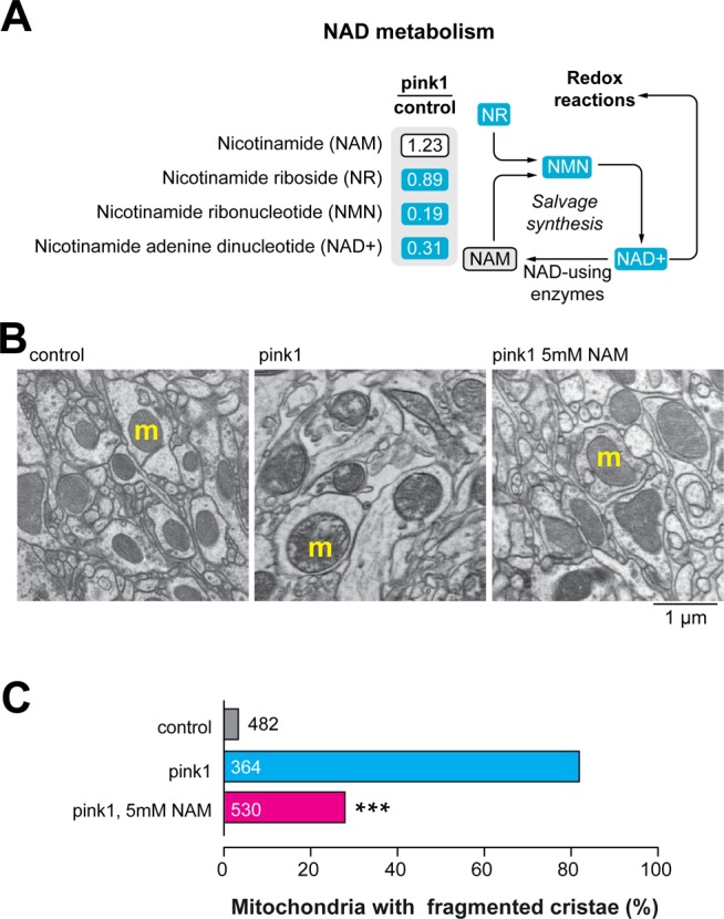
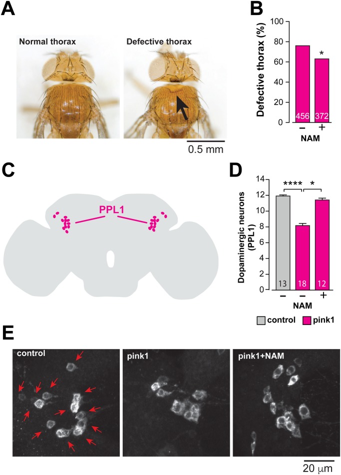
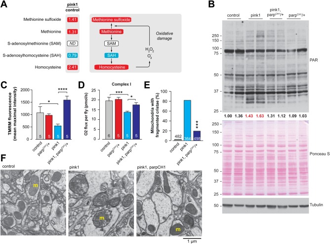
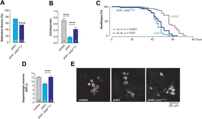
Familial forms of Parkinson's disease (PD) caused by mutations in PINK1 are linked to mitochondrial impairment. Defective mitochondria are also found in Drosophila models of PD with pink1 mutations. The co-enzyme nicotinamide adenine dinucleotide (NAD+) is essential for both generating energy in mitochondria and nuclear DNA repair through NAD+-consuming poly(ADP-ribose) polymerases (PARPs). We found alterations in NAD+ salvage metabolism in Drosophila pink1 mutants and showed that a diet supplemented with the NAD+ precursor nicotinamide rescued mitochondrial defects and protected neurons from degeneration. Additionally, a mutation of Parp improved mitochondrial function and was neuroprotective in the pink1 mutants. We conclude that enhancing the availability of NAD+ by either the use of a diet supplemented with NAD+ precursors or the inhibition of NAD+-dependent enzymes, such as PARPs, which compete with mitochondria for NAD+, is a viable approach to preventing neurotoxicity associated with mitochondrial defects.

Keywords: Drosophila; Mitochondria; NAD+; NAM; Niacin; Nucleotide metabolism; PARP; PINK1; Parkinson's disease.


Conflict of interest statement

Competing interests

The authors declare no competing or financial interests.

Figures

Fig. 1.
Fig. 1.
Dietary supplementation with NAM suppresses mitochondrial defects in pink1 mutant brains. (A) Loss of pink1 decreases NAD+ metabolite levels. Blue corresponds to metabolites that are significantly downregulated (P<0.05) compared to control. Statistical significance was determined using Welch's two-sample t-test (n=8). (B,C) An NAM-supplemented (5 mM) diet suppresses mitochondrial cristae fragmentation in pink1 mutant brains. (B) Ultrastructural analysis of the adult brains of pink1 mutants, showing mitochondria with fragmented cristae in neuropiles (m, mitochondria). Representative TEM micrographs of the indicated genotypes and treatments are shown. (C) Percentages of neuropile mitochondria exhibiting fragmented cristae normalised to the area are presented (asterisks, two-tailed chi-square test, 95% confidence intervals). Datasets labelled control and pink1 are also used in Fig. 3E. Genotypes: control: w1118, pink1: pink1B9.
Fig. 2.
Fig. 2.
An NAM-enhanced diet suppresses neurodegeneration in pink1 mutants. (A,B) Dietary supplementation with NAM (5 mM) rescues the thoracic defects of pink1 mutants. (A) Representative images of normal and defective thorax in pink1 mutants, the arrow points to a thoracic defect. (B) Quantification of the thoracic defects of pink1 mutants fed on a normal or 5 mM NAM-supplemented diet (asterisks, two-tailed chi-square test, 95% confidence intervals). (C-E) An NAM-enhanced diet rescues the loss of dopaminergic neurons in the PPL1 cluster of pink1 mutant flies. (C) Schematic diagram of an adult fly brain in the sagittal orientation, with PPL1 cluster neurons coloured magenta. (D) Quantification of PPL1 cluster neurons (mean± s.d.; asterisks, two-tailed unpaired t-test) and (E) representative images of anti-tyrosine hydroxylase staining showing cell bodies (red arrows) of PPL1 neurons of the indicated genotypes and treatments. Genotypes: control: w1118, pink1: pink1B9.
Fig. 3.
Fig. 3.
Parp mutation rescues mitochondrial function in pink1 mutants. (A) The levels of oxidative stress-related metabolites are increased in pink1 mutants. The metabolites indicated in red or blue are significantly upregulated or downregulated, respectively, compared to control (P<0.05). ND corresponds to a metabolite below detection threshold. The statistical significance for fold-changes was determined using Welch's two-sample t-test (n=8). (B) Protein PARylation is increased in pink1 mutants, and this increase is attenuated in pink1, ParpCH1/+ double mutants. Whole-fly lysates were analysed using the indicated antibodies. Tubulin was used as a loading control. Ponceau S staining was used to assess total protein load. Ratios of signal intensity between total PAR and total protein load (Ponceau S) are presented. Two biological replicates are shown for each genotype. Red text indicates ratios of samples with high level of PARylation. (C) Parp mutation protects against the loss of Δψm in pink1 mutants (mean±s.d.; asterisks, one-way ANOVA with Bonferroni's multiple comparison test). (D) Parp mutation increases complex I-mediated respiration in pink1 mutants (mean±s.d.; asterisks, one-way ANOVA with Bonferroni's multiple comparison test). Datasets in C and D labelled ‘control’ and ‘ParpCH1/+’ have been previously published in Lehmann et al. (2016), as data from these genotypes were obtained as a single experimental set before statistical analysis. (E,F) Parp mutation rescues mitochondrial cristae fragmentation in pink1 mutant brains. (E) Percentages of neuropile mitochondria exhibiting fragmented cristae normalised to the area are presented for the indicated genotypes (asterisks, two-tailed chi-square test, 95% confidence intervals). Datasets labelled control and pink1 are also used in Fig. 1C. (F) Representative TEM micrographs of the indicated genotypes are shown (m, mitochondria). Genotypes: control: w1118, pink1: pink1B9, pink1, parpCH1/+: pink1B9, ParpCH1/+.
Fig. 4.
Fig. 4.
Parp mutation rescues pink1 mutant phenotype. (A) Parp mutation rescues the thoracic defect (asterisks, two-tailed chi-square test, 95% confidence intervals), (B) climbing ability (mean±s.d.; asterisks, two-tailed unpaired t-test) and (C) increases survival of pink1 mutants (n=130 for control, n=114 for pink1, and n=106 for pink1, ParpCH1/+; asterisks, log-rank Mantel-Cox test). (D) Parp mutation rescues the loss of dopaminergic neurons in the PPL1 cluster of pink1 mutant flies (mean±s.e.m.; asterisks, one-way ANOVA with Bonferroni's multiple comparison test). (E) Representative images of anti-tyrosine hydroxylase-stained PPL1 cluster neurons are shown for the indicated genotypes. Genotypes: control: w1118, pink1: pink1B9, pink1, parpCH1/+: pink1B9, ParpCH1/+. Dataset in D labelled ‘control’ has been previously published in Lehmann et al. (2016), as data were obtained as a single experimental set before statistical analysis.

Similar articles

Cited by

References

    1. Bonifati V. (2007). Genetics of parkinsonism. Parkinsonism Relat. Disord. 13 Suppl. 3, S233-S241. 10.1016/S1353-8020(08)70008-7 - DOI - PubMed
    1. Celardo I., Martins L. M. and Gandhi S. (2014). Unravelling mitochondrial pathways to Parkinson's disease. Br. J. Pharmacol. 171, 1943-1957. 10.1111/bph.12433 - DOI - PMC - PubMed
    1. Clark I. E., Dodson M. W., Jiang C., Cao J. H., Huh J. R., Seol J. H., Yoo S. J., Hay B. A. and Guo M. (2006). Drosophila pink1 is required for mitochondrial function and interacts genetically with parkin. Nature 441, 1162-1166. 10.1038/nature04779 - DOI - PubMed
    1. Costa A. C., Loh S. H. Y. and Martins L. M. (2013). Drosophila Trap1 protects against mitochondrial dysfunction in a PINK1/parkin model of Parkinson's disease. Cell Death Dis. 4, e467 10.1038/cddis.2012.205 - DOI - PMC - PubMed
    1. de Castro I. P., Martins L. M. and Tufi R. (2010). Mitochondrial quality control and neurological disease: an emerging connection. Expert Rev. Mol. Med. 12, e12 10.1017/S1462399410001456 - DOI - PMC - PubMed

LinkOut - more resources