. 2017 Feb 15;6(2):187-199.
doi: 10.1242/bio.023218.

A developmental transcriptomic analysis of Pax1 and Pax9 in embryonic intervertebral disc development

Affiliations

A developmental transcriptomic analysis of Pax1 and Pax9 in embryonic intervertebral disc development

V Sivakamasundari et al. Biol Open. .

Abstract

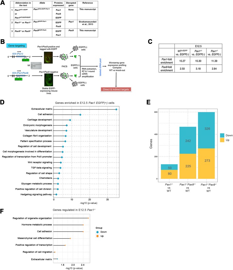
Pax1 and Pax9 play redundant, synergistic functions in the patterning and differentiation of the sclerotomal cells that give rise to the vertebral bodies and intervertebral discs (IVD) of the axial skeleton. They are conserved in mice and humans, whereby mutation/deficiency of human PAX1/PAX9 has been associated with kyphoscoliosis. By combining cell-type-specific transcriptome and ChIP-sequencing data, we identified the roles of Pax1/Pax9 in cell proliferation, cartilage development and collagen fibrillogenesis, which are vital in early IVD morphogenesis. Pax1 is up-regulated in the absence of Pax9, while Pax9 is unaffected by the loss of Pax1/Pax9 We identified the targets compensated by a single- or double-copy of Pax9 They positively regulate many of the cartilage genes known to be regulated by Sox5/Sox6/Sox9 and are connected to Sox5/Sox6 by a negative feedback loop. Pax1/Pax9 are intertwined with BMP and TGF-B pathways and we propose they initiate expression of chondrogenic genes during early IVD differentiation and subsequently become restricted to the outer annulus by the negative feedback mechanism. Our findings highlight how early IVD development is regulated spatio-temporally and have implications for understanding kyphoscoliosis.

Keywords: BMP; Intervertebral disc; Pax1; Pax9; Sox trio; TGF-B.


Conflict of interest statement

Competing interests

The authors declare no competing or financial interests.

Figures

Fig. 1.
Fig. 1.
Experimental workflow and differentially expressed genes in Pax1/Pax9 mutants. (A) Abbreviations of Pax1 and Pax9 EGFP-expressing mouse lines. (B) Diagrammatic representation of experimental workflow. (C) Pax1 and Pax9 fold enrichment in EGFP(+) cells compared to EGFP(−) cells from E12.5 embryos. (D) Bar chart of enriched GO terms in E12.5 Pax1 EGFP(+) cells. (E) Number of differentially expressed genes for the various genotype comparisons. (F) Bar chart of enriched GO terms for down-regulated and up-regulated genes in E12.5 Pax1−/− embryos. GOI, gene of interest; EGFP, enhanced green fluorescent protein; FACS, fluorescence activated cell sorting; GO, gene ontology; WT, wild type.
Fig. 2.
Fig. 2.
Gene ontology and Ingenuity Pathway Analyses (IPA) of differentially expressed genes in Pax1/Pax9 mutants. (A,B) Bar chart of enriched GO terms for down-regulated (A) and up-regulated genes (B) in E12.5 Pax1−/−Pax9−/− embryos. (C) Pax1−/−Pax9−/− embryos exhibited shortened tail phenotype compared to Pax1−/− embryos. Yellow arrows indicate tail tip. Fluorescence was seen in the IVD of E14.5 Pax1−/−Pax9+/+embryos in a regular metameric fashion. In the Pax1−/−Pax9−/− embryos, the fluorescing cells were mis-localized to the sides of the embryo with the notochord exposed in the middle (orange arrow). White arrows indicate Pax1/Pax9 lineage cells expressing EGFP in the IVD, detected by the presence of GFP expression. (D) Number of differentially expressed genes in E12.5 Pax1−/−Pax9−/− associated with the respective disease and disorder terms identified via IPA. (E) Genes differentially expressed in E12.5 Pax1−/−Pax9−/− that are associated with the respective axial skeletal defects, identified via IPA (top) and MGI phenotype (bottom). Genes down-regulated in Pax1−/−Pax9−/− respective to WT are in blue. GO, gene ontology; VC, vertebral column; IVD, intervertebral disc anlagen.
Fig. 3.
Fig. 3.
Gene dosage effect of Pax1/Pax9 on down-stream targets and regulation of cartilage development genes. (A-C) Differentially expressed genes that show: (A) Group1, gradual decrease or increase in expression with increasing loss of Pax1/Pax9 alleles; (B) Group 2, genes that require two copies of Pax9 to maintain their normal expression levels; and (C) Group 3, genes showing a significant change upon the loss of the last copy of Pax9. Mean trend of all genes in each subgroup is depicted as red line for respective groups; individual gene trends are shown as different colored dots. Enriched GO terms for each category indicated on the right of respective graphs. Genes with abnormal skeletal phenotype are shown in gray, of which direct Pax9 targets are in blue for the respective groups. (D) qPCR fold change of Pax1 and Pax9 in E13.5 WT, Pax1−/− and Pax9−/−. (E) Fold enrichment of selected cartilage development genes from microarray analysis of E12.5 Pax1−/−Pax9−/− versus Pax1−/−Pax9+/−. (F) qPCR fold-enrichment of cartilage development genes in E12.5 Pax1−/−Pax9−/− vs. Pax1+/−Pax9−/−. qPCR, quantitative PCR; FC, fold change; * significant FC ≥1.5, Student's t-test; error bars indicate s.e.m.
Fig. 4.
Fig. 4.
Pax9 direct in vivo targets and genes co-regulated by Pax, Sox, Bmp4 and TGF-B. (A) Pax9 binding site distribution in E12.5 WT vertebral column cells. (B) Gene ontology analyses of genes associated with Pax9 binding sites: MGI expression profile and mouse mutant phenotype. (C) De novo Pax9 motif discovered in in vivo Pax9 ChIP-Seq, that resembles the reported 3′ half site of Pax family of transcription factors. (D) Overlap of Pax9 binding associated targets with differentially expressed genes in the Pax1−/−Pax9−/− mutant. Overlap of genes regulated by single or two copies of Pax9 with Pax9 binding associated genes shown on right. (E) Pax9 binding peaks, visualized using IGV, at the TSS of Col2a1, Wwp2, Cbx2 and Hip1. Pax9 motif at the TSS identified via FIMO analysis is shown below as a red bar. (F) Overlap of genes regulated by Pax1/Pax9 and Sox5/Sox6 in the IVD anlagen. (G) Overlap of genes regulated by Pax1/Pax9, TGF-B pathway and Bmp4. TSS, transcriptional start site; IGV, Integrative Genomics Viewer.
Fig. 5.
Fig. 5.
IVD expression of Pax and Sox genes and proposed model. (A) Sagittal section of E13.5 WT vertebral column showing Pax1 expression in the IVD anlagen detected by SISH assay. (B) Mallory's tetrachrome staining of E15.5 WT vertebral column; IAF is stained blue and OAF is stained dark red. (C,D) Immunohistochemistry and Alcian blue staining of vertebral column showing (C) Pax1 and (D) Pax9 protein expression restricted to the OAF at E15.5. (E-I) Expression of Pax1, Pax9, Smad3, Tgfb3 and Bmp4 in the IVD anlagen at E12.5. (J-M) Expression of Pax1, Pax9, Sox5 and Sox9 in the IVD anlagen at E13.5. (N-R) Expression of Pax1, Pax9, Sox9 at E15.5 and Tgfb3 and Bmp4 at E14.5 in the IVD. (S) Proposed negative feedback loop mechanism between Pax, Sox, Bmp4 and TGF-B pathway at E12.5. Black arrows indicate the final site of expression at E14.5. Brackets indicate IVD region. VB, vertebral body; n, notochord; IVD, intervertebral disc; NP, nucleus pulposus; IAF, inner annulus fibrosus; OAF, outer annulus fibrosus; WT, wild type; SISH, sectioned in situ hybridization; D, dorsal; V, ventral.

Similar articles

Cited by

References

    1. Akiyama H., Chaboissier M.-C., Martin J. F., Schedl A. and de Crombrugghe B. (2002). The transcription factor Sox9 has essential roles in successive steps of the chondrocyte differentiation pathway and is required for expression of Sox5 and Sox6. Genes Dev. 16, 2813-2828. 10.1101/gad.1017802 - DOI - PMC - PubMed
    1. Aszódi A., Chan D., Hunziker E., Bateman J. F. and Fässler R. (1998). Collagen II is essential for the removal of the notochord and the formation of intervertebral discs. J. Cell Biol. 143, 1399-1412. 10.1083/jcb.143.5.1399 - DOI - PMC - PubMed
    1. Baffi M. O., Slattery E., Sohn P., Moses H. L., Chytil A. and Serra R. (2004). Conditional deletion of the TGF-beta type II receptor in Col2a expressing cells results in defects in the axial skeleton without alterations in chondrocyte differentiation or embryonic development of long bones. Dev. Biol. 276, 124-142. 10.1016/j.ydbio.2004.08.027 - DOI - PubMed
    1. Baffi M. O., Moran M. A. and Serra R. (2006). Tgfbr2 regulates the maintenance of boundaries in the axial skeleton. Dev. Biol. 296, 363-374. 10.1016/j.ydbio.2006.06.002 - DOI - PMC - PubMed
    1. Bailey T. L. and Machanick P. (2012). Inferring direct DNA binding from ChIP-seq. Nucleic Acids Res. 40, e128 10.1093/nar/gks433 - DOI - PMC - PubMed