. 2017 Feb;173(2):1164-1176.
doi: 10.1104/pp.16.01840. Epub 2016 Dec 23.

The Pentatricopeptide Repeat Protein EMB2654 Is Essential for Trans-Splicing of a Chloroplast Small Ribosomal Subunit Transcript

Affiliations

The Pentatricopeptide Repeat Protein EMB2654 Is Essential for Trans-Splicing of a Chloroplast Small Ribosomal Subunit Transcript

Nader Aryamanesh et al. Plant Physiol. 2017 Feb.

Abstract

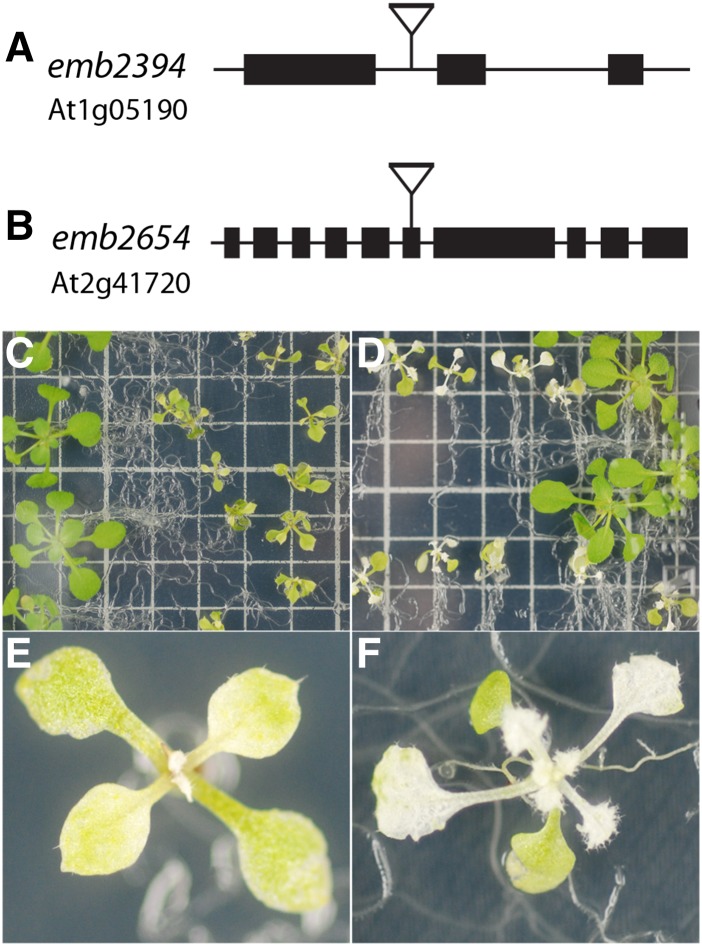
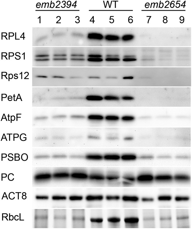
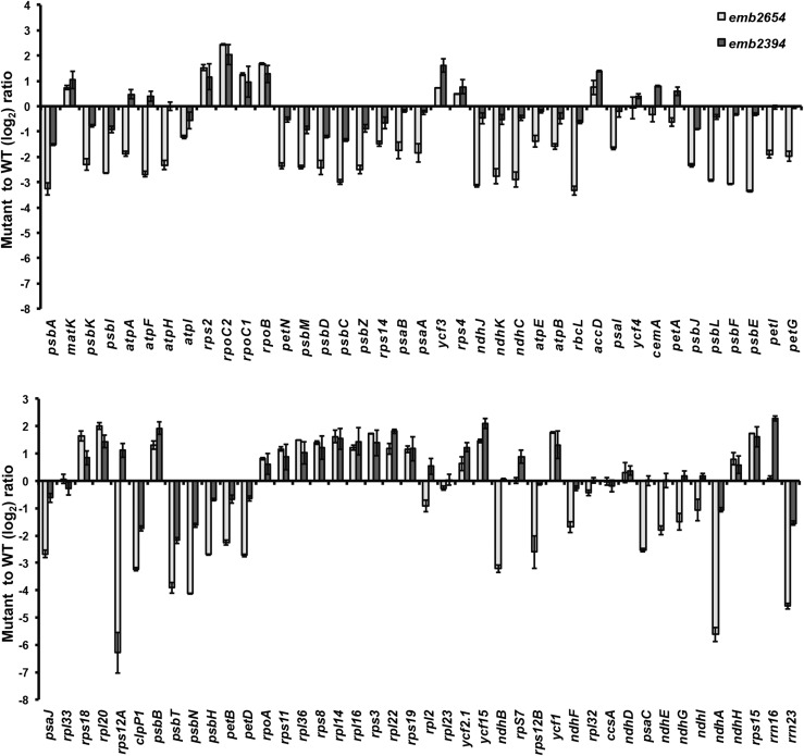
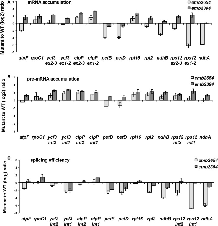
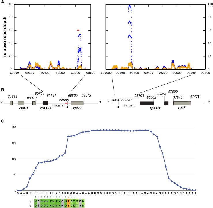
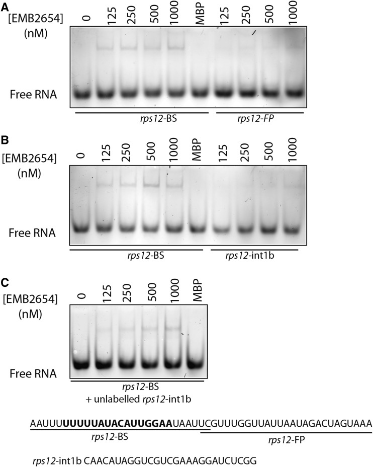
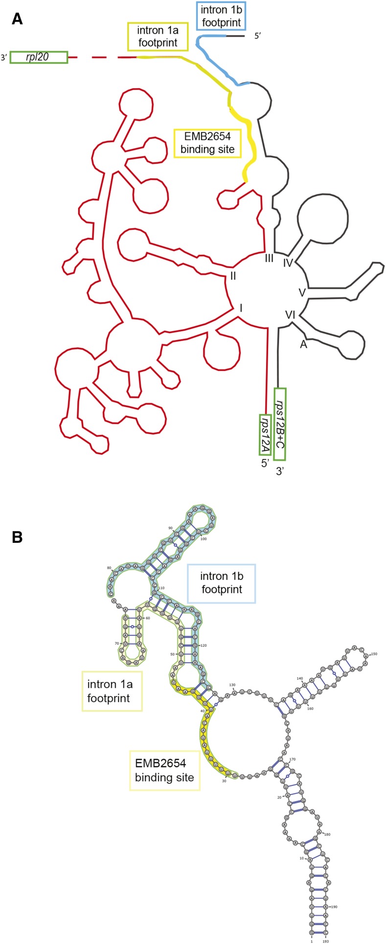
We report the partial complementation and subsequent comparative molecular analysis of two nonviable mutants impaired in chloroplast translation, one (emb2394) lacking the RPL6 protein, and the other (emb2654) carrying a mutation in a gene encoding a P-class pentatricopeptide repeat protein. We show that EMB2654 is required for the trans-splicing of the plastid rps12 transcript and that therefore the emb2654 mutant lacks Rps12 protein and fails to assemble the small subunit of the plastid ribosome, explaining the loss of plastid translation and consequent embryo-lethal phenotype. Predictions of the EMB2654 binding site match a small RNA "footprint" located on the 5' half of the trans-spliced intron that is almost absent in the partially complemented mutant. EMB2654 binds sequence specifically to this target sequence in vitro. Altered patterns in nuclease-protected small RNA fragments in emb2654 show that EMB2654 binding must be an early step in, or prior to, the formation of a large protein-RNA complex covering the free ends of the two rps12 intron halves.


Figures

Figure 1.
Figure 1.
Gene models and phenotypes of the partially complemented mutants. A and B, The positions of the T-DNA insertions in emb2394 (A) and emb2654 (B) are shown. C to F, The emb2394 (C and E) and emb2654 (D and F) mutants expressing their respective wild-type proteins under control of the seed-specific ABI3 promoter. Large green seedlings in each case are wild-type siblings. Both mutant lines display yellowish green cotyledons and yellow to white leaves with increasingly severe phenotypes until development ceases. Seedlings were grown on half-strength Gamborg B5 medium. Squares on the grid are 1 × 1 cm.
Figure 2.
Figure 2.
Western-blot analysis of chloroplast proteins in emb2654 and emb2394. Immunoblots of total proteins from partially complemented seedlings were probed using antibodies raised against various plastid proteins. Several acrylamide gels (three biological replicates for each genotype) were run with identical loadings and blotted onto PVDF membranes. RPL4, RPS1, ATPG (subunit of the ATP synthase), PSBO (subunit of photosystem II), and PC (plastocyanin), are encoded by the nuclear genome. Rps12, PetA (subunit of the cytochrome b6/f), AtpF, and RbcL are encoded by the plastid genome. Due to very different RbcL quantities between wild type (WT) and mutants (RbcL bands are visualized on a stain-free acrylamide gel), the loading was adjusted by comparing nonvarying background bands on gels and ACT8 (actin) was used as a nonplastid control.
Figure 3.
Figure 3.
Plastid transcript levels in emb2654 and emb2394 seedlings. Genome-wide qRT-PCR was performed on chloroplast transcripts from the partially complemented seedlings from both lines (measurements shown here as log2 ratios of gene expression in mutant samples compared to that of phenotypically normal siblings grown in parallel). Both lines display a general accumulation of transcripts related to transcription and translation (transcribed by nuclear-encoded phage-type RNA polymerase), with the noticeable exception of rps12A in emb2654, and a general decrease of the transcripts encoding subunits of the photosynthetic apparatus (transcribed by PEP). The values are means of two biological replicates (bars indicate se).
Figure 4.
Figure 4.
RT-qPCR analysis of intron-containing plastid transcripts in emb2654 and emb2394 seedlings. Transcript levels are compared to phenotypically normal siblings (wild type [WT]) grown in parallel. RT-qPCR was carried out using two sets of primers: One set was designed to specifically amplify spliced RNA (A) and the other to specifically amplify unspliced RNA (B). C, Splicing efficiency as the log2 ratio of spliced to unspliced transcripts in the mutants compared to the wild type. The values are means of three biological replicates for emb2654 and two replicates for emb2394 (bars indicate se).
Figure 5.
Figure 5.
RNA-seq analysis of putative “footprints” in the rps12 intron halves. A, RNA-seq was performed on gel-purified 15- to 50-nucleotide RNA fragments from partially complemented emb2654 seedlings (orange) and wild-type siblings (blue). The plots indicate the relative read depth at each nucleotide in the rps12 intron halves. Read depth has been normalized to the average depth across each intron (excluding the region of the putative footprint in each case). Data from three biological replicates are shown. The predicted EMB2654 binding site is shown by a red bar. B, Arrangement of the rps12 genes on Arabidopsis chloroplast DNA. The first exon is in rps12A, which is in the same transcription unit as rpl20 and clpP1. The second and third exons are in rps12B, located approximately 30 kb away and cotranscribed with ndhB and rps7. The position of the predicted EMB2654 binding site is indicated by a red bar, and RNA-seq footprints by black stars. Genome coordinates are indicated. C, Alignment of EMB2654 to its predicted binding site in rps12 intron 1a. The amino acids at the fifth and last positions in each PPR motif are aligned with the RNA sequence. Combinations that correlate with the aligned base (Barkan et al., 2012) are shaded in dark green. Combinations where only the fifth residue correlates with the aligned nucleotide are shaded in light green, combinations of unknown affinity are shaded in gray, and combinations that significantly anticorrelate with the aligned nucleotide are shaded in orange. The blue trace indicates the mean read depth observed in the RNA-seq analysis for wild-type samples in this region, showing the shape and extent of the footprint.
Figure 6.
Figure 6.
Binding of EMB2654 to sequences within rps12 introns 1a and 1b. REMSA was performed with recombinant EMB2654 protein and RNA oligonucleotides labeled with fluorescein. The rps12-BS probe includes the predicted EMB2654 binding site, while the rps12-int1b probe contains the intron 1b footprint. Numbers above the images refer to the concentration of EMB2654-MBP fusion (nm), except for the lane MBP, in which 1,000 nm of maltose binding protein was used as a control. A and B, No competitor present. C, Competition experiment: EMB2654 binding to rps12-BS in the presence of 10 nm unlabeled rps12-intb probe. The gels were scanned at 488 nm (excitation wavelengths for fluorescein) detected through a 520-nm band-pass filter. EMB2654 shows detectable binding to the rps12-BS probe from 125 nm upwards, but no significant binding to the rps12-FP and rps12-int1b probe.
Figure 7.
Figure 7.
Predicted structure of the rps12 intron. A, Sketch of the Arabidopsis rps12 intron, using as a model the structure proposed for the tobacco intron by Kohchi et al. (1988). The Arabidopsis and tobacco sequences are aligned in Supplemental Figure S1. Both the intron 1a footprint (greenish yellow), comprising the EMB2654 binding site (yellow) and intron 1b footprint (blue) are marked. B, Predicted secondary structure of rps12 intron 1 domain III showing the potential interactions between the two intron footprints. This prediction was obtained using RNAcofold (Lorenz et al., 2011) and drawn using VARNA (http://varna.lri.fr/).

References

    1. Asakura Y, Barkan A (2006) Arabidopsis orthologs of maize chloroplast splicing factors promote splicing of orthologous and species-specific group II introns. Plant Physiol 142: 1656–1663 - PMC - PubMed
    1. Asakura Y, Bayraktar OA, Barkan A (2008) Two CRM protein subfamilies cooperate in the splicing of group IIB introns in chloroplasts. RNA 14: 2319–2332 - PMC - PubMed
    1. Babiychuk E, Vandepoele K, Wissing J, Garcia-Diaz M, De Rycke R, Akbari H, Joubès J, Beeckman T, Jänsch L, Frentzen M, et al. (2011) Plastid gene expression and plant development require a plastidic protein of the mitochondrial transcription termination factor family. Proc Natl Acad Sci USA 108: 6674–6679 - PMC - PubMed
    1. Barkan A. (1993) Nuclear mutants of maize with defects in chloroplast polysome assembly have altered chloroplast RNA metabolism. Plant Cell 5: 389–402 - PMC - PubMed
    1. Barkan A. (2004). Intron splicing in plant organelles. In Daniell H., Chase C.D., eds., Molecular Biology and Biotechnology of Plant Organelles. Springer, Dordrecht, The Netherlands, pp 295–322.

Publication types

MeSH terms

Substances

LinkOut - more resources