. 2017 Feb 10;292(6):2531-2541.
doi: 10.1074/jbc.M116.760256. Epub 2016 Dec 23.

Phosphatidylserine Stimulates Ceramide 1-Phosphate (C1P) Intermembrane Transfer by C1P Transfer Proteins

Affiliations

Phosphatidylserine Stimulates Ceramide 1-Phosphate (C1P) Intermembrane Transfer by C1P Transfer Proteins

Xiuhong Zhai et al. J Biol Chem. .

Abstract

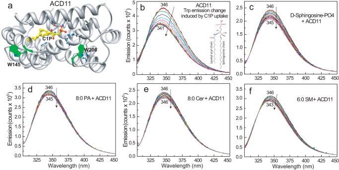
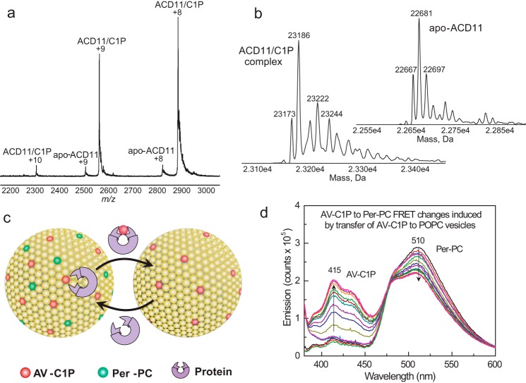
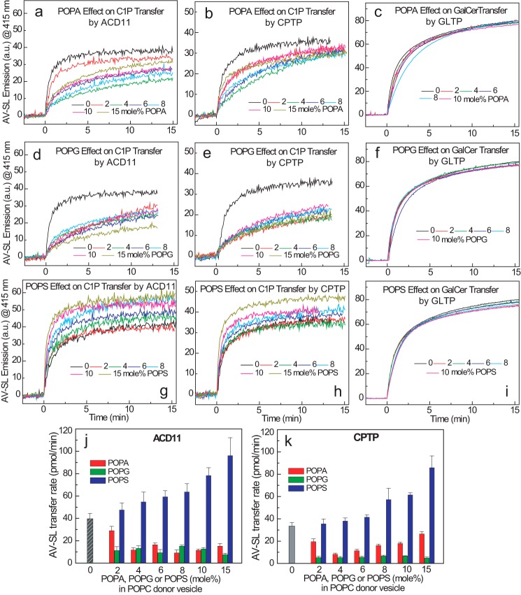
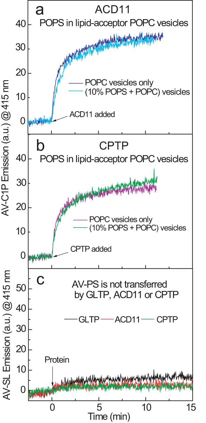
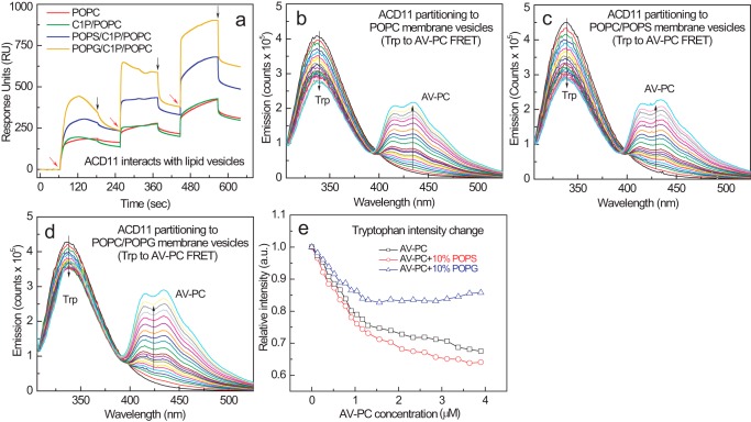
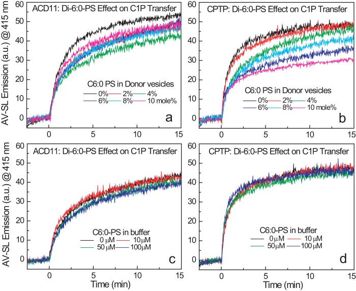
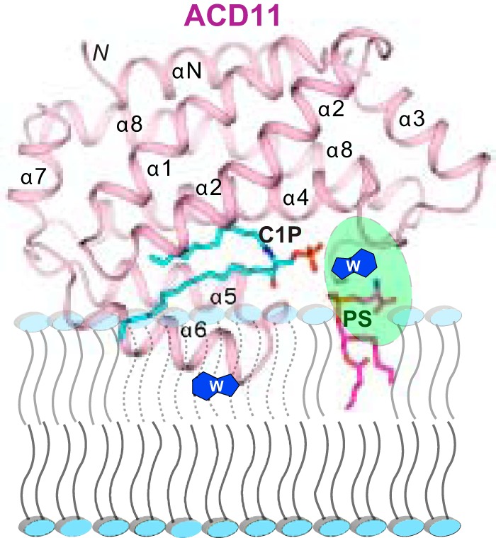
Genetic models for studying localized cell suicide that halt the spread of pathogen infection and immune response activation in plants include Arabidopsis accelerated-cell-death 11 mutant (acd11). In this mutant, sphingolipid homeostasis is disrupted via depletion of ACD11, a lipid transfer protein that is specific for ceramide 1-phosphate (C1P) and phyto-C1P. The C1P binding site in ACD11 and in human ceramide-1-phosphate transfer protein (CPTP) is surrounded by cationic residues. Here, we investigated the functional regulation of ACD11 and CPTP by anionic phosphoglycerides and found that 1-palmitoyl-2-oleoyl-phosphatidic acid or 1-palmitoyl-2-oleoyl-phosphatidylglycerol (≤15 mol %) in C1P source vesicles depressed C1P intermembrane transfer. By contrast, replacement with 1-palmitoyl-2-oleoyl-phosphatidylserine stimulated C1P transfer by ACD11 and CPTP. Notably, "soluble" phosphatidylserine (dihexanoyl-phosphatidylserine) failed to stimulate C1P transfer. Also, none of the anionic phosphoglycerides affected transfer action by human glycolipid lipid transfer protein (GLTP), which is glycolipid-specific and has few cationic residues near its glycolipid binding site. These findings provide the first evidence for a potential phosphoglyceride headgroup-specific regulatory interaction site(s) existing on the surface of any GLTP-fold and delineate new differences between GLTP superfamily members that are specific for C1P versus glycolipid.

Keywords: Arabidopsis thaliana; lipid trafficking; lipid-protein interaction; membrane biophysics; phosphatidic acid; phosphatidylglycerol; phosphatidylserine; protein-lipid interaction; sphingolipid.


Conflict of interest statement

The authors declare that they have no conflicts of interest with the contents of this article

Figures

FIGURE 1.
FIGURE 1.
C1P binding to ACD11 in solution. a, structure of ACD11 (13) showing the location of intrinsic Trp (W145 and W206) with respect to bound C1P (Protein Data Bank entry 4NTI). b, ACD11 Trp emission change induced by C1P uptake. N-Octanoyl-C1P was added stepwise (C1P step concentration = 0.08 μm in EtOH (1 μl)) to ACD11 (1 μm) stirring in buffer (sodium phosphate (pH 6.6) containing 150 mm NaCl) with 5-min incubation times between injections. The vertical arrow indicates response to increasing C1P concentration. Increments beyond the sixth addition (∼0.48 μm C1P) induce little change in Trp emission intensity or wavelength maximum (λmax blue shift), yielding a saturable binding response. c, same as b, but lipid titrant is sphingosine 1-phosphate; d, same as b, but lipid titrant is phosphatidic acid; e, same as b, but lipid titrant is ceramide; f, same as b, but lipid titrant is sphingomyelin.
FIGURE 2.
FIGURE 2.
Surface electrostatics surrounding the sphingolipid liganding site in ACD11, CPTP, and GLTP and sphingolipid intermembrane transfer measurement using a FRET-based assay. a, ESI-MS analysis of ACD11·C1P complex. Direct infusion under nondenaturing conditions results in two main positive charge states (+8 and +9) for the ACD11·N-octanoyl C1P complex. Traces of apo-ACD11 also are evident for the +8 and +9 charge states. The m/z label (x axis) represents mass divided by charge number. b, the transformed spectra indicate molecular masses of 22,681 Da for apo-ACD11 and 23,186 Da for the ACD11·N-octanoyl C1P complex, confirming a single binding site for C1P. c, schematic for in vitro measurement of SL intermembrane transfer via FRET loss. In the SL source vesicle, the AV-SL fluorescence signal (energy donor; red dots) is low due to FRET involving perylenoyl-PC (energy acceptor; green dots; nontransferable lipid). Loss of FRET occurs when AV-SL is removed by protein (catalytic amount) and transferred to the excess (10-fold) POPC receiver vesicles, resulting in a time-dependent increase in AV-SL emission signal. d, spectral emission changes for AV-SL/Per-PC energy transfer pair showing FRET loss induced by transfer protein activity. AV-C1P and Per-PC are at 1 mol % in SL source vesicles. Black, red, and green traces, successive scans of SL source vesicles (2.5-min intervals) showing signal stability; blue, cyan, and magenta traces, successive scans (2.5-min intervals) after the addition of excess POPC receiver vesicles; other spectral traces show the time-dependent emission changes in AV and Per (2-min intervals) after injection of transfer protein (2 μg).
FIGURE 3.
FIGURE 3.
Anionic phosphoglycerides exert differing effects on sphingolipid transfer by ACD11, CPTP, or GLTP. In each panel, traces represent AV-SL emission intensity measured at 415 nm as a function of time resulting from the loss of AV-SL/Per-PC FRET as AV-SL is transferred to POPC vesicles. Shown are POPA effects (0–15 mol %) (a–c) and POPG effects (0–15 mol %) (d–f) on ACD11, CPTP, and GLTP (2 μg), respectively. Per-PC (nontransferable) along with AV-C1P or AV-GalCer is present at 1 mol % in the SL source vesicles at time 0. g–i, POPS effects (0–15 mol %) on ACD11, CPTP, and GLTP (2 μg), respectively. Per-PC along with AV-C1P or AV-GalCer is present at 1 mol % in the SL source vesicles at time 0. j and k summarize the C1P transfer rate changes of ACD11 and CPTP induced by POPS, POPA, and POPG. The C1P transfer rates are expressed as pmol/min transferred from SL source to POPC vesicles as a function of different amounts of anionic phosphoglycerides (0–15 mol %) present in the SL source vesicles for ACD11 (a) and CPTP (b). Red, POPA; green, POPG; blue, POPS; gray, control. Error bars, S.D.
FIGURE 4.
FIGURE 4.
POPS in acceptor vesicles does not stimulate C1P transfer and POPS itself is not transferred. Traces for a and b represent AV-C1P emission intensity measured at 415 nm as a function of time resulting from the loss of AV-SL/Per-PC FRET as AV-SL is transferred to POPC vesicles. The presence of POPS (10 mol %) in POPC receiver (acceptor) vesicles (10-fold excess) does not stimulate AV-C1P transfer by ACD11 (a) or CPTP (b). c, traces represent AV-PS emission intensity measured at 415 nm as a function of time resulting from the loss of AV-PS/Per-PC FRET. The AV-PS transfer to POPC receiver (acceptor) vesicles (10-fold excess) by ACD11, CPTP, and GLTP is virtually nil.
FIGURE 5.
FIGURE 5.
ACD11 partitioning to POPC vesicles containing POPS or POPG. a, surface plasmon resonance assessments of ACD11 adsorption and desorption membrane vesicles of differing lipid composition. POPC (red trace), POPC/C1P (95:5; green trace), POPC/C1P/POPS (80:5:15; blue trace), and POPC/C1P/POPG (80:5:15; orange trace) vesicles were prepared and adsorbed to a Sensor Chip L1 as described under “Experimental Procedures.” ACD11 injections of 46, 144, and 433 nmol into the 2 μl/min flow stream are indicated by red arrows; switches to buffer wash are shown by black arrows. b–d, FRET involving ACD11 intrinsic Trp (energy donor) and AV-PC (energy acceptor) in POPC vesicles enabled assessment of ACD11 partitioning to the membrane surface. ACD11 (0.5 μm) was titrated stepwise with POPC vesicles containing 2 mol % AV-PC to produce 0.13 μm incremental increases. b, control POPC vesicles lacking anionic phosphoglyceride; c, POPC vesicles containing 10 mol % POPS; d, POPC vesicles containing 10 mol % POPG; e, ACD11 partitioning curves to membrane vesicles consisting of POPC, POPC + POPS, or POPC + POPG.
FIGURE 6.
FIGURE 6.
Soluble PS effects on AV-C1P transfer by ACD11 and CPTP. Traces in each panel represent AV-SL emission intensity measured at 415 nm as a function of time resulting from loss of AV-SL/Per-PC FRET as AV-SL is transferred to lipid acceptor POPC vesicles. a and b, effect of dihexanoyl-PS (0 to 10 mol %) on AV-C1P transfer by ACD11 and CPTP (2 μg; ∼0.1 nmol), respectively, when the dihexanoyl-PS is included as a lipid-donor vesicle component. c and d, effect of dihexanoyl-PS on AV-C1P transfer by ACD11 and CPTP (2 μg; ∼0.1 nmol), respectively, when the dihexanoyl-PS is included in the buffer (2.5-ml volume) at the indicated concentrations. The critical micelle concentration of dihexanoyl-PS is ∼700 μm.
FIGURE 7.
FIGURE 7.
Model for putative PS-induced enhancement of ACD11. The model shows how the PS acyl chains remain embedded in the membrane while the PS headgroup (magenta) interacts with a putative surface site in ACD11 (ribbon representation) to help orient the C1P binding site to improve uptake of the C1P binding site and the membrane interaction region of ACD11.

Similar articles

Cited by

References

    1. Hannun Y. A., and Obeid L. M. (2008) Principles of bioactive lipid signalling: lessons from sphingolipids. Nat. Rev. Mol. Cell Biol. 9, 139–150 - PubMed
    1. Berkey R., Bendigeri D., and Xiao S. (2012) Sphingolipids and plant defense/disease: the “death” connection and beyond. Front. Plant Sci. 3, 68. - PMC - PubMed
    1. Hla T., and Dannenberg A. J. (2012) Sphingolipid signaling in metabolic disorders. Cell Metab. 16, 420–434 - PMC - PubMed
    1. Maceyka M., and Spiegel S. (2014) Sphingolipid metabolites in inflammatory disease. Nature 510, 58–67 - PMC - PubMed
    1. D'Angelo G., Polishchuk E., Di Tullio G., Santoro M., Di Campli A., Godi A., West G., Bielawski J., Chuang C.-C., van der Spoel A. C., Platt F. M., Hannun Y. A., Polishchuk R., Mattjus P., and De Matteis M. A. (2007) Glycosphingolipid synthesis requires FAPP2 transfer of glucosylceramide. Nature 449, 62–67 - PubMed

MeSH terms

Associated data