. 2017 Jan;29(1):54-69.
doi: 10.1105/tpc.16.00623. Epub 2016 Dec 23.

WIND1 Promotes Shoot Regeneration through Transcriptional Activation of ENHANCER OF SHOOT REGENERATION1 in Arabidopsis

Affiliations

WIND1 Promotes Shoot Regeneration through Transcriptional Activation of ENHANCER OF SHOOT REGENERATION1 in Arabidopsis

Akira Iwase et al. Plant Cell. 2017 Jan.

Abstract

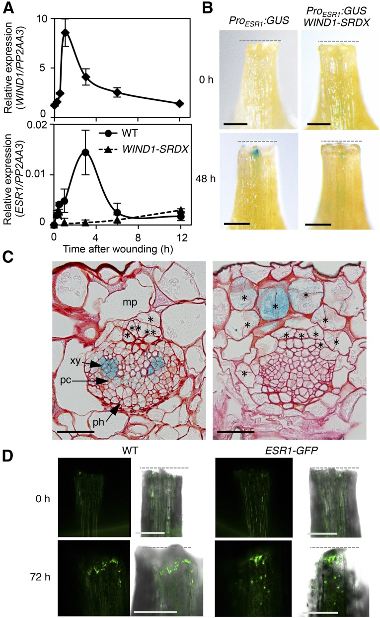
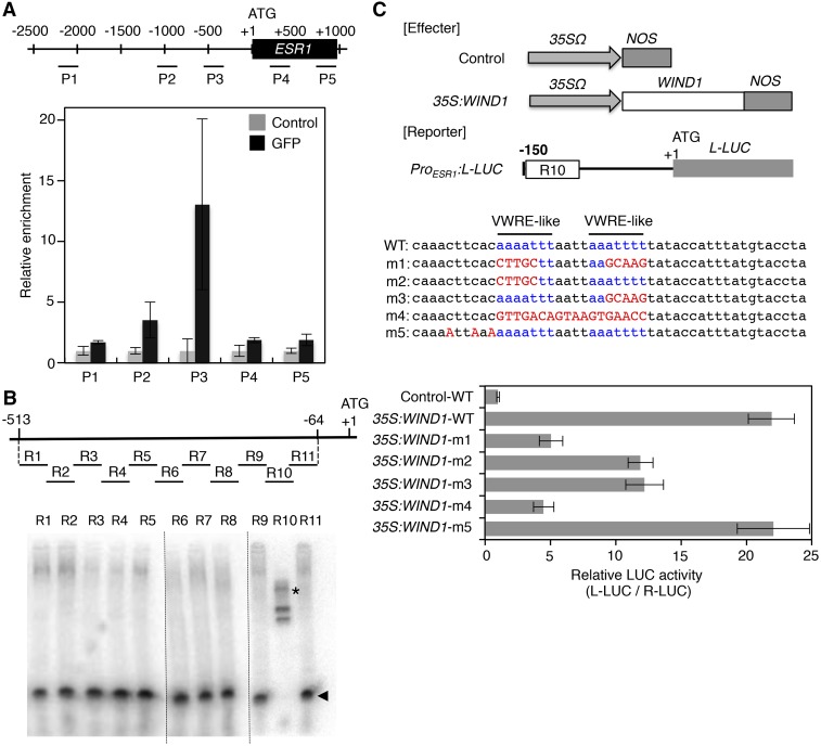
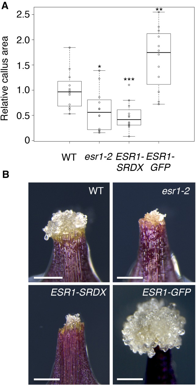
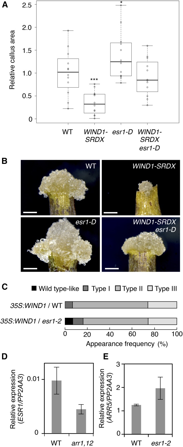
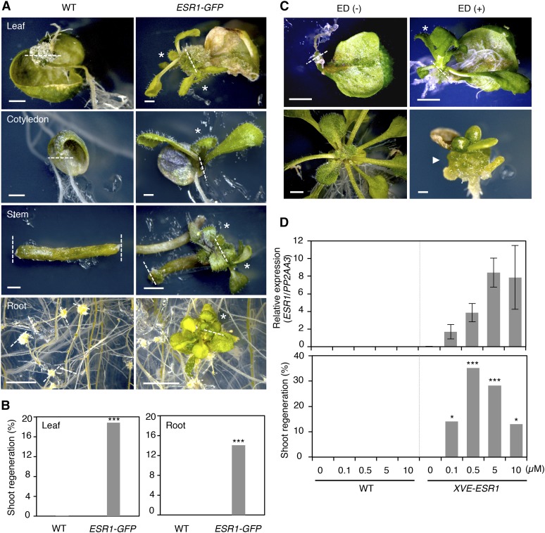
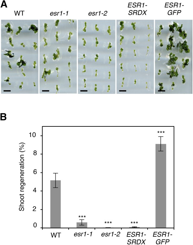
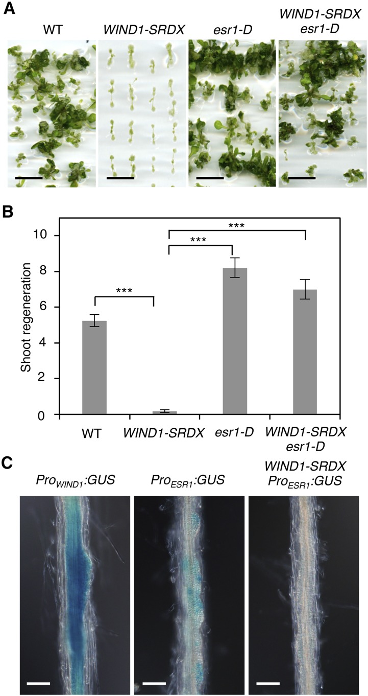
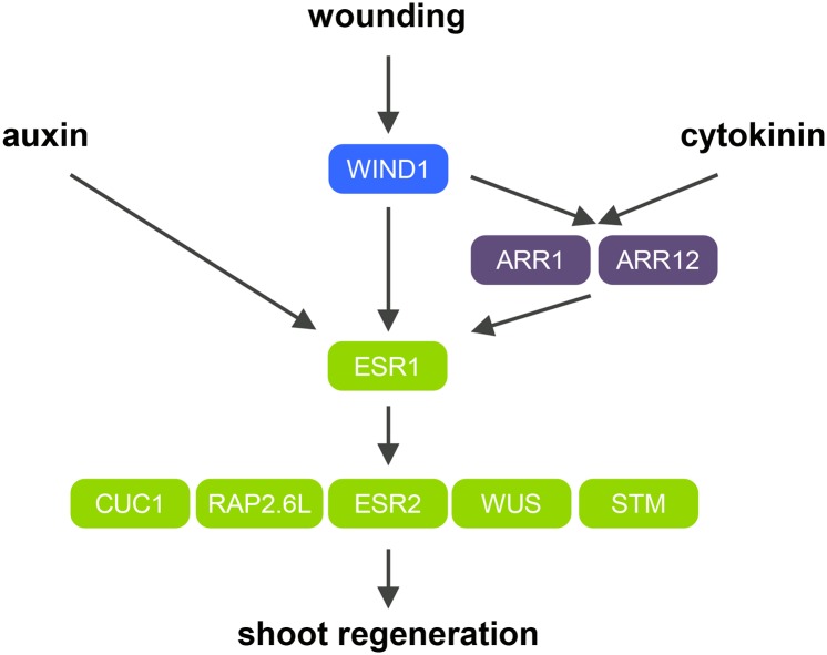
Many plant species display remarkable developmental plasticity and regenerate new organs after injury. Local signals produced by wounding are thought to trigger organ regeneration but molecular mechanisms underlying this control remain largely unknown. We previously identified an AP2/ERF transcription factor WOUND INDUCED DEDIFFERENTIATION1 (WIND1) as a central regulator of wound-induced cellular reprogramming in plants. In this study, we demonstrate that WIND1 promotes callus formation and shoot regeneration by upregulating the expression of the ENHANCER OF SHOOT REGENERATION1 (ESR1) gene, which encodes another AP2/ERF transcription factor in Arabidopsis thaliana The esr1 mutants are defective in callus formation and shoot regeneration; conversely, its overexpression promotes both of these processes, indicating that ESR1 functions as a critical driver of cellular reprogramming. Our data show that WIND1 directly binds the vascular system-specific and wound-responsive cis-element-like motifs within the ESR1 promoter and activates its expression. The expression of ESR1 is strongly reduced in WIND1-SRDX dominant repressors, and ectopic overexpression of ESR1 bypasses defects in callus formation and shoot regeneration in WIND1-SRDX plants, supporting the notion that ESR1 acts downstream of WIND1. Together, our findings uncover a key molecular pathway that links wound signaling to shoot regeneration in plants.


Figures

Figure 1.
Figure 1.
Wounding Activates ESR1 Expression in a WIND1-Dependent Manner. (A) RT-qPCR analysis of WIND1 (upper panel) and ESR1 (lower panel) expression after wounding. First and second leaves of 14-d-old wild-type seedlings were cut and leaf explants were cultured on phytohormone-free MS medium. WIND1 expression peaks at 1 h after wounding, and ESR1 expression peaks at 3 h. The induction of ESR1 is strongly suppressed in ProWIND1:WIND-SRDX (WIND1-SRDX) plants. Expression levels are normalized against those of PP2AA3. Data are mean ± se (n = 3, biological replicates). (B) Induction of the ESR1 promoter activity at the wound sites of leaf petioles. Leaf explants of ProESR1:GUS and ProESR1:GUS WIND1-SRDX plants were cultured on MS medium. ESR1 activation is compromised in ProESR1:GUS WIND1-SRDX plants. Dashed lines mark wound sites. Representative images of petioles at 0 and 48 h after wounding are shown. (C) Transverse section of ProESR1:GUS petioles close to wound sites at 48 h after wounding. GUS staining is found in several cell types, xylem parenchyma cells, procambium cells (left panel), and mesophyll cells (right panel) that have started to undergo cell division. Sections were counterstained by safranin O. Asterisks mark new division planes. mp, mesophyll cells; xy, xylem cells; pc, procambium cells; ph, phloem cells. (D) Nuclear accumulation of ESR1-GFP fusion proteins within the epidermal cells near wound sites at 72 h after wounding. Note that wound stress produces strong green autofluorescence in both wild-type and ProESR1:ESR1-GFP (ESR1-GFP) plants, but these signals are found mostly at the cut edge or cytoplasm. Bars = 300 µm in (B) and (D) and 50 µm in (C).
Figure 2.
Figure 2.
WIND1 Directly Binds the ESR1 Promoter and Activates Its Expression. (A) Chromatin immunoprecipitation of WIND1-GFP fusion proteins on the ESR1 locus. Quantitative PCR analysis, using P1-P5 primers designed within the promoter and coding sequence of ESR1, shows the strongest enrichment of WIND1-GFP using P3 primers designed around −500 bp upstream of the translational start site. The black box represents the coding sequence of ESR1 and +1 ATG indicates the translational start site. Black lines mark the relative distance from the translational start site. Data are normalized against input DNA and shown as a relative enrichment of DNA immunoprecipitated with rabbit serum (control). Data are mean ± se (n = 3, technical replicates). (B) EMSAs of MBP-WIND1-His6 protein’s binding to the ESR1 promoter in vitro. Upper panel shows the position of ∼50-bp DNA probes, designated as R1 to R11, that cover −513 to −64-bp nucleotides of the ESR1 promoter. Arrowheads and asterisks show free and shifted DNA probes, respectively. Dashed lines separate results obtained in three different experiments. Note that the band shifts only with the R10 probe, indicating that MBP-WIND1-His6 binds −153 to −104 bp upstream of the translational start site. (C) WIND1-induced transient activation of the ESR1 expression in Arabidopsis culture cells. Upper panel shows the effector constructs, control and 35S:WIND1, and the reporter construct, ProESR1:L-LUC. For the effector constructs, gray arrows mark 35SΩ, the cauliflower mosaic virus 35S promoter with the tobacco mosaic virus omega translation amplification sequence, and gray boxes mark NOS, the Agrobacterium tumefaciens nopaline synthase transcriptional terminator. The white box marks the WIND1 coding sequence. For the reporter construct, the black bar represents the 150-bp promoter sequence of ESR1 with the R10 sequence marked with a white box. The gray box represents the coding sequence of L-LUC, encoding a firefly luciferase gene, and +1 ATG indicates the translational start site. The middle panel shows the wild-type R10 sequence with two VWRE-like motifs marked in blue and m1 to m5 mutations marked in red. Bottom panel shows the ESR1 promoter activity as judged by the L-LUC activity relative to R-LUC, Renilla luciferase. Cobombardment of 35S:WIND1 and ProESR1:L-LUC activates the ESR1 promoter. Note that abolishing each of the two VWRE-like motifs by m2 or m3 mutation results in reduced ESR1 induction and abolishing both motifs by m1 or m4 mutation has additive effects, indicating that both motifs are required for activation of ESR1 by WIND1. Data are mean ± se (n = 6, technical replicates).
Figure 3.
Figure 3.
ESR1 Promotes Callus Formation at Wound Sites. (A) Callus formation at wound sites of wild-type, esr1-2, ProESR1:ESR1-SRDX (ESR1-SRDX), and ESR1-GFP leaf explants. Leaf explants were cultured on phytohormone-free MS medium, and callus phenotypes were scored at 8 d after wounding. Box plots represent the distribution of projected callus area (n = 12 per genotype). Statistical significance against the wild type was determined by a Student's t test (***P < 0.001, **P < 0.01, and *P < 0.1). (B) Callus generated at wound sites of wild-type, esr1-2, ESR1-SRDX, and ESR1-GFP leaf explants. Representative images at 8 d after wounding are shown. Bars = 500 µm.
Figure 4.
Figure 4.
ESR1 Functions Downstream of WIND1 in Wound-Induced Callus Formation. (A) Callus formation at wound sites of wild-type, ProWIND1:WIND1-SRDX (WIND1-SRDX), esr1-D, and WIND1-SRDX esr1-D leaf explants. Leaf explants were cultured on phytohormone-free MS medium, and callus phenotypes were scored at 8 d after wounding. Box plots represent the distribution of projected callus area (n = 12 per genotype). Statistical significance against wild-type was determined by a Student's t test (***P < 0.001 and *P < 0.1). (B) Callus generated at wound sites of wild-type, WIND1-SRDX, esr1-D, and WIND1-SRDX esr1-D leaf explants. Note that ectopic induction of ESR1 rescues the callus formation deficiency in WIND1-SRDX explants. Representative images at 8 d after wounding are shown. Bars = 250 µm. (C) The esr1-2 mutation partly suppresses WIND1-induced callus formation in T1 seedlings grown on MS medium. Phenotypic severity was scored according to Figure 2 in Iwase et al. (2011a). T1 plants showing weak, intermediate, and strong callus formation are classified as type I, type II, and type III plants, respectively (n = 104 for the wild type; n = 43 for esr1-2). (D) The arr1 arr12 mutation partially suppresses the ESR1 expression after wounding. (E) The esr1-2 mutation does not suppress the ARR5 expression after wounding. First and second leaves of 14-d-old wild-type, arr1 arr12, and esr1-2 seedlings were cut and leaf explants were cultured on phytohormone-free MS medium. Expression levels are normalized against those of the PP2AA3 gene. Data are mean ± se (n = 3, biological replicates).
Figure 5.
Figure 5.
ESR1 Promotes Shoot Regeneration at Wound Sites. (A) Induction of shoot regeneration at wound sites of ESR1-GFP explants. Wild-type and ESR1-GFP explants were cultured on phytohormone-free MS medium for 50 d (leaves), 30 d (cotyledons and inflorescence stems), and 40 d (roots). Dashed lines represent wound sites and asterisks mark regenerating shoots. (B) Quantitative analysis of shoot regeneration at wound sites. Leaf and root explants were cultured on phytohormone-free MS medium for 55 d. Data are shown as frequency (%) of explants regenerating shoots (n = 646 for wild-type leaves, 170 for ESR1-GFP leaves, 576 for wild-type roots, and 634 for ESR1-GFP roots). Statistical significance was determined by a proportion test (***P < 0.001). (C) Induction of shoot regeneration at wound sites of XVE-ESR1 leaf explants. Top panel: XVE-ESR1 leaf explants were cultured on phytohormone-free MS medium in the absence (–) or presence (+) of 10 µM 17β-estradiol (ED). Bottom panel: Unwounded XVE-ESR1 plants develop callus in the presence of 10 µM ED. The dashed line represents wound sites and an asterisk marks regenerating shoots. An arrowhead marks callus developing from hypocotyls in intact XVE-ESR1 plants. Bars = 1 mm in (A) and (C). (D) The frequency of shoot regeneration positively correlates with the level of ESR1 expression in XVE-ESR1 plants. ESR1 expression and shoot regeneration were quantified at 6 and 16 d, respectively, after the application of 0.1 to 10 μM β-estradiol. Expression levels are normalized against those of PP2AA3. Expression data are mean ± se (n = 3, biological replicates). Shoot regeneration is quantified as the frequency (%) of explants regenerating shoots (n = 50 per β-estradiol concentration). Statistical significance was determined by a proportion test (***P < 0.001 and *P < 0.1).
Figure 6.
Figure 6.
ESR1 Promotes Shoot Regeneration in Vitro. (A) Shoot regeneration of wild-type, esr1-1, esr1-2, ESR1-SRDX, and ESR1-GFP root explants in vitro. Root explants were cultured on CIM for 4 d and transferred to SIM. Representative images of root explants cultured on SIM for 18 d are shown. Bars = 5 mm. (B) Quantitative analysis of shoot regeneration phenotypes. Regeneration phenotypes are scored as the number of regenerating shoots per explant. Data are mean ± se (n ≥ 30 per genotype). Statistical significance was determined by a Student’s t test (***P < 0.001).
Figure 7.
Figure 7.
ESR1 Is Required for the Transcriptional Activation of Shoot Regeneration Regulators in Vitro. (A) Incubation of leaf (top panel) and root (bottom panel) explants on B5 medium containing both kinetin and 2,4-D strongly enhances the ESR1 promoter activity at wound sites. ProESR1:GUS explants were freshly prepared on MS medium and transferred to B5 medium with or without 0.1 mg/L kinetin and 0.5 mg/L 2,4-D. Representative images at 0 and 48 h after incubation are shown. Note that kinetin or 2,4-D alone does not activate the ESR1 promoter in both leaf and root explants. Bars = 500 µm. (B) RT-qPCR analysis of ESR1 expression and other shoot regeneration regulators in wild-type and esr1-2 root explants cultured on CIM and SIM. Total RNA was extracted from root explants freshly prepared on MS medium (MS 0), cultured on CIM for 4 d (CIM 4), cultured on CIM for 4 d, and subsequently on SIM for 7 or 16 d (SIM 7 or SIM 16). Expression levels are normalized against those of PP2AA3. Data are mean ± se (n = 3, biological replicates).
Figure 8.
Figure 8.
ESR1 Functions Downstream of WIND1 in in Vitro Shoot Regeneration. (A) Shoot regeneration of wild-type, WIND1-SRDX, esr1-D, and WIND1-SRDX esr1-D root explants in vitro. Root explants were cultured on CIM for 4 d and transferred to SIM. Representative images of root explants cultured on SIM for 21 d are shown. (B) Quantitative analysis of shoot regeneration phenotypes. Regeneration phenotypes are scored as a number of regenerating shoots per explant. Data are mean ± se (n ≥ 50 per genotype). Statistical significance was determined by a Student’s t test (***P < 0.001). Note that ectopic induction of ESR1 is sufficient to rescue the shoot regeneration phenotypes in WIND1-SRDX explants. (C) Induction of WIND1 and ESR1 promoter activities in callus developing from root explants on SIM. Root explants from ProWIND1:GUS, ProESR1:GUS, and ProESR1:GUS WIND1-SRDX plants were cultured on CIM for 4 d and transferred to SIM. Note that the promoter activity of ESR1 is strongly reduced by dominant repression of WIND1. Representative images at 1 d after transfer to SIM are shown. Bars = 500 µm in (A) and 100 µm in (C).
Figure 9.
Figure 9.
A Schematic Model Describing How the WIND1-ESR1 Pathway Promotes Shoot Regeneration in Arabidopsis. Local wound stress induces WIND1 expression at wound sites. WIND1 directly binds the ESR1 promoter and activates its expression. WIND1 also activates ESR1 indirectly through enhancing the B-type ARR-mediated cytokinin signaling. ESR1 expression is synergistically enhanced by exogenously supplied auxin and cytokinin to further boost shoot regeneration in vitro. ESR1 is required for the upregulation of key shoot regulators such as CUC1, RAP2.6L, ESR2, WUS, and STM to promote shoot regeneration.

Similar articles

Cited by

References

    1. Aida M., Ishida T., Fukaki H., Fujisawa H., Tasaka M. (1997). Genes involved in organ separation in Arabidopsis: an analysis of the cup-shaped cotyledon mutant. Plant Cell 9: 841–857. - PMC - PubMed
    1. Aida M., Ishida T., Tasaka M. (1999). Shoot apical meristem and cotyledon formation during Arabidopsis embryogenesis: interaction among the CUP-SHAPED COTYLEDON and SHOOT MERISTEMLESS genes. Development 126: 1563–1570. - PubMed
    1. Aida M., Beis D., Heidstra R., Willemsen V., Blilou I., Galinha C., Nussaume L., Noh Y.S., Amasino R., Scheres B. (2004). The PLETHORA genes mediate patterning of the Arabidopsis root stem cell niche. Cell 119: 109–120. - PubMed
    1. Argyros R.D., Mathews D.E., Chiang Y.H., Palmer C.M., Thibault D.M., Etheridge N., Argyros D.A., Mason M.G., Kieber J.J., Schaller G.E. (2008). Type B response regulators of Arabidopsis play key roles in cytokinin signaling and plant development. Plant Cell 20: 2102–2116. - PMC - PubMed
    1. Atta R., Laurens L., Boucheron-Dubuisson E., Guivarc’h A., Carnero E., Giraudat-Pautot V., Rech P., Chriqui D. (2009). Pluripotency of Arabidopsis xylem pericycle underlies shoot regeneration from root and hypocotyl explants grown in vitro. Plant J. 57: 626–644. - PubMed

Publication types

MeSH terms