. 2017 Mar 1;220(Pt 5):856-867.
doi: 10.1242/jeb.146845. Epub 2016 Dec 23.

Evolution of vocal patterns: tuning hindbrain circuits during species divergence

Affiliations

Evolution of vocal patterns: tuning hindbrain circuits during species divergence

Charlotte L Barkan et al. J Exp Biol. .

Abstract

The neural circuits underlying divergent courtship behaviors of closely related species provide a framework for insight into the evolution of motor patterns. In frogs, male advertisement calls serve as unique species identifiers and females prefer conspecific to heterospecific calls. Advertisement calls of three relatively recently (∼8.5 Mya) diverged species - Xenopus laevis, X. petersii and X. victorianus - include rapid trains of sound pulses (fast trills). We show that while fast trills are similar in pulse rate (∼60 pulses s-1) across the three species, they differ in call duration and period (time from the onset of one call to the onset of the following call). Previous studies of call production in X. laevis used an isolated brain preparation in which the laryngeal nerve produces compound action potentials that correspond to the advertisement call pattern (fictive calling). Here, we show that serotonin evokes fictive calling in X. petersii and X. victorianus as it does in X. laevis As in X. laevis, fictive fast trill in X. petersii and X. victorianus is accompanied by an N-methyl-d-aspartate receptor-dependent local field potential wave in a rostral hindbrain nucleus, DTAM. Across the three species, wave duration and period are strongly correlated with species-specific fast trill duration and period, respectively. When DTAM is isolated from the more rostral forebrain and midbrain and/or more caudal laryngeal motor nucleus, the wave persists at species-typical durations and periods. Thus, intrinsic differences within DTAM could be responsible for the evolutionary divergence of call patterns across these related species.

Keywords: Central pattern generator; Communication; Evolution; Motor; Vocalization; Xenopus.


Conflict of interest statement

Competing interests

The authors declare no competing or financial interests.

Figures

Fig. 1.
Fig. 1.
Species-specific advertisement call patterns in the Xenopus laevis clade. (A) The X. laevis clade includes four species: X. laevis, X. petersii, X. victorianus and X. poweri, estimated to have diverged from their most recent common ancestor 8.5 Mya (Furman et al., 2015). (B–D) Representative male advertisement calls in X. laevis, X. petersii and X. victorianus consist of a series of sound pulses (trills) with characteristic temporal patterns and rates (Hz). (B) Oscillogram (intensity versus time) of the X. laevis biphasic advertisement call. The slow trill (∼30 Hz, blue) alternates with the fast trill (∼60 Hz, yellow). (C) Oscillogram of the biphasic X. petersii call. A short (i.e. 3–4 pulses) slow trill (blue) is followed by either (i) two fast pulses (yellow) or (ii) a single sound pulse. (D) Oscillogram of the monophasic X. victorianus call. The call consists of short trills with either (i) three or (ii) two fast pulses (yellow). (E) Frequency histograms depict pulse rate distribution for each species. In each species, a minimum is present at ∼50 Hz, separating fast trill and slow trill rates.
Fig. 2.
Fig. 2.
Comparison of the temporal features of in vivo calling. Boxplots illustrate the 25th and 75th percentiles; the horizontal line is the median, whiskers were calculated using Tukey's method and depict the most extreme data value that is not an outlier. Red indicates X. laevis (in vivo n=5), green X. petersii (in vivo n=5) and blue X. victorianus (in vivo n=5). *P<0.01, **P<0.0001 [generalized linear models (GLMs) with gamma distribution]. (A) Slow pulse rates of in vivo calls do not differ significantly between X. laevis and X. petersii (P>0.05). (B) Fast pulse rates of in vivo calls do not differ significantly between X. laevis and X. petersii (P>0.05) or between X. victorianus and X. petersii (P>0.05). Xenopus laevis fast rate is significantly slower than that of X. victorianus. (C) Call duration differs significantly between all species. (D) Call period differs significantly between all species.
Fig. 3.
Fig. 3.
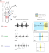
Serotonin application to the in vitro brain induces patterned laryngeal nerve activity across species. (A) A schematic view of the isolated male hindbrain viewed from the dorsal surface; anterior is up. (B–D) In response to serotonin bath application, patterned bouts of activity – compound action potentials (CAPs) – produced by vocal motor neurons in the laryngeal motor nucleus (LMN), can be recorded from the laryngeal nerve. In vitro patterns correspond to advertisement call patterns (Fig. 1) and are termed fictive calls. During the fast trill of a fictive call, a local field potential (LFP) can be recorded from nucleus DTAM of the rostral hindbrain. Laryngeal motor neurons are located in the posterior LMN (pink) and project to the larynx via the laryngeal nerve (4th root of cranial nerve IX-X). Interneurons in the anterior LMN (red) project to the LMN and DTAM. DTAM interneurons (red) provide monosynaptic, excitatory input to laryngeal motor neurons. Axons from DTAM cross the midline to innervate the contralateral DTAM. (B–D) Left: representative laryngeal nerve activity bouts in X. laevis, X. petersii and X. victorianus following bath application of serotonin to the isolated brain. Right: single fictive calls with individually labeled slow (blue) and fast (yellow) trills. (B) Bouts of activity recorded from the X. laevis laryngeal nerve include alternating slow and fast CAPs. (C) Bouts of activity recorded from the X. petersii laryngeal nerve also include alternating slow and fast CAPs; call duration and period are shorter than in X. laevis. (D) The in vitro laryngeal nerve activity pattern produced by X. victorianus includes only fast CAPs.
Fig. 4.
Fig. 4.
Comparison of the temporal features of in vitro laryngeal nerve activity. Boxplots illustrate the 25th and 75th percentiles; the horizontal line is the median, whiskers were calculated using Tukey's method and depict the most extreme data value that is not an outlier. Red indicates X. laevis (in vitro n=5), green X. petersii (in vitro n=6) and blue X. victorianus (in vitro n=3). *P<0.01, **P<0.0001 (GLMs with gamma distribution). (A) Slow pulse rates of in vivo calls do not differ significantly between X. laevis and X. petersii (P>0.05). (B) Fast pulse rates of in vivo calls do not differ significantly between X. laevis and X. petersii (P>0.05) or between X. victorianus and X. petersii (P>0.05). Xenopus laevis fast rate is significantly slower than that of X. victorianus. (C) Call duration differs significantly between all species. (D) Call period differs significantly between all species.
Fig. 5.
Fig. 5.
A LFP wave in DTAM corresponds to fast trill in all species. (A) Representative extracellular recording in DTAM (gray) and resulting LFP wave [blue, low-pass filtered at 5 Hz for X. laevis (n=5) and 20 Hz for X. petersii (n=6) and X. victorianus (n=2)] in response to serotonin application coincides with the fast trill (yellow) of the (B) fictive call simultaneously recorded from the laryngeal nerve (black). Wave onset and offset are depicted by open circles. (C) The DTAM LFP begins before the start of each fast trill and ends after the trill stops. The fast trill onset is subtracted from the corresponding wave onset, resulting in a negative number across species (median±s.d.: X. laevis: −160.0±115.8 ms, X. petersii: −59.2±13.6 ms, X. victorianus: −47.6±10.0 ms). The fast trill offset is subtracted from the wave offset, resulting in a positive value across species (X. laevis: 106.7±73.4 ms after, X. petersii: 49.4±8.3 ms, X. victorianus: 28.3±12.2 ms). The y-axis bins are percentage of total events and arrowheads indicate group medians. (D) Linear regression analysis of mean fictive fast trill duration versus each animal's corresponding mean DTAM LFP wave duration. Asterisks depict mean values of individual animals (R2=0.947, slope=0.6618, P<0.0001). (E) Linear regression analysis of inter-call period versus each animal's corresponding wave period. Asterisks depict mean values of individual animals (R2=0.991, slope=1.0028, P<0.0001).
Fig. 6.
Fig. 6.
The DTAM LFP wave is NMDA dependent. (A) Representative laryngeal nerve (black, top) and DTAM recordings (bottom, gray) following serotonin application in vitro in X. laevis (n=1), X. petersii (n=3) and X. victorianus (n=2). Fast trill is highlighted in yellow. (B) Laryngeal nerve (top) and DTAM (bottom) activity are abolished by 500 µmol l−1 APV added 15 min prior to serotonin application. (C) Laryngeal nerve (top) and DTAM (bottom) activity are restored following 1 h washout and re-application of serotonin.
Fig. 7.
Fig. 7.
Species-specific LFP wave duration and period are independent of connection to the caudal hindbrain. (A) Representative DTAM recording (gray) and LFP wave (blue) evoked by serotonin application in vitro. (B) Representative DTAM recording and LFP wave following transection caudal to DTAM that eliminates connections with the LMN. (C) Boxplot (as described for Fig. 2) illustrating LFP wave duration prior to [X. laevis (n=5), X. petersii (n=6) and X. victorianus (n=2)] and following caudal hindbrain transection [X. laevis (n=5), X. petersii (n=5) and X. victorianus (n=2, plotted as individual means)]. The darker shade represents intact brains (I) and the lighter shade, transected brains (T). Plus signs denote outliers. LFP wave durations are not significantly altered by transections (P>0.05, GLM). Xenopus laevis wave duration differs significantly from that of X. petersii and X. victorianus (P<0.0001 for each comparison, GLM). Means±s.d.: X. laevis: 444.2±29.6 ms intact, 465.3±28.9 ms transected; X. petersii: 108.7±7.3 ms intact, 118.0±2.3 ms transected; X. victorianus: 119.8, 105.4 ms intact, 113.2, 121.1 ms transected. (D) Boxplot illustrating LFP wave period prior to and following hindbrain transection. LFP wave periods are not significantly changed by transections (P<0.05, GLM). Xenopus laevis wave period differs significantly from that of X. petersii and X. victorianus (P<0.0001 for each comparison, GLM). Means±s.d.: X. laevis: 1115.6±188.8 ms intact, 1016.2±71.4 ms transected; X. petersii: 695.7±157.5 ms intact, 733.9±194.4 ms transected; X. victorianus: 372.9, 396.6 ms intact, 442.5, 463.3 ms transected.

References

    1. Albersheim-Carter J., Blubaum A., Ballagh I. H., Missaghi K., Siuda E. R., McMurray G., Bass A. H., Dubuc R., Kelley D. B., Schmidt M. F. et al. (2016). Testing the evolutionary conservation of vocal motoneurons in vertebrates. Respir. Physiol. Neurobiol. 224, 2-10. 10.1016/j.resp.2015.06.010 - DOI - PMC - PubMed
    1. Bongianni F., Mutolo D., Cinelli E. and Pantaleo T. (2014). Neural mechanisms underlying respiratory rhythm generation in the lamprey. Respir. Physiol. Neurobiol. 224, 1-10. 10.1016/j.resp.2014.09.003 - DOI - PubMed
    1. Brahic C. J. and Kelley D. B. (2003). Vocal circuitry in Xenopus laevis: telencephalon to laryngeal motor neurons. J. Comp. Neurol. 464, 115-130. 10.1002/cne.10772 - DOI - PMC - PubMed
    1. Browaldh N., Bautista T. G., Dutschmann M., and Berkowitz R. G. (2015). The Kölliker-Fuse nucleus: a review of animal studies and the implications for cranial nerve function in humans. Eur. Arch. Otorhinolaryngol. 273, 3505-3510. 10.1007/s00405-015-3861-9 - DOI - PubMed
    1. Bumbarger D. J., Riebesell M., Rödelsperger C. and Sommer R. J. (2013). System-wide rewiring underlies behavioral differences in predatory and bacterial-feeding nematodes. Cell 152, 109-119. 10.1016/j.cell.2012.12.013 - DOI - PubMed

Publication types