. 2017 Feb;469(2):263-277.
doi: 10.1007/s00424-016-1929-z. Epub 2016 Dec 24.

Differential distribution and functional impact of BK channel beta1 subunits across mesenteric, coronary, and different cerebral arteries of the rat

Affiliations

Differential distribution and functional impact of BK channel beta1 subunits across mesenteric, coronary, and different cerebral arteries of the rat

Guruprasad Kuntamallappanavar et al. Pflugers Arch. 2017 Feb.

Abstract

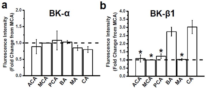
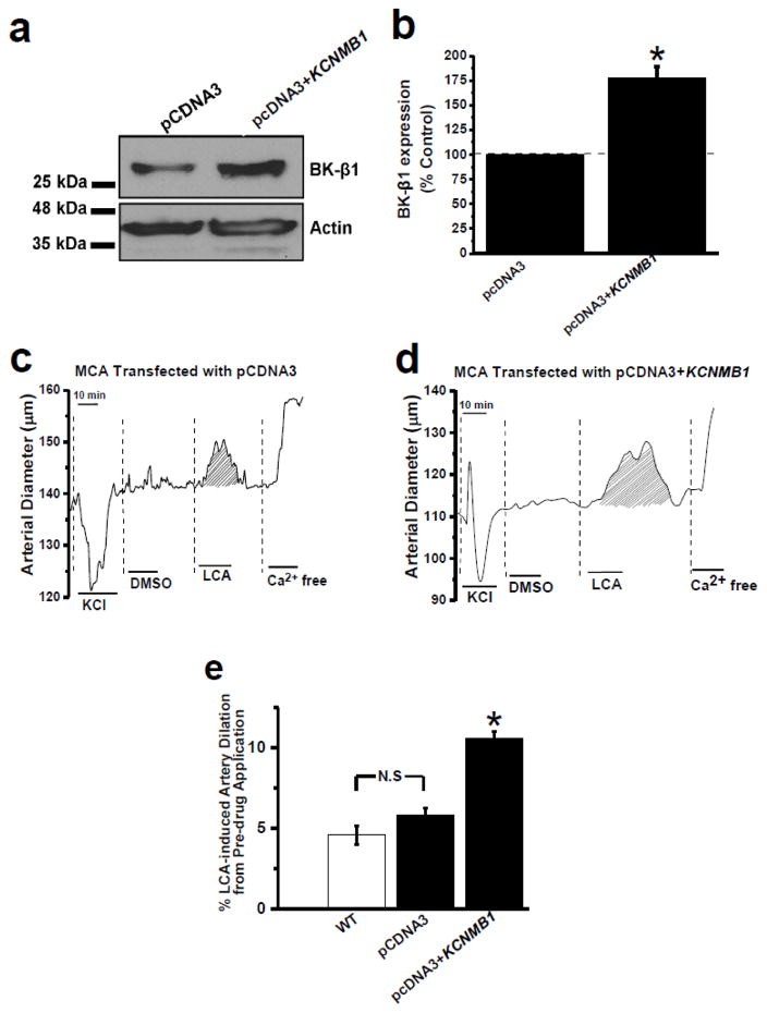
Large conductance, Ca2+i- and voltage-gated K+ (BK) channels regulate myogenic tone and, thus, arterial diameter. In smooth muscle (SM), BK channels include channel-forming α and auxiliary β1 subunits. BK β1 increases the channel's Ca2+ sensitivity, allowing BK channels to negatively feedback on depolarization-induced Ca2+ entry, oppose SM contraction and favor vasodilation. Thus, endothelial-independent vasodilation can be evoked though targeting of SM BK β1 by endogenous ligands, including lithocholate (LCA). Here, we investigated the expression of BK β1 across arteries of the cerebral and peripheral circulations, and the contribution of such expression to channel function and BK β1-mediated vasodilation. Data demonstrate that endothelium-independent, BK β1-mediated vasodilation by LCA is larger in coronary (CA) and basilar (BA) arteries than in anterior cerebral (ACA), middle cerebral (MCA), posterior cerebral (PCA), and mesenteric (MA) arteries, all arterial segments having a similar diameter. Thus, differential dilation occurs in extracranial arteries which are subjected to similar vascular pressure (CA vs. MA) and in arteries that irrigate different brain regions (BA vs. ACA, MCA, and PCA). SM BK channels from BA and CA displayed increased basal activity and LCA responses, indicating increased BK β1 functional presence. Indeed, in the absence of detectable changes in BK α, BA and CA myocytes showed an increased location of BK β1 in the plasmalemma/subplasmalemma. Moreover, these myocytes distinctly showed increased BK β1 messenger RNA (mRNA) levels. Supporting a major role of enhanced BK β1 transcripts in artery dilation, LCA-induced dilation of MCA transfected with BK β1 complementary DNA (cDNA) was as high as LCA-induced dilation of untransfected BA or CA.

Keywords: BK β1 subunit; Cerebral artery myocyte; KCNMB1; Lithocholate; MaxiK channel; Vascular smooth muscle; Vasodilation.


Figures

Fig. 1
Fig. 1. Dilatation by lithocholate, a BK β1 ligand, is highest in basilar and coronary arteries
(a) Representative arterial diameter trace from rat MCA pressurized in vitro. After development of myogenic tone, the artery was probed with 60 mM KCl to test maximal contraction. After KCl washout, the artery chamber was perfused with PSS that contained vehicle (DMSO, 0.1% v/v). Then, artery was probed with 45 μM LCA, which constitutes EC50 for LCA activation of β1-containing BK channels [15]. Vertical lines underscore the beginning of perfusion with any given solution. The shadowed area shows the AUC corresponding vasodilation by LCA, which is fully reversible. An identical protocol was used with ACA, PCA, BA, MA and CA. (b) Bar graph demonstrating that LCA-induced dilation of BA and CA is drastically larger that evoked on ACA, MCA, PCA or MA; n=3–7, *Different from BA, P<0.05; ACA, anterior cerebral artery; MCA, middle cerebral artery; PCA, posterior cerebral artery; BA, basilar artery; MA, mesenteric artery; CA, coronary artery; LCA, lithocholate.
Fig. 2
Fig. 2
Representative BK channel recordings at single-channel resolution obtained from inside-out membrane patches from rat ACA (a), MCA (b), PCA (c), BA (d), MA (e) and CA (f) myocytes. These records show that LCA-induced increase in channel steady-state activity (NPo) is larger in BA and CA myocytes than in myocytes isolated from another arteries. Vm= −40 mV; Ca2+i=3 μM. Recordings were obtained before (top trace on each panel), during (middle trace) and after (bottom trace) patch exposure to 45 μM LCA. Arrows indicate the baseline (all channels closed); ACA, anterior cerebral artery; MCA, middle cerebral artery; PCA, posterior cerebral artery; BA, basilar artery; MA, mesenteric artery; CA, coronary artery; LCA, lithocholate.
Fig. 3
Fig. 3. Lithocholate-induced increase in BK channel activity is significantly larger in myocytes from coronary and basilar than in myocytes from another arteries
Bar graph showing averaged NPo ratios in presence and absence of 45 μM LCA from ACA, MCA, PCA, BA, MA and CA myocyte BK channels; n=5–10, *Different from BA, P<0.05; ACA, anterior cerebral artery; MCA, middle cerebral artery; PCA, posterior cerebral artery; BA, basilar artery; MA, mesenteric artery; CA, coronary artery; LCA, lithocholate; N, number of functional channels present in the membrane patch; Po, single channel open probability.
Fig. 4
Fig. 4. Steady-state BK channel activity in basilar and coronary artery is higher than in myocytes isolated from other intracranial arteries and from mesenteric artery
(a) Averaged Po/Po max-V curves of BK channels in myocytes isolated from MA, CA and the different branches of the Willis’ circle. (b) Bar graph showing averaged Vhalf values. N=3–4; *p<0.05, different from myocytes from BA. ACA, anterior cerebral artery; MCA, middle cerebral artery; PCA, posterior cerebral artery; BA, basilar artery; MA, mesenteric artery; CA, coronary artery; Po, single channel open probability.
Fig. 5
Fig. 5
Representative images of individual myocytes isolated from different cerebral arteries. Myocytes were subjected to immunofluorescence staining against (a) BK channel-forming α (green) and (b) accessory β1 (red) subunits. ACA, anterior cerebral artery; MCA, middle cerebral artery; PCA, posterior cerebral artery; BA, basilar artery; MA, mesenteric artery; CA, coronary artery.
Fig. 6
Fig. 6. BK α protein levels in myocyte membranes are similar across the different arteries whereas BK β1 subunit levels in the plasmalemma-subplasmalemma region are significantly higher in basilar and coronary arteries than in other arteries
Averaged fluorescence intensity (fold difference compared to MCA) of BK α (a) and BK β1 (b) subunit-associated signal in myocytes from intracranial arteries, MA and CA. Each bar value represents an average of 22–74 myocytes isolated from 2–3 different rats for each vascular bed. *p<0.05, when compared to expression levels in BA myocytes. ACA, anterior cerebral artery; MCA, middle cerebral artery; PCA, posterior cerebral artery; BA, basilar artery; MA, mesenteric artery; CA, coronary artery.
Fig. 7
Fig. 7. BK β1 mRNA transcript level is higher in basilar and coronary arteries than in other arteries
Bar graph showing BK β1 mRNA levels measured using quantitative real-time RT-PCR across the different intracranial arteries, MA and CA. mRNA level in a given artery was normalized to expression of an endogenous control gene, i.e., Tata Binding Protein (TBP) in the same artery. Then, normalized mRNA expression levels in any given artery were further normalized to mRNA expression levels in MCA. n=8–11; *p<0.05, different from expression level in BA. ACA, anterior cerebral artery; MCA, middle cerebral artery; PCA, posterior cerebral artery; BA, secondary branches of basilar artery; MA, quaternary branches of mesenteric artery; CA, coronary artery.
Fig. 8
Fig. 8. Overexpression of BK β1 protein in middle cerebral artery significantly increases the dilatory response of this artery to lithocholate
(a) Representative Western blots showing expression of BK β1 protein in arteries transfected with “empty” vector (pcDNA3; left lane) or pcDNA3+BK-β1 cDNA insert (right lane). Actin levels were used as control. (b) Electroporation of MCA with pcDNA3+cDNA BK β1 leads to significantly higher levels of BK β1 protein when compared to control (pcDNA3). (c) Diameter trace from de-endothelialized, pressurized rat MCA transfected with “empty” vector (pcDNA3). (d) Diameter trace from de-endothelialized, pressurized MCA transfected with BK β1 cDNA. (e) Averaged data showing that in rat MCA transfected with BK β1 cDNA, application of 45 μM LCA evokes an arterial dilation that is significantly higher than that from arteries transfected with empty vector (pcDNA3). n=4–5; *P<0.05, when compared to arteries transfected with pcDNA3 alone; MCA, middle cerebral artery; LCA, lithocholate.

Similar articles

Cited by

References

    1. Amberg GC, Bonev AD, Rossow CF, Nelson MT, Santana LF. Modulation of the molecular composition of large conductance, Ca(2+) activated K(+) channels in vascular smooth muscle during hypertension. The Journal of clinical investigation. 2003;112:717–724. doi: 10.1172/jci18684. - DOI - PMC - PubMed
    1. Amberg GC, Santana LF. Downregulation of the BK channel beta1 subunit in genetic hypertension. Circulation research. 2003;93:965–971. doi: 10.1161/01.RES.0000100068.43006.36. - DOI - PubMed
    1. Arab L, Liebeskind DS. Tea, flavonoids and stroke in man and mouse. Archives of biochemistry and biophysics. 2010;501:31–36. doi: 10.1016/j.abb.2010.03.015. - DOI - PubMed
    1. Bannister JP, Adebiyi A, Zhao G, Narayanan D, Thomas CM, Feng JY, Jaggar JH. Smooth muscle cell alpha2delta-1 subunits are essential for vasoregulation by CaV1.2 channels. Circulation research. 2009;105:948–955. doi: 10.1161/circresaha.109.203620. - DOI - PMC - PubMed
    1. Bao L, Cox DH. Gating and ionic currents reveal how the BKCa channel’s Ca2+ sensitivity is enhanced by its beta1 subunit. The Journal of general physiology. 2005;126:393–412. doi: 10.1085/jgp.200509346. - DOI - PMC - PubMed

Publication types

MeSH terms

Substances

LinkOut - more resources