Review
. 2017 Feb;147(2):119-147.
doi: 10.1007/s00418-016-1526-4. Epub 2016 Dec 23.

Eukaryotic protein glycosylation: a primer for histochemists and cell biologists

Affiliations
Review

Eukaryotic protein glycosylation: a primer for histochemists and cell biologists

Anthony Corfield. Histochem Cell Biol. 2017 Feb.

Abstract

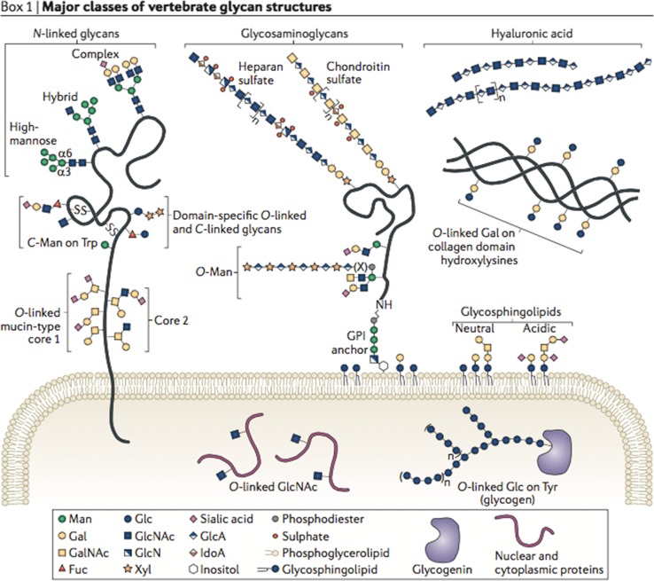
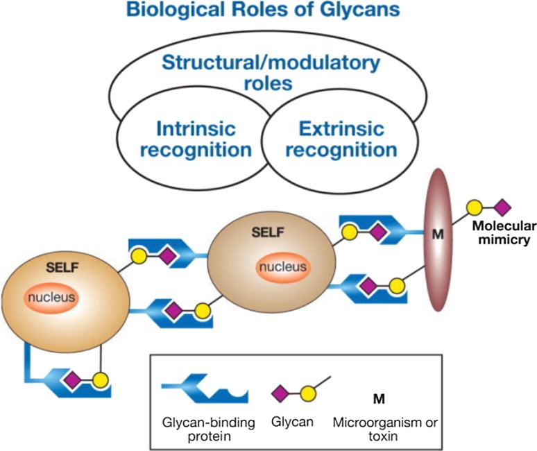
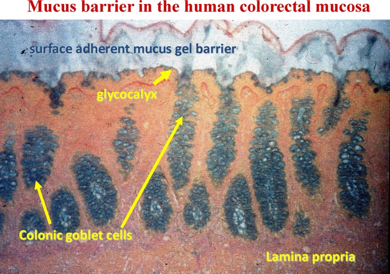
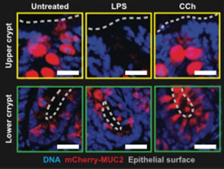
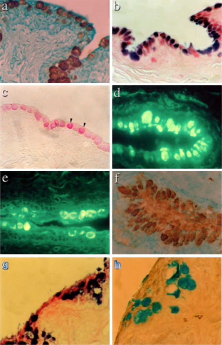
Proteins undergo co- and posttranslational modifications, and their glycosylation is the most frequent and structurally variegated type. Histochemically, the detection of glycan presence has first been performed by stains. The availability of carbohydrate-specific tools (lectins, monoclonal antibodies) has revolutionized glycophenotyping, allowing monitoring of distinct structures. The different types of protein glycosylation in Eukaryotes are described. Following this educational survey, examples where known biological function is related to the glycan structures carried by proteins are given. In particular, mucins and their glycosylation patterns are considered as instructive proof-of-principle case. The tissue and cellular location of glycoprotein biosynthesis and metabolism is reviewed, with attention to new findings in goblet cells. Finally, protein glycosylation in disease is documented, with selected examples, where aberrant glycan expression impacts on normal function to let disease pathology become manifest. The histological applications adopted in these studies are emphasized throughout the text.

Keywords: Eukaryocyte; Glycans; Glycoprotein; Glycosylation; Histochemistry; Mucin.


Figures

Fig. 1
Fig. 1
Classes of vertebrate glycan structures. Membrane and secreted proteins have N-glycan, GlcNAc to asparagine as oligomannose, complex or hybrid forms, or O-glycans linked through GalNAc to serine/threonine with eight core structures and extension. Glycosaminoglycans have a core linkage tetrasaccharide to protein, with subsequent disaccharide repeats and characteristic sulphation patterns. They may be secreted, transmembrane or GPI-anchored. Hyaluronan is not linked to a protein. O-Mannosyl residues may be extended. O-Glucose and O-fucose are found in EGF domains of some proteins. C-Mannose is attached to protein tryptophan side chains. Single β-O-GlcNAc is found on many cytosolic and nuclear proteins. The collagen disaccharide is linked to hydroxylysine and through galactose. Glycogen is linked through glucose unit to a tyrosine in glycogenin. Glycosphingolipids contain glycans linked to a ceramide carrier; from Moremen et al. (2012), with permission
Fig. 2
Fig. 2
mPAS detection of sialic acids in human colon. Mucus stored in goblet cell thecae. Staining of the colonic mucosa with the mild periodic acid-Schiff reaction stains non-O-acetylated sialic acids and demonstrates the location of the mucus prior to secretion; from Corfield (2011), with permission
Fig. 3
Fig. 3
Biological roles of glycans. A general classification of the biological roles of glycans is presented, emphasizing the roles of organism proteins in the recognition of glycans; from Varki and Lowe (2009), with permission
Fig. 4
Fig. 4
Intestinal mucus barrier. Mucosal sample stained histochemically with Alcian Blue and Van Gieson counterstain after stabilizing the mucus gel layer by cryostat and molten agar. The image shows the secreted gel layer, glycocalyx, goblet cells and lamina propria from human colon; from Pullan et al. (1994), with permission
Fig. 5
Fig. 5
Sentinel goblet cells in the human colon. Goblet cells responsive to Toll-like receptor ligands (TLR ligands) are located in the upper crypt. Cryosections in colonic explants treated with TLR ligands and visualized by confocal microscopy. Red MUC2; blue DNA. Upper crypt (yellow boxes) or lower crypt (green boxes). A dashed grey line shows the epithelial surface. Scale bars 20 mm From Birchenough et al. Science 352:1535–1542 (2016). Reprinted with permission from the American Association for the Advancement of Science (AAAS)
Fig. 6
Fig. 6
Major glycan and glycoconjugate classes of human sperm glycocalyx. Monosaccharides are coded by coloured symbols shown in the figure. Proteins and lipids are grey, except cholesterol, and the lipids of glycosphingolipids. Mammals synthesize most glycans with a dozen different monosaccharide-building blocks; some of these monosaccharides can be further modified by sulphation and/or acetylation; from Tecle and Gagneux (2015), with permission
Fig. 7
Fig. 7
Mucin expression in the human cornea and conjunctiva. Schematic of the location of mucins in the corneal and conjunctival epithelium. The membrane-associated mucins MUC1, MUC4 and MUC16 at the apical cell membrane glycocalyx, and the secreted mucin MUC5AC in goblet cell vesicles; from Gipson (2004), with permission
Fig. 8
Fig. 8
Loss of MUC2 and sulpho-Lewisa in ulcerative colitis. Detection of MUC2 glycoprotein by immunostaining with the Lum2–3 antibody for normal (a) and clinically severe UC (c). The same specimens were also tested for MUC2 mRNA for normal (b) and UC (d). Immunostaining for MUC2 in mucosa adjacent to an ulcer (e) showed reduced staining compared with normal intact mucosa (f). Detection of sulpho-Lea with the F2 antibody showed localization throughout the mucosa, including the mucus gel layer (g). Sulpho-Lea staining was preserved in mild colitis (i), but was depleted at the luminal surface and upper crypts in severe colitis (h). In situ hybridization samples were counterstained with toluidine blue and immunohistological sections with haematoxylin; from Longman et al. (2006), with permission
Fig. 9
Fig. 9
Depletion of goblet cells in necrotizing enterocolitis. The trefoil factor family peptide TFF3 is shown with immunostaining (a) in normal neonatal colon (×10 magnification), with staining of all goblet cells and the lumen (arrow). Normal neonatal colon at ×40 magnification shows a granular pattern of TFF3 in goblet cells (b). Reduction of goblet cells in a patient with NEC is shown at ×10 (c) and ×40 (d) magnification and reveals empty goblet cells (arrows in d), in particular at the surface epithelium; from Vieten et al. (2005), with permission
Fig. 10
Fig. 10
Histology of normal and KCS canine conjunctival tissues. Tissue sections from normal canine conjunctiva were stained with the anti-MUC5AC antibody 21M1 (a); Alcian Blue/PAS (b); mild-PAS (c); SNA for α(2–6)-linked sialic acids (d); WGA (e) and antibody TKH2 against sialyl-Tn (sialyl-α(2–6)GalNAc) (f). Magnification is ×40 in all cases. Histology of KCS canine conjunctival tissues. Tissue sections from animals with KCS stained with periodic acid-Schiff/Alcian Blue (g) and high iron diamine/Alcian Blue (h). Magnification is ×40 in both cases. Taken from Corfield et al. (2005) with permission

References

    1. Aebi M. N-Linked protein glycosylation in the ER. Biochim Biophys Acta. 2013;1833:2430–2437. doi: 10.1016/j.bbamcr.2013.04.001. - DOI - PubMed
    1. Agawa S, Jass JR. Sialic acid histochemistry and the adenoma–carcinoma sequence in colorectum. J Clin Pathol. 1990;43:527–532. doi: 10.1136/jcp.43.7.527. - DOI - PMC - PubMed
    1. Albenberg LG, Lewis JD, Wu GD. Food and the gut microbiota in inflammatory bowel diseases: a critical connection. Curr Opin Gastroenterol. 2012;28:314–320. doi: 10.1097/MOG.0b013e328354586f. - DOI - PMC - PubMed
    1. Albert TK, Laubinger W, Müller S, Hanisch FG, Kalinski T, Meyer F, Hoffmann W. Human intestinal TFF3 forms disulfide-linked heteromers with the mucus-associated FCGBP protein and is released by hydrogen sulfide. J Proteome Res. 2010;9:3108–3117. doi: 10.1021/pr100020c. - DOI - PubMed
    1. Alemka A, Corcionivoschi N, Bourke B. Defense and adaptation: the complex inter-relationship between Campylobacter jejuni and mucus. Front Cell Infect Microbiol. 2012;2:15. doi: 10.3389/fcimb.2012.00015. - DOI - PMC - PubMed

LinkOut - more resources