Review
. 2017 Mar;40(3):181-193.
doi: 10.1016/j.tins.2016.11.006. Epub 2016 Dec 22.

Secondary Motor Cortex: Where 'Sensory' Meets 'Motor' in the Rodent Frontal Cortex

Affiliations
Review

Secondary Motor Cortex: Where 'Sensory' Meets 'Motor' in the Rodent Frontal Cortex

Florent Barthas et al. Trends Neurosci. 2017 Mar.

Abstract

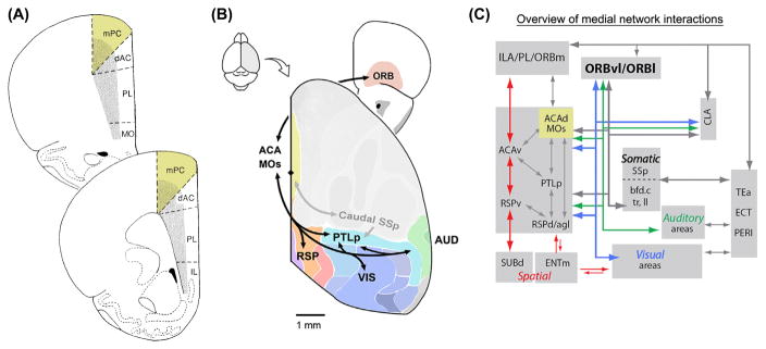
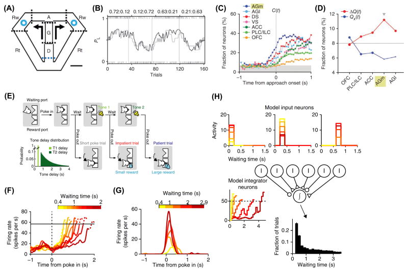
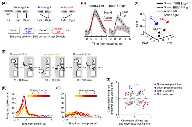
In rodents, the medial aspect of the secondary motor cortex (M2) is known by other names, including medial agranular cortex (AGm), medial precentral cortex (PrCm), and frontal orienting field (FOF). As a subdivision of the medial prefrontal cortex (mPFC), M2 can be defined by a distinct set of afferent and efferent connections, microstimulation responses, and lesion outcomes. However, the behavioral role of M2 remains mysterious. Here, we focus on evidence from rodent studies, highlighting recent findings of early and context-dependent choice-related activity in M2 during voluntary behavior. Based on the current understanding, we suggest that a major function for M2 is to flexibly map antecedent signals such as sensory cues to motor actions, thereby enabling adaptive choice behavior.

Keywords: action selection; motor planning; prefrontal cortex; rodents; sensorimotor transformation; voluntary behavior.


Figures

Figure 1
Figure 1. Afferent and efferent connectivity
(A) Coronal view of rat mPFC subdivisions including M2, labeled as medial precentral cortex (mPC) in this diagram. Other mPFC regions are the dorsal anterior cingulate cortex (dAC), prelimbic cortex (PL), and infralimbic cortex (IL). (B) The medial subnetwork, a cluster of connected cortical regions identified in a study of mouse brain connectivity. Sensory regions including visual cortex (VIS), auditory cortex (AUD) and caudal primary somatosensory cortex (SSp) are connected to association regions including the retrosplenial (RSP), parietal (PTLp), anterior cingulate/secondary motor (ACA, MOs), and orbital areas (ORB). (C) Summary of interactions between the medial subnetwork and prefrontal regions including infralimbic (ILA), and prelimbic (PL) areas, parahippocampal structures including dorsal subiculum (SUBd), medial entorhinal area (ENTm), and other regions including claustrum (CLA), temporal association area (TEa), ectorhinal area (ECT), and perirhinal area (PERI). Part A adapted from [17]. Parts B and C adapted from [8].
Figure 2
Figure 2. Choice-related activity in M2 is early
(A) Onset of choice signals in M2: In each trial, the rat goes through 5 stages: delay (D), go (G), approach to reward (A), reward (Rw), and return (Rt). At the end of go stage, the animal has a choice between two arms. Each arm is assigned to reward with a certain probability. The probabilities change after a block of ~40 trials. (B) Probability of choosing left (gray line) over a behavioral session. Actual performance is compared to a reinforcement-learning model (black line). Top: reward probabilities for the two arms. (C) Time courses of neural signals for the upcoming choice for five frontal cortical and two striatal regions. M2 is denoted as the medial agranular cortex (AGm). AGl, lateral agranular cortex. DS, dorsal striatum. VS, ventral striatum. ACC, anterior cingulate cortex. PLC/ILC, prelimbic/infralimbic cortex. OFC, orbitofrontal cortex. (D) Regional specificity for coding of action value Qx(t) or decision value ΔQx(t) during the last 1 s of the delay stage. The triangle indicates a significant difference between the two fractions (χ2-test, P < 0.05). (E) Cortical circuit mechanisms: The rat initiates a trial with a nose poke. Waiting longer results in a larger reward. Inset, distribution of waiting times. (F) Example M2 neuron with ramp-to-threshold activity. (G) Example M2 neuron with transient predictive activity. (H) An integrator model. Inset, the waiting time histogram generated using the model. Parts (A – D) adapted from [45]. Parts (E – H) adapted from [62].
Figure 3
Figure 3. Choice-related activity in M2 is task-specific
(A) Context specificity: The mouse made left or right licks in response to an auditory cue. Trials were organized into blocks, each with a distinct set of stimulus-response contingencies. When performance reached a criterion, a new block began with different contingencies. (B) Example M2 neuron with context-dependent activity. Gray shading, 95% confidence intervals. (C) Neuronal circuit trajectories were calculated from the trial-averaged activity of a 56-cell ensemble using demixed principal component analysis. PC, principal component. (D) Action specificity: The waiting task (Fig. 2E) implemented with interleaving blocks involving different operant actions: nose-poke and lever-press. (E) Example M2 neuron with nose-poke-specific predictive activity. (F) The cell from (E) during lever-press trials. (G) Summary of nose-poke- and lever-press-specific predictive activities. Each circle represents one neuron. Parts (A – C) adapted from [44]. Parts (D – G) adapted from [62].

Similar articles

Cited by

References

    1. Brecht M. Movement, confusion, and orienting in frontal cortices. Neuron. 2011;72:193–196. - PubMed
    1. Paxinos G, Franklin K. The mouse brain in stereotaxic coordinates: compact second edition. San Diego: Academic; 2003.
    1. Lein ES, et al. Genome-wide atlas of gene expression in the adult mouse brain. Nature. 2006;445:168–176. - PubMed
    1. Donoghue JP, Wise SP. The motor cortex of the rat: cytoarchitecture and microstimulation mapping. J Comp Neurol. 1982;212:76–88. - PubMed
    1. Brecht M, et al. Organization of rat vibrissa motor cortex and adjacent areas according to cytoarchitectonics, microstimulation, and intracellular stimulation of identified cells. J Comp Neurol. 2004;479:360–373. - PubMed

Publication types